02.12.2019
After visiting the dental office, sometimes patients discover that the hole does not heal for a long time after tooth extraction. It bleeds a lot and hurts for unknown reasons. The bleeding cannot be stopped and prevents normal eating. It is rough food that often leads to injury to the socket, and when it festeres, an unpleasant odor begins to emanate from the mouth. Re-infection of surrounding tissues, development of inflammation or alveolitis of the socket may occur, as complications after tooth extraction. It is important to find out why the postoperative process prevents the wound from healing quickly and what are the provoking factors?
Why perforation occurs and how to avoid it
Such consequences can be avoided by properly planning removal using a CT scanner and following gentle removal protocols .
There is a group of patients with the anatomy of roots whose apices penetrate into the sinus, and if such patients are treated as a standard removal protocol, perforation and all associated complications will certainly occur. Only this is visible in the image created by a computer tomograph in a special ENT mode, and this is not possible in any clinic. In our Center this can be done with the latest SIRONA-SIEMENS equipment and the GALILEOS diagnostic software package.
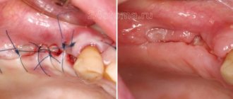
Having studied the CT image, the surgeon must be prepared for an emergency situation. If perforation does occur, it is important to perform immediate microsurgical closure in a sterile operating room to avoid the development of an inflammatory process.
Even an experienced doctor without a good image on a modern expensive tomograph may not notice the details and skip the patient to a regular dental surgeon, not pay attention to the anatomical features and not carry out the necessary actions. The patient will find out about this later by the characteristic symptoms:
- passage of air, whistling and squelching in the socket of the extracted tooth;
- foamy, bloody or yellowish nasal discharge;
- strange, unreasonable organic odors in the nose and mouth,
- change in voice timbre (nasality).
If you do not carry out timely emergency closure of the anastomosis after tooth extraction, the hole itself may never heal . The gum tissues heal, epithelialize, and shrink, but the bones in the area of such a fistula between the nose and mouth never grow together due to the difference in the growth periods of the bone and gum, the gum will instantly take up all the free space, the slowly growing bone simply will not have time to fill the defect.
Within 2-3 weeks, a thin fistulous tract forms from the oral cavity to the sinus. In this case, the diameter of the hole is reduced naturally due to the formation of scar tissue; the symptoms of perforation may temporarily disappear or not appear. But this does not prevent infection from entering the sinus due to food entering the sinus.
As a result, the sinus becomes inflamed, signs of unilateral sinusitis appear, which should confuse a thinking patient (sometimes there are coincidences, but sinusitis-sinusitis, which comes as an accompaniment of the flu or a cold, is always bilateral).
Other causes of perforation
The remaining 5% of clinical cases of sinus perforation injuries occur for the following reasons::
- Not ideal endodontic treatment of tooth canals - the therapist treated the canals, was in a hurry, used excessive force when filling the canal, and under the influence of a rotating canal filler with pressure, the filling material fell beyond the tooth root. We often encounter cases where even fragments of endodontic instruments are sometimes found in the thickness of the filling material, which jam in the canal and tear into pieces, leaving metal fragments in different places of the tooth root;
- Punitive sinus lifting - an inexperienced or rude doctor does not feel the density of the tissues, breaks through the Schneiderian membrane, the bone material is forced into the gap and enters the sinus;
- Author's implantation - it happens that due to the lack of skills in performing a sinus lift and the desire to restore the missing bone, a decision is made - the implant is placed in the residual bone that exists, without bone grafting. Result: the implant completely fails or partially comes out into the sinus.
A fragment of an endodontic instrument in a tooth canal
Alveolitis
Inflammation of the walls of the socket.
- In the initial stage of alveolitis, an intermittent aching pain appears in the socket, which intensifies while eating;
- The general condition is not disturbed, the body temperature is normal;
- The tooth socket is only partially filled with a loose, disintegrating blood clot.
With further development of inflammation:
- The pain intensifies and becomes constant;
- Transmitted to the ear, temple, corresponding half of the head;
- The general condition worsens, malaise and fever appear.
Why you should entrust your treatment to the ENT Department of Dentistry
ENT dentistry combines two areas of medical services and treatment options - otolaryngology and dentistry . This is a modern format for the clinic’s work on comprehensive rehabilitation in the treatment of traumatic and inflammatory processes in the upper jaw, penetrating into the maxillary sinus or passing along its borders.
The Center for Private Dentistry “Doctor Levin” has been specializing in providing care to patients with combined ENT and dental pathology for many years. Medical and surgical treatment programs are carried out by candidates of medical sciences, maxillofacial surgeons with ENT training.
Statistics for the 20 years of work of the Center and for the last 10 years of work of the Department, unfortunately, are inexorable. The main providers of patients with complications are our colleagues who are caught in the dead end of an unsuccessful treatment plan. We are grateful to our colleagues who do not hesitate to send us patients with ENT complications, despite the reputational damage. It is always possible to avoid a dramatic outcome if you stop treatment in time.
Sharp edges of the alveoli
Socket pain can be caused by protruding sharp edges of the socket, which injure the mucous membrane located above them. Pain appears 1-2 days after tooth extraction , when the edges of the gums above the socket begin to approach each other.
- The pain intensifies during chewing and when touching the gums;
- When you feel the hole with your finger, a protruding sharp edge of the bone is detected, and a sharp pain occurs.
This pain can be distinguished from the pain of alveolitis by the absence of inflammation in the socket area and the presence of an organizing blood clot in it.
How we treat
Treatment is carried out only comprehensively and strives to be carried out in one visit , simultaneously:
- Hygienic cleaning and dental treatment We prepare the oral cavity for sterile surgical work. We treat compromised roots. Operations are performed only in a sanitized oral cavity to avoid re-infection.
- The operation is carried out in the surgical department, subject to all conditions of sterile operating rooms. Ultrasound removes inflammatory processes, removes roots that provoked or supported inflammation, eliminates all inflammatory elements, removes cysts, polyps, mucoceles, and foreign bodies.
- Orthopedics Temporary crowns or any other orthopedic elements are fixed to mask the work performed. We do everything to avoid sending the patient home without teeth!
We do not welcome radical and punitive surgery!
Rest assured that after surgery we will never keep you in the hospital unless absolutely necessary.
Levin Dmitry Valerievich Chief physician and founder of the Doctor Levin center
Associated symptoms
The fistula is impossible to miss. Its end looks like a tumor that is filled with pus. It can be red, yellow or gray. The surrounding tissues are usually inflamed.
Possible symptoms also include:
- bad breath,
- increase in body temperature,
- pain when eating, brushing teeth,
- foreign body sensation
- itching and burning of gums.
If these symptoms appear, you should consult your doctor.
Hospitalization
The day hospital at our Center is a post-operative support service for patients. Necessary if the patient has concomitant cardiac issues, such as hypertension, arrhythmia, AOS, etc.
Gentle ultrasound surgical protocols for low-impact PiezoSurgery operations in combination with microscopic surgery performed by operating teams consisting of pairs of the most experienced surgeons at our Center allow you to carry out treatment so delicately that you will not need mandatory hospitalization, which is so popular in Moscow hospitals.
All operations are performed only in medicated sleep, without pain or nervous overload.
The treatment will be carried out with respect for your personal time, in a short time and at a comfortable time, the operating teams and anesthesiology department work seven days a week and holidays.
Toothache after caries treatment
Constant pain after filling can occur for many reasons. A common cause of discomfort is the application of an insulating lining made of glass ionomer cement (GIC) and filling on the same day with a composite. The polymerization times of GIC and composite differ, which can result in displacement of the gasket and its pressure on the pulp.
If the pain is short-term in nature as a reaction to cold, sour or sweet and quickly passes after removing the irritant, it may be due to the fact that during the treatment there was a mechanical effect on the pulp. After 2-3 days, the pulp will return to normal functioning and the pain will stop.
If you experience pain when biting, you need your dentist to grind the filling to fit your bite.
Complications of a fistula on the gum
If the pathological process is started, the following consequences may occur:
- Tooth loss.
- Damage to the soft tissues of the face.
- Infection of the periosteum and death of blood vessels, as a result of which you can lose several teeth at once.
It should be borne in mind that signs of a fistula do not appear immediately. Especially if the reason for its appearance is a medical error. At first, the pathological process is hidden and asymptomatic. Therefore, if a patient has doubts about the doctor’s qualifications during a dental procedure, after the procedure he should be especially attentive to the condition of the oral cavity and, at the slightest suspicion of the development of purulent inflammation, seek medical help.
Reasons for appearance
It is not precisely determined why osteophytes appear on the gums. But factors contributing to the disease include:
- Hereditary predisposition;
- frequent inflammation, purulent processes leading to atrophy, deformation of the jaw bone and nearby tissues;
- injuries of the dental system, especially accompanied by fractures of the facial part of the skull with improper reposition of bone fragments;
- complex tooth extraction;
- advanced periodontitis, periodontal disease;
- bite pathologies;
- congenital abnormalities of the jaw.
Often the pathology appears in childhood or adolescence. Also, the appearance of jaw growths may be associated with dysfunction of the endocrine system.
What is a fistula on the gum
When inflammation develops due to the presence of a dental disease or due to poor quality treatment, a cavity appears near the root of the tooth filled with pus mixed with blood. A fistula is a formation that is a canal between this cavity and the surface of the gum. It has access to the oral cavity. In other words, this is a kind of tube through which pus flows out from the source of infection.
A fistula looks like a small hole in the gum located at the base of the tooth. The color of the formation differs from healthy tissue. It may be pink or red in color. Healthy teeth are not affected by infection.
The fistula occurs gradually and can be detected at an early stage. First, a swelling appears on the gum, then a formation resembling an abscess or pimple. After this, the seal is opened, and purulent or mucous contents come out of it. After some time, the wound scars. However, the canal does not disappear anywhere, so several times a year there are exacerbations with the formation of a swelling and its opening.
Causes of fistula on the gum
The main cause of a fistula is a chronic purulent process in the soft or bone tissues of the oral cavity. An infection (most often bacterial) can enter the periodontium (a complex of collagen fibers and connective tissues located in the crevice-like space between the cementum of the tooth root and the alveolar plate) due to:
- advanced caries,
- pulpitis,
- perforation of the tooth root,
- chronic periodontitis,
- incorrectly performed or unfinished dental treatment (poor installation of a filling),
- delayed growth of wisdom teeth,
- inflammation of the cyst.
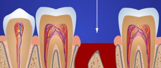
The most common dental procedure after which a fistula develops is root canal filling. Of course, if it is carried out poorly. Canal filling is usually carried out in the treatment of pulpitis, periodontitis, and also as a preparatory procedure for dental prosthetics. The most common dental mistake is filling not to the top of the root. However, even if the canal is sealed to its entire length, pores may form if the filling material is not placed conscientiously and tightly. In both cases, an infection can develop in the resulting voids, which will extend beyond the tooth and cause an inflammatory process.
Also, during cleaning of the canals before filling them with instruments, the root of the tooth may be damaged (such damage is called perforation).