Alveolitis is an inflammation of the socket (alveoli) left after tooth extraction. This pathology does not always develop; its development depends on many factors. The disease is characterized by severe pain in the area of the hole formed after surgery, general weakness, fever, headache, enlarged submandibular lymph nodes, bad breath and other unpleasant symptoms.
Alveolitis is not only physically painful, but also a dangerous disease. In the absence of proper treatment for several days, the inflammatory process can result in limited osteomyelitis, purulent melting of the jaw bone, and then surgical intervention will be required again.
With timely diagnosis of pathology and proper sanitation of the socket, the treatment prognosis is favorable. The main thing is to detect the symptoms of the disease in time and begin to treat it.
Reason 1: the bleeding does not stop
Heavy bleeding is the most common complication. Usually occurs in people with bleeding disorders. Before removing a tooth, no tests are required; it is for this reason that the likelihood of this particular complication is high. If you know about your problem, be sure to tell your doctor. In this case, the specialist may prescribe medications to stop the bleeding or apply sutures. If the blood does not stop or suddenly “gushes out like a fountain” shortly after the operation, immediately go to the hospital or call an ambulance.
Injuries during tooth extraction
They are considered a complication, most often appear on the upper jaw, the highest risk is when removing premolars and molars. If removal is difficult, there is a risk of perforation of the floor of the maxillary sinus. It becomes damaged, and a hole appears in the bony septum between the nasal and oral cavities. This may be due to the anatomical features of the jaw structure (there is no bone septum or the roots of the teeth being removed are located close to the bottom of the sinus). Another possible cause is chronic inflammation in the area of the tooth root, due to which the bone septum becomes thinner and destroyed. Such an injury is eliminated immediately. If this is not done, liquid may enter the nose when swallowed or chewed. If such a symptom appears, you should contact your dentist so as not to provoke inflammation.
What to do if the tooth is not completely removed?
If a piece of a tooth or part of a root remains after removal, and the doctor did not notice it, then sooner or later the symptoms described above will appear. What to do if a tooth is pulled out and a fragment remains? First of all, there is no need to panic. A regular x-ray can detect the presence of foreign bodies, including remaining fragments. In most cases, tooth removal does not take much time and is performed using standard surgical techniques. If the case is complex, an incision of the mucosa may be required. Today there are advanced surgical treatment techniques, such as laser surgery. Thanks to this, in difficult cases it is possible to reduce the invasiveness of the intervention. Be that as it may, for a qualified dentist, coping with the complication will not pose any great difficulties. The main thing is to contact him in time.
Many people are interested in whether the tooth root can come out on its own. If after removal part of the root remains in the gum, then you definitely shouldn’t count on it. Sometimes a tooth fragment may come out on its own, but there is no need to hope for this either. Moreover, you should not try to remove a tooth fragment from the socket yourself. You will most likely hurt yourself even more. If you notice remains of a tooth in the hole, go to the doctor immediately.
Tooth root fracture
A tooth root fracture can occur due to: • an error when choosing a tool for tooth extraction; • violation of the rules for the rational use of tools; • features of the anatomical structure of the tooth (long, thin curved roots, root divergence).
Prevention of tooth root fracture
Prevention of root fracture comes down to following the recommendations described above. The first dislocation movement should be made towards the thinner and less durable alveolar wall. Subsequent stages of tooth dislocation should be carried out with smooth back-and-forth movements with a gradual increase in the applied force, mainly in the direction in which the tooth being moved encounters less resistance. It is advisable to plan a tooth extraction operation taking into account X-ray data. If, based on the analysis of radiographs, it is determined that the tooth being removed has diverging curved roots, then to prevent a root fracture, it is better to start the operation by crossing the interroot adhesions with a fissure bur, and then sequentially remove each part of the tooth using an elevator or forceps.
Treatment of tooth root fracture
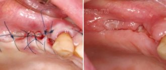
If a root fracture occurs, you should proceed to an atypical tooth extraction operation: • detachment of the mucoperiosteal flap; • access to the root through a burr hole in the area of the vestibular wall of the alveolar process (alveolar part of the lower jaw); • dislocation of the root with an elevator and pushing through the alveolus; • filling the alveoli with an osteotropic drug; • fixation of the mucoperiosteal flap and bringing together the edges of the gums above the alveolus of the extracted tooth with sutures.
Should I treat or remove my wisdom tooth?
Wisdom tooth removal
Late eruption of a wisdom tooth and the lack of a “prepared” place for it can lead to unpleasant consequences of varying severity, including caries of the eight itself and the teeth adjacent to it, pericoronitis, displacement and damage to nearby healthy teeth, malocclusion, inflammation of the gums, periostitis and much more. other.
It is technically possible to cure such a tooth, but sometimes it is very difficult. Therefore, dentists act as follows: if the erupting wisdom tooth does not lead to any pathologies, it is left in place. Sometimes it can even be useful in the future, for example, as an abutment tooth in bridge prosthetics.
If the figure eight erupts with problems and, moreover, turns into an impacted tooth, then they try to remove it, and sometimes this requires serious surgical intervention with cutting the gums and drilling the tooth out of the bone tissue (if it has not erupted or has only partially erupted).
Treatment of alveolitis
When the socket becomes inflamed, the main thing is to eliminate the source of infection, prevent the development of inflammation and preserve the dentition. In order to alleviate the patient’s condition, the dentist uses the following therapy methods:
- Mechanical cleansing of the hole, washing out purulent residues with a solution of nitrofural or hydrogen peroxide.
- Anesthesia of the hole. The pain syndrome is relieved with the help of local applications with anesthetics and analgesics. To do this, the doctor applies the lotion for half an hour and then removes it to prevent the proliferation of microbes in the area. The dentist will advise the patient to repeat the procedure several times a day. Taking painkillers orally is not recommended.
- Taking antibiotics. In the presence of concomitant diseases, alveolitis is treated with antibiotics.
With the right approach, the signs of alveolitis subside 2-3 days after the start of treatment. If therapy was not started on time, residual pain may drag on for 2–3 weeks.
With the permission of the dentist, additional treatment of alveolitis with folk remedies is possible:
- Sage rinses. To prepare the solution, brew a large spoonful of dry sage in 250 ml of boiling water, leave the mixture for an hour, wrapping the container in a towel. After this, the liquid should be filtered and used for rinsing.
- Gargling with chamomile flowers. To prepare the composition, brew a large spoonful of chamomile flowers in a glass of water for 15 minutes, insulating the container with a towel. The infusion should be strained and rinsed your mouth up to 12 times a day.
- Poplar buds. To prepare, take half a glass of buds, pour into a glass container and pour in 500 ml of vodka. The product should be infused for 10 days in the dark and cool, then filtered, soaked in cotton swabs and applied to the inflamed area.
- A soda rinse solution can also be an effective addition in the treatment of alveolitis. You should take a large spoonful of powder in a glass of warm water or mix soda with water to obtain a paste-like mass, which then needs to be used to treat the hole.
- Burdock leaves. To prepare a medicinal decoction of burdock leaves, you need to pour 20 grams of raw material with 2 glasses of water, and then simmer the mixture over low heat for about 40 minutes. The resulting solution should be cooled and filtered, and then used for rinsing.
- Aspen bark. Pour 1 tablespoon of crushed aspen bark into one glass of boiling water. It is important to leave the solution in an airtight container for 3 hours, and then use 100 ml of strained warm liquid at least 3 times a day.
- Anise infusion. To prepare the infusion, pour 1 tablespoon of anise into 200 ml of boiling water, and then keep in a thermos for 50 minutes. The liquid should be filtered and used to rinse 3 times a day.
Symptoms
This complication has very pronounced symptoms.
- Pain.
Many people perceive pain as a standard consequence of tooth extraction. This is partly logical, but over time the pain does not stop and only gets stronger. This is a clear sign of a complication. - Swelling and inflammatory process.
A tooth fragment injures soft tissues, causing swelling and inflammation. The longer you delay treatment, the stronger the inflammation. - A characteristic coating in the area of the hole.
Appears at a later stage, when the body tries to fight the inflammatory process. - Pus and bad breath.
A late stage complication that requires immediate intervention.
Complications during tooth extraction
They occur rarely and may be associated with the actions of the surgeon and the condition of the dental system:
- damage to adjacent teeth or artificial crowns. The appearance of chips, fractures, other mechanical defects, weakening, loosening is possible. More often it occurs when working with molars, access to which can be complicated, due to crowding. Requires additional treatment;
- incomplete removal. After resection, a root fragment may remain in the bone. If the area is close to a nerve or there is a higher risk of additional injury, the dentist may leave it. Doctors at the DentoSpas clinic recommend atraumatic removal to prevent such situations from arising;
- removal of a section of the alveolar ridge. If the forceps were applied to the bone tissue surrounding the tooth, it can be removed along with it. This results in a serious bone defect. To remove it, plastic surgery is performed, protective membranes and bone tissue blocks are installed;
- jaw fracture. The risk of such a complication is increased with a weak jaw bone structure. Its changes may be associated with osteoporosis and other diseases.
Classification
Depending on the nature of the healing of the hole, dentists distinguish several main forms of dental alveolitis:
- Serous. The initial stage of the disease usually appears 2–3 days after tooth extraction. This form is characterized by continuous pain that worsens while eating. Although the patient does not yet complain of feeling unwell, his lymph nodes are not enlarged, but he already feels that the disease is progressing.
- Purulent. If the serous form of alveolitis is not treated, the disease turns into a purulent form. Most often it is diagnosed 6–7 days after tooth extraction. The painful sensations can no longer be ignored, the pain intensifies, radiating to the ear or temple. Exploring the affected area also causes severe pain. Purulent alveolitis is characterized by a dirty gray coating inside the socket, significant swelling around the wound, thickened alveolar process and other problems. The patient's general health deteriorates significantly. Lymph nodes enlarge and become painful on palpation. Often the patient cannot even eat or open his mouth.
- Hypertrophic. At this stage, the symptoms of the disease subside. The patient notes a decrease in body temperature, improved well-being and decreased pain. However, at the hypertrophic stage, dangerous tissue proliferation occurs, which is clearly visible upon examination. When touched, pus is released from the inflamed area, and the mucous membrane acquires a bluish tint.
Ask a Question
Treatment of alveolitis in STOMA clinics
Surgeons at STOMA clinics are required to examine patients and give recommendations on how to care for the hole formed after tooth extraction. If problems arise after extraction, the patient can always call the clinic and clarify whether inflammation has really begun and what to do to reduce pain.
At any time after removal, the patient can contact our clinics. The attending surgeon will examine him and prescribe appropriate therapy.
Feel free to call us at any time and consult about your condition. The sooner we start treating alveolitis, the lower the risk of complications.
Contraindications
- acute diseases of the cardiovascular system;
- high blood pressure numbers;
- infectious processes in the body;
- menstruation period in women, since blood clotting time is increased, which threatens bleeding after extraction;
- acute, severe diseases of the central nervous system;
- pregnancy, lactation period;
- purulent-septic processes in the oral cavity, which provokes sepsis; mental disorders.
The possibility of extracting a diseased tooth appears after treatment of these pathologies and stabilization of the patient’s condition. In case of an emergency, the doctor performs removal in a patient with concomitant diseases. Severe pain syndrome is the cause of exacerbation of chronic pathology.
Recommendations after surgery
- Do not eat or drink water for 6-7 hours after surgery;
- reduce physical activity, avoid visiting saunas and swimming pools for 4-5 weeks to prevent the development of bleeding;
- take analgesics to relieve pain in the early period;
- regularly take antibiotics prescribed for a course of 7 days;
- rinse your mouth with an antiseptic solution after eating;
- follow a gentle diet, avoid solid and irritating foods;
- stop smoking - nicotine provokes bleeding.
Following the doctor's recommendations reduces wound healing time and prevents the development of complications.
Complicating factors
The extraction procedure itself is ordinary and highly predictable, given the level of modern dentistry and the technological equipment of clinics. Nevertheless, even such manipulation has complications, especially if you take it lightly. It is worth taking into account that each clinical case is individual: if in one case tooth extraction is not very difficult, then in another the dental surgeon has to apply all his skills and use advanced technologies and equipment in treatment. It is no coincidence that the price lists of clinics include the item “Complicated tooth extraction”: you have to pay more for this service. Experts identify several factors that can lead to complications, including fragments in the socket.
- Wisdom teeth.
Because of their remoteness, default eights are quite difficult to remove. They often grow incorrectly or do not fully erupt, which increases the risk of complications during removal. - Retention.
A tooth that has not fully erupted, when only part of the crown is visible above the gum or is completely hidden in the soft tissues. - Dystopia.
The tooth erupts at the wrong degree and abuts its neighbors. - The tooth is severely damaged due to trauma.
In such a situation, when removed, it may crumble, and some of the fragments will remain in the hole.
- Simple tooth extraction “all inclusive” - 2500 rub.
- Removal of a tooth with periodontal disease - 1500 rub
- Removal of a permanent tooth (complicated) - resorcinol formalin / bur separation machine / wisdom tooth - 4000 rub
- Removal of impacted (dystopic) wisdom tooth - 5500 rub
- Removal of an impacted (dystopic) wisdom tooth using an ultrasonic device - 7500 rub
Tooth extraction is a full-fledged operation, after which certain unpleasant consequences may arise, caused both by the behavior of the patient himself and by factors beyond his control. Complications can also arise during the operation, since the removal of some teeth can be very difficult: due to the large size of the root or strong bone tissue, it is necessary to make incisions, which, after a successful operation, are stitched. In any case, there is no need to worry, since unprotected tissues in the postoperative period are under maximum influence of microbes, which can result in inflammation.
Alveolitis
Very often, after tooth extraction, a complication such as alveolitis occurs. This problem occurs when a blood clot necessary for healing has not formed at the site of the extracted tooth. In this case, the hole becomes defenseless against external influences, as a result of which an inflammatory process often develops in it .
The key symptom of this complication is pain after tooth extraction (of varying degrees of severity). Pain may occur after 2-3 days. At the same time, the mucous membrane of the gum swells, the edges of the socket become inflamed, there is no blood clot in the tooth socket and the socket is possibly filled with food debris. The patient may have a fever, and sometimes there is pain when swallowing. The hole itself becomes covered with a dirty gray coating that emits an unpleasant odor. Along with these symptoms, the patient often feels general malaise, enlarged lymph nodes, slight swelling, fever, and pain in the area of the extracted tooth.
The main causes of alveolitis
Alveolitis is a disease that is not associated with the introduction of infection into the tooth socket as a result of working with an unsterile instrument. The disease develops with the participation of those microbes that are normally found in the oral cavity of every person.
So, teeth are usually removed due to the fact that chronic foci of inflammation are localized in the area of their roots that cannot be eliminated by conservative methods.
Therefore, the socket of the extracted tooth is primarily infected, and the concentration of microorganisms in it is quite high. If a person is healthy and all immune systems function normally, then the microflora is suppressed and the hole heals without complications. In the event that there are any local or general malfunctions in the mechanisms of the body's reactivity, the likelihood of developing inflammatory complications in the socket increases significantly.
Thus, the following local and general causes may contribute to the development of alveolitis:
- long-term existence of chronic inflammatory foci with frequent exacerbations, as well as exacerbation of the chronic inflammatory process;
- traumatic removal, when conditions arise for the destruction of the created barrier and the penetration of microflora deep into the tissues;
- absence of a blood clot in the socket of the extracted tooth (the clot did not form, or the patient did not follow the doctor’s instructions and the clot was removed; this usually happens when the patient is inattentive to the doctor’s recommendations and diligently rinses the tooth socket);
- general changes in the body due to stress, recent colds (infectious or viral) diseases, the presence of chronic diseases (mainly endocrine), especially in the stage of decompensation, general physical exhaustion, etc.
Treatment of this complication after tooth extraction
Treatment consists of relieving inflammation with local and general means. Sometimes it is sufficient to simply thoroughly rinse the hole with antiseptic solutions and then treat it with a special aseptic ointment or paste. Then, with the help of antibiotics and vitamins, general anti-inflammatory therapy is carried out. But sometimes treatment is delayed up to 1.5 - 2 weeks. In some cases, for this complication, physiotherapy or laser therapy may be prescribed.
Socket bleeding
One of the most common complications after tooth extraction is alveolar bleeding, which can occur immediately after surgery, within the next hour, day, and sometimes more than a day after tooth extraction.
Main causes of bleeding
- Early alveolar bleeding can be caused by the use of adrenaline: when it stops its action, a short dilation of the blood vessels occurs, which causes bleeding.
- Late socket bleeding may occur due to violation of the doctor’s recommendations in the postoperative period - mainly as a result of external disturbance of the socket of the extracted tooth.
- Local causes of socket bleeding include various physical injuries in the area of the socket of an extracted tooth: damage to the gums, fracture of part of the alveolus or interradicular septum, development of inflammation in the area of the extracted tooth, damage to blood vessels in the palate and under the tongue.
- The general causes of the appearance of alveolar bleeding are most often associated with various concomitant diseases of the patient (leukemia, scarlet fever, jaundice, sepsis, hypertension, etc.).
Treatment of this complication after tooth extraction
The effectiveness of stopping alveolar bleeding depends on how correctly the causes and source of bleeding were identified.
- If blood comes from the soft tissue of the gums, then sutures are placed on the edges of the wound.
- If blood comes from a vessel in the wall of the tooth socket, then first apply local cold in the form of an ice pack, then tightly squeeze the bleeding vessel and place a tampon soaked in a special hemostatic agent into the socket, which is removed no earlier than 5 days.
- If local measures do not help, dentists turn to general hemostatic agents that increase blood clotting.
Paresthesia
Much less often, after tooth extraction, a complication such as paresthesia may occur, which is caused by nerve damage during the process of tooth extraction. The main symptom of paresthesia is numbness in the tongue, chin, cheeks and lips. Paresthesia, as a rule, is a temporary phenomenon, disappearing within 1-2 days to several weeks.
Treatment of paresthesia is carried out through therapy with vitamins B and C, as well as injections of dibazole and galantamine.
Changing the position of adjacent teeth after tooth extraction
After tooth extraction, defects can often form in the jaw, and neighboring teeth begin to tilt towards the formed defect, and the antagonist tooth from the opposite jaw begins to move towards the defect, which leads to disruption of the chewing process. At the same time, the chewing load sharply increases, the usual state of the jaws is disrupted and bite deformation develops, which can greatly affect the general condition of the teeth. In this case, it is recommended to replace the extracted tooth with an artificial one using bridges, implants, or removable partial dentures.
All kinds of injuries that occurred during tooth extraction
Often, when removing the second premolar and molars of the upper jaw, perforation of the bottom of the maxillary sinus , resulting in communication between the oral cavity and the nasal cavity through the sinus.
The reasons are as follows:
(subject to the correct and careful actions of the doctor)
- anatomical features: the roots of the above teeth are close to the bottom of the sinus, and in some cases the bony septum is completely absent;
- the presence of a chronic inflammatory focus at the apex of the tooth, which destroys the already thinned bone plate.
If, after the removal of premolars or molars of the upper jaw, a message still appears, the doctor must use one of the known methods to eliminate it during the same visit.
There is only one contraindication:
The presence of a purulent inflammatory process in the sinus (acute purulent maxillary sinusitis). If the message is not diagnosed and eliminated in time, the patient feels liquid and liquid food entering the nose. In this case, you should consult a doctor as soon as possible. If you delay visiting a doctor, a chronic inflammatory process will inevitably develop in the sinus, which will require more serious and technically complex treatment.
Potential complications during the tooth extraction procedure include:
- Damage to adjacent teeth. Adjacent teeth or dentures (eg, crowns, bridges, implants) next to the extracted tooth can sometimes be damaged during the procedure. Adjacent teeth may become broken, split into pieces, or weakened during the extraction of the tooth or teeth, sometimes requiring more time from the dentist.
- Tooth fracture . The tooth may break during the extraction process, making the procedure more difficult and requiring more time and effort to complete the extraction. You may have to remove the tooth in parts. By the way, the process of removing a tooth in parts can cause complications after tooth extraction.
- Incomplete tooth extraction. A small portion of the tooth root may be left in the jawbone. Although this may increase the risk of infection, sometimes the dentist will choose not to try to extract it because removal may be too dangerous, for example if it is very close to a nerve.
- Jaw fracture. Patients with weak jaw bone structure (such as older women with osteoporosis) may be at risk for a jaw fracture. Even if the actual tooth extraction procedure is completed smoothly without any problems, there are cases of complications during the recovery period. Most often, a fracture of the jaw (in the lower jaw) occurs during the removal of “wisdom teeth” and in the upper jaw – a separation of the tubercle of the upper jaw.
- Removal of part of the alveolar ridge - occurs when a tooth is removed incorrectly, when forceps are placed directly on the bone surrounding the tooth and the tooth is removed along with it. In this case, an extensive bone and cosmetic defect occurs (especially in the anterior-frontal area). This problem can only be solved with the help of plastic surgery using artificial bone tissue and special protective membranes.
- Removal of a baby tooth and the germ of a permanent tooth - Occurs due to the carelessness or lack of professionalism of the doctor. When a baby tooth is removed (very often the roots of the tooth are missing, since they dissolve before changing teeth), the doctor begins to look for them in the tooth socket and perceives the germ of a permanent tooth as the roots of a baby tooth.
Remember the main thing: you should trust your doctor and actively participate in the treatment yourself, i.e. unquestioningly and carefully follow all recommendations. And if you suspect the development of complications, do not delay and do not hesitate to consult a doctor again.
Diagnostics
The main symptom of the disease is the appearance of acute pain, which does not subside either 24 hours or 2–3 days after tooth extraction. Sometimes a dentist can identify chronic alveolitis during routine oral examinations. In this case, an empty socket without granulation tissue appears in place of the impacted tooth. The bone is already visible at the bottom of the hole.
The dentist will be able to determine the presence of tissue changes during the examination; radiography and radiovisiography of the affected area may also be prescribed.