Indications
Most dental procedures are performed under local anesthesia.
Its use is mandatory in the following cases:
- Treatment of pulpitis (first visit)
- Periodontal operations (closed and open curettage, gingivoplasty, elimination of gum recession, guided tissue regeneration)
- Prosthetics of vital (living) teeth with fixed structures (crowns, inlays, onlays, bridges)
- Tooth extraction, implantation, bone tissue augmentation, tooth-preserving operations (apex resection, root amputation, hemisection), making incisions for periostitis, pericoronitis, opening abscesses and phlegmons, removing cysts and tumors
In some situations, anesthesia is mainly used, but patients with reduced sensitivity can do without it:
- Treatment of caries, wedge-shaped defects, non-carious lesions, periodontitis
Procedures that most often do not require anesthesia, but particularly sensitive patients may require:
- Professional oral hygiene
- Restoration of pulpless teeth (both fillings and crowns or inlays)
- Removable prosthetics (if the gag reflex is increased, there may be a need for mucosal anesthesia)
Anesthesia is not recommended:
- Oral examination
- Preventive measures (fluoride varnish applications, fissure sealing)
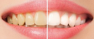
- Teeth whitening
- Orthodontic treatment
Children often tolerate tooth preparation for caries much easier than an injection into the gum. Therefore, you should not necessarily give an injection on your first visit before starting treatment. It makes more sense to try drilling without anesthesia and switch to it only if the child complains of pain. Compassionate parents, do not worry that your child may experience hellish pain and refuse further treatment. Dentin sensitivity in children is less than in adults. The likelihood of getting psychological trauma from an injection is higher.
Diagnostic local anesthesia is used to identify the diseased tooth. When several adjacent teeth are severely damaged, it is difficult for both the patient and the doctor to determine the source of pulpal pain. In order not to depulpate them all, you can give one anesthesia and wait. If the pain has passed, it should be treated first; if not, anesthetize the next tooth and check the reaction.
Paramedian access
With the paramedian approach, only the ligamentum flavum is in the path of the needle. Because this approach offers a larger window, it is advantageous in those patients who cannot extend their back sufficiently, eg the elderly, obese patients or women in late pregnancy (Fig. 4.17). There is in vitro evidence that an angled approach to the dura mater results in less CSF leakage than a right-angle approach.
| The needle (or conductor) is inserted 1-2 cm laterally to the upper border of the spinous process, inferior to the selected interspinous space, i.e. if the L 3/4 interval is selected, then the needle is inserted laterally to the upper border of the spinous process of L4 (Fig. 4.18). The needle is directed medially and cranially to the middle of the selected space. Thus, the interspinous ligament is not affected and the first rigid resistance that is felt is due to the ligamentum flavum. The needle is then advanced into the subarachnoid space. If contact with the bone is felt, then this is probably the lower border of the lamina of the upper vertebra and it is necessary to make the angle of entry less steep (Fig. 4.14). |
Types of local anesthesia in dentistry
Superficial (application) anesthesia
It is carried out by spraying, irrigating or lubricating the mucous membrane with a topical anesthetic (most often lidocaine). The main advantage is that it is painless (no injection is required). However, it is only effective for soft tissue anesthesia. It does not reduce tooth sensitivity at all. Therefore, the use of only superficial anesthesia in the treatment of small carious cavities or ultrasonic teeth cleaning is unjustified.
Often used to reduce pain before an injection.
Application anesthesia helps patients with an increased gag reflex (when taking impressions, for example).
To remove mobile baby teeth in children, superficial anesthesia is sometimes sufficient, but if the roots have not yet completely resolved, then such anesthesia is not enough - infiltration anesthesia is necessary.
Lubricating with ointment is preferable to spraying with a spray because it is easier to control the area of application. When splashed, the anesthetic can get onto the soft palate, creating discomfort and additional anxiety for the patient in terms of difficulty breathing (choking).
Infiltration anesthesia
It is carried out by injecting an anesthetic drug into the place whose sensitivity needs to be reduced. The anesthetic solution from the injection point spreads to nearby tissues (including dental nerves), temporarily eliminating pain and other sensations. When treating teeth, infiltration anesthesia is applied to the projection of the apexes (where the nerves enter the canals of the tooth). This type of anesthesia is preferred for the upper and lower anterior teeth. For lower chewing teeth, infiltration anesthesia is ineffective (due to the thick and dense bone tissue in this area).
The safest injection method of all. Possible minor complications include hematomas (bruises) and stomatitis at the injection site.
When anesthetizing single-rooted teeth (incisors and canines), one injection on the cheek side is sufficient. Multi-rooted teeth (molars and some premolars) require injections on both sides: buccal (labial, vestibular) and palatal (lingual on the lower jaw). On the palatal side, the injection is more painful than on the buccal side (since the mucous membrane of the palate is denser, and the injected solution injures the soft tissues more).
With periostitis, abscess and other acute inflammatory processes in soft tissues, the effectiveness of infiltration anesthesia decreases, and the pain of the injection increases. In such situations, conduction anesthesia is indicated.
Conduction anesthesia
It consists of blocking the sensitivity of the entire nerve trunk by introducing an anesthetic to the place where it exits the jaw to the surface. In this case, several teeth can be treated at once.
There are the following subtypes of conduction anesthesia in dentistry: mandibular and torusal (the lower teeth on one side are anesthetized), mental (lower premolars, canines and partially incisors), palatal (upper teeth on the palatal side), incisive (upper incisors and canines on the palatal side) , tuberal (upper molars on the buccal side), infraorbital (upper premolars and canines on the labial side).
Conduction anesthesia is not always successfully performed by the dentist. You have to do it again from time to time. Due to the technical complexity of the implementation: the doctor does not see the nerve trunk, the needle is brought to the average location of its location in the anatomical atlas, and a particular patient may have individual (quite different from the standard) anatomical features. The introduction of an anesthetic at a considerable distance from a large nerve does not allow achieving a complete block of sensitivity.
Even worse is a direct needle hit on a nerve during conduction anesthesia. In addition to sharp pain (as patients describe it - “electric shock”), subsequent loss of sensitivity is possible. Most often, mandibular or torus anesthesia is performed, so paresthesia of the tongue and lower lip (one half) is most common. Tingling, loss of taste, and discomfort may last for several weeks or months. Usually, sensitivity returns on its own without any manipulation, but in rare cases, paresthesia remains for life.
Therefore, conduction anesthesia is not recommended when safer infiltration can be used.
Intraligamentary anesthesia
It is carried out by injecting the drug into the periodontal ligament (located between the alveolus and gum on one side and the tooth on the other). To completely remove the tooth, several injections are required around the perimeter. Only one tooth is numbed. Effective for all teeth, but extremely painful. It is recommended to use it after preliminary infiltration anesthesia.
If the anesthetic is administered excessively, necrosis of the periodontal ligament (with subsequent tooth loss) is possible. If anesthesia is used to remove a tooth, this problem is irrelevant.
Intrapulpal anesthesia
It is carried out by injecting an anesthetic directly into the pulp of the tooth in case of pulpitis. This injection is very painful, so it is given only after preliminary infiltration, conduction or intraligamentary anesthesia. The tooth is still pre-drilled (otherwise you won’t be able to get to the pulp). The effectiveness is close to 100%, the analgesic effect does not last long, but usually this period of time is enough to remove the pulp.
Apart from pain, there are no complications.
Intraosseous anesthesia
It is achieved by introducing an anesthetic into the bone tissue surrounding the tooth after its perforation. Preliminary infiltration or conduction anesthesia is required. Due to the technical complexity of implementation, it is rarely used. Only if other types of local anesthesia have failed. About 90% effective, can block multiple teeth.
Median access
Having selected the appropriate interspinous space, firmly hold the skin on one of the adjacent protrusions with the second and third fingers of the non-dominant hand. This will prevent the skin from moving as the needle passes through it (Fig. 4.9).
| Fig.4.9 |
The needle is directed slightly cephalad and placed equidistant between the two protrusions of the lumbar vertebrae. The bevel of the needle should follow a line so as to separate the longitudinal fibers of the dura mater, and not cut them off (Fig. 4.10).
| Fig.4.10. The effect that the direction of the needle bevel has on the hole left in the dura mater. If the bevel of the needle is oriented so that it is located across the fibers of the dura mater (right), then the needle cuts these fibers and leaves a hole. If the bevel of the needle separates the fibers (left), it causes less damage and results in a much smaller hole. |
As the needle advances, the different ligaments are identified by "feel": the intervertebral ligament is stiffer than the subcutaneous tissues, and the ligamentum flavum is even stiffer. Sometimes, at the time of puncture of the dura mater, a barely perceptible click is felt (Fig. 4.11). The stylet is removed and the needle pavilion is examined for the presence of CSF (Fig. 4.12). If after a sufficient period of time nothing appears, the needle is inserted further 0.5 cm. If after 2-3 cm from the surface of the skin the needle encounters bone, this is probably the bony plate of the lower vertebra (Fig. 4.13) and the needle should be redirected more cephalad. To do this, a thin spinal needle must be pulled back into the subcutaneous tissue before being redirected and reinserted.
| Fig.4.11. Needle directions. Anatomical Models Erling Worm Skole, MD |
| Fig.4.12. The needle pavilion is checked for the presence of CSF. |
| Fig.4.13, Fig.4.14, Fig.4.15. Needle directions. Anatomical Models Erling Worm Skole, MD |
If the initial insertion path is too cephalad, the needle may impinge on the superior vertebra and a more caudal approach will be required (Fig. 4.14).
If the bone is felt deep in the tissues of the back, then it is most likely the anterior wall of the spinal canal (Fig. 4.15). In this case, the stylet is removed and the needle is slowly withdrawn, while at the same time the needle pavilion is checked for the presence of CSF.
The guidewire is typically used for 25 gauge needles and finer. It is injected until the ligamentum flavum is passed through. The spinal needle is then passed through it into the subarachnoid space (Fig. 4.16).
| Fig.4.16. The spinal needle passes through the epidural needle. Anatomical Models Erling Worm Skole, MD |
If contact with bone occurs before the spinal canal is reached, this indicates that the guidewire was not inserted correctly. The needle must be brought back into the guidewire, which is then itself withdrawn and redirected, usually more cephalad.
If CSF does not appear in the needle pavilion, the following possibilities should be considered:
- Not enough time has passed for the CSF to appear. When using 29 gauge needles for patients in the lateral position, this wait may take 60-70 seconds. Although aspiration is often recommended, its value is controversial. However, removing the stylet may act like aspiration, leaving a drop of CSF in the clear plastic sheath of the needle.
- The bevel of the needle is not completely in the subarachnoid space. Try to insert the needle another 1-2 mm deep and rotate it 90 - 180 degrees.
- The needle is not in the subarachnoid space and must be reinserted. Before doing this, pull the needle out slowly as the tip of the needle may be anterior to the subarachnoid space (Fig. 4.15).
Do not administer local anesthetic until the CSF is clearly identified and freely drained.
In difficult cases, decide as quickly as possible whether to seek help from a more experienced specialist or abandon this technique. Frequently repeated needle insertion is unacceptable. In addition to discomfort, it can cause multiple puncture holes in the dura mater and possible neurological impairment.
Instruments for local anesthesia
The injection requires a syringe, a needle and a local anesthetic solution.
For several decades now, in dentistry, instead of disposable syringes, a reusable carpule syringe has been used. A disposable anesthetic cartridge and a disposable needle are inserted into it. They are used once, and the metal syringe itself is sterilized after each patient. Reuse of an incompletely used carpule is prohibited, since during the injection a reverse flow of blood or other liquid through the needle into the carpule is possible (there is a risk of infection of the next patient).
A special syringe gun is available for intraligamentary anesthesia. The same needles and carpules are inserted into it as into a carpule syringe. It allows you to more accurately dose the volume of anesthetic for a given type of anesthesia (but it is also possible to perform intraligamentary anesthesia with a regular carpule syringe).
The thickness of the needles used in carpule syringes is 0.3-0.5 mm. This is much thinner than disposable syringes (therefore the injection is much less painful). Length – 8-30 mm. For mandibular and torusal anesthesia, longer and thicker needles are used than for infiltration. To carry out intrapulpal and intraligamentary anesthesia, the needle can be bent (it does not break).
Carpula is a sealed glass cartridge with a rubber plunger. In dentistry, in most cases, the anesthetic solution, in addition to the anesthetic drug itself, contains a vasoconstrictor - a vasoconstrictor component that prevents the rapid elimination of the anesthetic through the general bloodstream. This is adrenaline (epinephrine). Its concentration is negligible - 1:100000 or 1:200000. When manually drawing such a mixture into a disposable syringe, add 1 drop of adrenaline to the anesthetic solution. However, the size of a drop is such a relative value that the concentration of this very active component can differ tens of times in different syringes. This creates many complications, even life-threatening situations for the patient.
The introduction of carpules with precise industrial dosages of components has greatly reduced the number of such complications. However, it should be noted that different manufacturers have different attitudes towards maintaining strict dosages of their own carpules. For the product of the Russian pharmaceutical industry, Brilocaine (manufacturer: Bryntsalovskiy Ferein), the anesthetic effect of two capsules from one package can be radically different: from complete absence of pain relief to super strong (“my legs froze,” according to the patient). Although the packaging states exactly the same ingredients as imported Ultracain, Ubistezin or Septanest.
Patient preparation
Spinal anesthesia can only be performed where a complete set is available
equipment for anesthesia. Intravenous infusions and monitoring of blood pressure, heart rate and electrocardiogram should be started. The patient must be placed in the desired position and landmarks noted. Every precaution must be taken to ensure sterile anesthesia conditions, at a minimum, sterile rubber gloves are worn. The skin must be wiped with a cloth containing a suitable antiseptic solution and sterile swabs must be used. Once the selected interspinous space has been identified, a “lemon peel” may rise over it after a small injection of local anesthetic. This is not as necessary as with an epidural block because the spinal needles (or their guidewires) are thinner and sharper, making insertion less painful.
Anatomical landmarks
(Fig.4.7)
- A line connecting the iliac crests, which are located at the level of the fourth lumbar vertebra.
- Spinous processes of the lumbar vertebrae.
- For lumbosacral approach, the spinous process of the fifth lumbar vertebra and the posterior superior iliac spine
| Fig.4.7. Anatomical landmarks for identifying the spinous processes of the vertebrae. 1.Superior view of the iliac cross. 2. Posterior superior iliac spine. |
Local anesthetics
In Russia, 4 types of anesthetics are most widely used: novocaine, lidocaine, articaine and mepivacaine.
Novocaine (procaine) was synthesized in 1905 and became widespread throughout the world as the first non-narcotic anesthetic. It is a basic reference point - all subsequent anesthetics are compared in terms of effectiveness and toxicity with novocaine, whose indicators are taken as one. After the introduction of lidocaine, it lost popularity in developed countries. There is a high frequency of allergic reactions to novocaine.
Lidocaine was invented in 1943. Its effectiveness turned out to be 4 times higher than that of novocaine (with only twice the toxicity). Widely distributed throughout the world (1st place in the number of injections among anesthetics in the USA). However, like novocaine, it has a relatively high percentage of allergic reactions (including the development of anaphylactic shock). In addition, it is often used with increased concentrations of the vasoconstrictor 1:50000 and 1:25000, which increases its effectiveness, but increases the number of complications from the cardiovascular system. Indicated for pregnant women - FDA category B (see article Use of local anesthetics for dental treatment during pregnancy; safety for women in labor).
Articaine was synthesized in 1969. It began to be used in Germany, where it was registered under the commercial name "Ultracaine". This name of the drug is now no less popular than articaine, although it represents the product of only one, “Septanest”, “Alfacaine” and several other commercial names - this is the same as “Ultracaine”. The most common local anesthetic in Europe and Russia (it was approved in the USA only in 2000, 10 years later than here). 5 times more effective than novocaine. 1.5 times more toxic. According to the FDA classification, it is classified as category C.
Mepivacaine was developed in 1957. It is equivalent in effectiveness to lidocaine and inferior to articaine. It is noteworthy that, despite category C, it is often used for pregnant patients, due to the permission of non-adrenaline release forms (lidocaine and articaine carpules are sold only with a vasoconstrictor). Although in fact it is not the first choice drug for expectant mothers (see the article Is it possible to do local anesthesia during pregnancy?).
Adrenaline , also known as epinephrine, is not a local anesthetic, but is used in the vast majority of cases for dental injections. By narrowing the blood vessels, it helps preserve the anesthetic depot at the injection site, reduces its toxic effect on the body, and also reduces bleeding (which improves visibility during surgical procedures). Its use in pregnant women and patients with cardiovascular diseases (paroxysmal ventricular tachycardia, atrial fibrillation) is undesirable. Use with caution in patients with arterial hypertension, diabetes mellitus, and hyperthyroidism.
In addition to the anesthetic and vasoconstrictor, the solution may contain preservatives (methylparaben) and adrenaline stabilizers (sodium pyrosulfite). Both methylparaben and sodium pyrosulfite (metabisulfite) have a high frequency of allergic reactions, including the most dangerous - anaphylactic shock. This risk is significantly higher than that of the anesthetics themselves (and in principle there cannot be an allergic reaction to adrenaline). Therefore, methylparaben was completely abandoned in carpules - it is needed only when using large containers for preserving an unused solution after opening the ampoule. Sulfites are necessary to prevent the oxidation of adrenaline - they cannot be abandoned in carpules with a vasoconstrictor. Therefore, anesthesia without adrenaline is recommended for patients with multivalent allergies. The frequency (up to 5%) of sulfites provoking an attack of bronchial asthma is high, so anesthesia with adrenaline is also not recommended for asthmatics.
Lumbosacral approach
This is also a good method for patients with limited spinal mobility. The lumbosacral space is the largest in the vertebral column, but the spinous processes of the fifth lumbar vertebra close the space and usually prevent needle access with a midline approach. Although Taylor's original description of this approach included identification of the posterior superior iliac process, it is often not palpable and it is much easier to use the spinous process of the fifth lumbar vertebra as an anchor point.
The spinal needle is inserted 2 cm lateral and 2 cm below the spinous process of the fifth lumbar vertebra at the level of the first sacral process (Fig. 4.19 and 4.20). It is directed medially and cranially forward, but deep to the fifth lumbar vertebra. It should reach the ligamentum flavum in the midline. The needle should be positioned very close to a plane parallel to the posterior surface of the sacrum. If there is contact with bone, it is probably the lamina of the fifth vertebra and the needle in this case should be directed more caudally.
| Fig.4.19. Lumbosacral approach | Fig. 4.20 1. Spinous process 2. Lumbosacral space (ligamentum flavum) 3. Spinous process 4. Posterior superior iliac spine |
Prolonged spinal anesthesia (PSA) was introduced by Dean in 1907, who left the spinal needle in place for the duration of the operation. In 1940, Lemmon proposed the use of a flexible needle with a rubber tube and a special mattress for the patient in order to overcome the problems associated with needle trauma and needle breakage. Tuohy (1944) used an N4 ureteral catheter inserted through a 15-gauge needle into the subarachnoid space. Since that time, the diameter of the needles and catheters used for PSA has decreased. 17 or 18 gauge epidural needles are used to insert 19G and 20G epidural catheters. Although prolonged spinal anesthesia has certain advantages, it has not achieved the popularity of continuous epidural block due to the significantly high incidence of post-dural puncture headaches (vide infra).
Local anesthesia in pediatric dentistry
Local anesthesia is not recommended for young children under 2-4 years of age. Even if it is possible to fraudulently persuade a child to take an injection, after it, as a rule, he will no longer open his mouth for further treatment. Up to 6-7 years of age, the optimal method is infiltration anesthesia (including for the treatment of lower teeth). At this age, the lower jaw is not yet so dense and there is no need for conduction anesthesia. Of the drugs for children, the optimal choice would be articaine with a low adrenaline content of 1:200000 - since long-term manipulations are still contraindicated for children (they quickly get tired of the treatment), there is no need for long-term pain relief for many hours.
Patient position
The patient can be either in a lateral position, lying on his side, or in a sitting position (Fig. 4.8a and b). The spine should be arched as much as possible. The lateral position allows for minimal movement before and after the procedure. This position is also less likely to cause postural hypotension as anesthesia progresses. In the sitting position, the patient sits on the operating table with his legs supported (Fig. 4.8b). This position has the advantage that it provides higher CSF pressure, thus reducing the time until CSF appears in the needle pavilion, which is important when very thin needles are used. This position is also more preferable for obese patients when determining the midline is difficult. For sacral spinal nerve blocks (saddle block), the sitting position is preferred. Sometimes spinal anesthesia is performed in a tilted “jackknife” position (Fig. 48c). When the patient is in an inclined position, the lumbosacral and paramedian approach is often used.
| A | b |
| V | |
| Fig.4.8. Patient positions. a.Lateral position lying down. b. Sitting position. c. “Folding knife” position. | |
The effectiveness of local anesthesia
The success of deep anesthesia depends on the anesthetic, the concentration of the vasoconstrictor, the type of anesthesia, the dose of the drug, the qualifications of the dentist and the individual response of the patient. 4% articaine with an adrenaline concentration of 1:100000 is the most effective. Conduction anesthesia provides better pain relief than infiltration anesthesia, but requires a more highly qualified doctor. (However, even the most experienced specialists have a certain percentage of failures). The patient's agitated panic state and prolonged pain tolerance for several days before the visit reduce the effectiveness of local anesthesia. Alcohol and drugs - even more so.
Development of microcatheters
32-gauge polyamide catheters were chosen for their physical characteristics and lack of biological reactivity. Initial research was promising but also alarming. Lack of tensile strength and entanglement were the main problems. The corrosion-resistant steel stiletto with a diameter of 0.0076 mm was first used in the traditional way, i.e. was removed after catheter placement. However, leaving the stylet inside (and further bonding it to the polyamide) significantly improved its tensile strength and reduced curl problems (Figure 4.22). The stylet also made the catheter radiopaque.
Dosage
The volume of one carpule is 1.7-1.8 ml. This amount is enough for most manipulations within one or two teeth. When treating a larger number of teeth (especially if they are located far from each other), several carpules and injections into different parts of the oral cavity are required.
A second injection of anesthetic into the same place is carried out if the first one is unsuccessful or after some time, when long-term treatment has not yet been completed and the anesthesia begins to wear off. The introduction of the same drug can help if conduction anesthesia is ineffective the first time. With other types of anesthesia, it is necessary to change the anesthetic itself to a more powerful one. It is impossible to increase the volume of the injected solution indefinitely - in case of an overdose, a toxic reaction occurs. For articaine with adrenaline and lidocaine with adrenaline, the maximum dose is 7 mg/kg body weight. One carpule (1.7-1.8 ml) contains 34-36 mg of 2% lidocaine or 68-72 mg of 4% articaine. Therefore, for a person weighing 70 kg, the maximum number of carpules at one time is: 14 for 2% lidocaine and 7 for 4% articaine.
Recommendations for choosing catheters
There are no clear recommendations. The main goal is to select a catheter that will keep the incidence of post-puncture headaches (PDPH) at an acceptable level. Smaller catheters can be used in older patients, although larger catheters are easier to use and are not associated with a high incidence of PDPH in this age group (Table 4.3).
Table 4.3. Catheter Size Recommendations
| Age | Caliber |
| <50 | 32 |
| 50-70 | 27,28 |
| >70 | 19, 20, 24, 27, 28 |
Coming time
Intrapulpal and intraligamentary anesthesia begins to take effect within a few seconds. Application, infiltration, intraosseous - after 1-5 minutes. Conduction anesthesia is the most variable - from instant pain relief at the same second (if the needle hits the nerve) to half an hour. Sometimes patients, rising from the dental chair after completion of treatment, claim that “only now the freezing has really taken hold.”
Soft tissues are anesthetized before teeth. If the lip or tongue is already “frozen,” the teeth may still remain sensitive, and their premature preparation will be painful.
Validity
The duration of anesthesia also depends on the anesthetic, the concentration of the vasoconstrictor, the type of anesthesia, the dose of the drug, the qualifications of the dentist and the individual response of the patient. Conduction anesthesia lasts up to 2-3 hours or more. Some patients note that the anesthesia wears off completely only in the evening (if the treatment was carried out in the morning). But this is with a strong anesthetic, a high concentration of adrenaline, a significant dosage, and close contact with the nerve trunk. In other situations, conduction anesthesia may not last even one hour. Infiltration anesthesia lasts 1 hour or less. Other types are even smaller.
Methodology
After performing a spinal puncture, the loop of the catheter is held in one hand while the tip is passed through the needle (Fig. 4.23). Assistance is required to insert 27-32 gauge catheters (Fig. 2.24). As the catheter passes through the bevel of the needle, a slight increase in resistance is often felt. The catheter should be advanced 2-3 cm beyond the tip of the needle, using the markings on the catheter as a guide. Avoid inserting an excessively long catheter as this may cause entanglement.
| Rice. 4.23. The end of the catheter is passed through the needle | Fig.4.24. Method of inserting a smaller catheter for continuous spinal anesthesia. A threading aid is inserted into the needle sheath to strengthen the catheter as it passes through the needle sheath. |
A common problem is difficulty in advancing the catheter into the subarachnoid space despite good CSF flow from the needle. The reason for this is usually unclear, but maneuvers such as turning the needle or passing the catheter while withdrawing the needle slowly can help resolve the problem. If these manipulations do not produce results, the entire procedure may have to be repeated at another interspinous space, keeping in mind that this may increase the likelihood of post-puncture headache.
Once the catheter is inserted, the needle is carefully removed without removing the catheter, which is gently pushed inward as the needle is withdrawn. No attempts to remove the catheter back through the needle are allowed, as this may cause part of the catheter to come off. CSF aspiration to confirm correct catheter placement can be done through a 19- to 28-gauge catheter, but not through a 32-gauge catheter. However, if no difficulties were encountered when inserting this very thin catheter, the correct placement can be considered confirmed. Once the needle and stylet (all catheters except polyamide catheters have a stylet that must be removed) are removed, the catheter is secured to the patient's back.