Blood clotting disorders are a group of pathological conditions characterized by malfunctions in the hemostatic system. It consists of keeping blood in a liquid state, stopping bleeding in cases where the walls of blood vessels have been damaged, and also dissolving blood clots that have already fulfilled their function. The reasons may be different: as a rule, they arise due to a lack of one or several factors at once or the appearance of their inhibitors in the blood. They provoke a decrease in the ability of blood to clot and the development of serious and potentially life-threatening diseases.
The Department of Hematology at CELT offers diagnosis and treatment of blood clotting disorders in Moscow. We employ leading domestic hematologists, whose qualifications are sufficient to accurately diagnose and carry out effective treatment according to international standards. They are helped in this by a powerful diagnostic and treatment base and modern gentle techniques. You can find out the approximate prices for our services in the price list in the “Services and Prices” section of our website. We recommend checking the numbers with our information line operators.
General information
The blood coagulation system is a complex sequential cascade of reactions occurring in the body that is aimed at stopping bleeding. The coagulation process is an important protective reaction of the body, thanks to which a constant volume of circulating blood is maintained. homeostasis system involves many components, the main ones of which are shown in the figure below.
Clotting factors are in an inactive state in the blood. If a blood vessel is injured, the coagulation process begins and all factors are sequentially activated and ensure the formation of a clot. The direct coagulation process itself is associated with the conversion of the fibrinogen protein (factor I) into insoluble fibrin.
Coagulopathy is a disease, or rather a group of diseases or conditions, which are based on a blood clotting disorder. The clinical sign of coagulopathy is bleeding. Acquired coagulopathies are the most common syndromes. In this case, poor blood clotting can be caused by pathology of different parts of the coagulation system: fibrin , platelets or blood clotting factors . If any part of this system does not function or is missing, the person will have prolonged bleeding and develop a critical condition. Among coagulopathies there are congenital conditions and diseases ( hypofibrinogenemia (factor I deficiency), afibrinogenemia , hemophilia A , von Willebrand disease , hemophilia B ) and acquired syndromes that occur in various septic conditions and diseases of the kidneys and liver.
What to do if blood doesn't clot well
If you have the symptoms listed above, you should definitely consult a doctor and undergo an examination. At the moment of bleeding itself, first aid should be provided in accordance with general recommendations, based on the location and type of injury. If necessary, call an ambulance.
Diagnostics
To diagnose blood clotting, the doctor first reviews the patient's medical history. To do this, he will ask questions about your health problems and medications you are taking. You need to answer something like this list of questions:
- What are the accompanying symptoms?
- How often does bleeding occur?
- How long does the bleeding last?
- What were you doing before the bleeding started (for example, were you sick, took medications)?
Basic tests to check blood clotting :
- A complete blood count to check the blood loss at the time the test was taken and the number of red and white blood cells.
- Platelet aggregation test, which shows the extent to which platelets are able to adhere to each other.
- Measuring bleeding time to see how quickly blood vessels clog after pricking a finger with a feather.
Treatment options for poor blood clotting
Treatment for a bleeding disorder is based on the causes that cause it. If possible, the diseases that caused the disorder, such as cancer or liver disease, are immediately treated. Additional treatments include:
- Taking vitamin K by injection;
- Drugs aimed at improving clotting function;
- Transfusion of frozen donor blood plasma or donor platelets;
- Other drugs, including hydroxyurea (Droxia, Hydrea), and oprelvequin (Neumega) to treat platelet-related disorders.
Treatment of consequences caused by blood loss
Iron supplements
If there is significant blood loss, the doctor may prescribe medications containing iron to replenish its amount in the body. Low iron levels can lead to iron deficiency anemia, which is accompanied by feelings of weakness, shortness of breath and dizziness. One of the most common and accessible drugs in this case is Hematogen. In addition to treatment with iron supplements, blood transfusions may be required.
Blood transfusion
During this procedure, as most people know, blood loss is compensated with the help of donor blood. Donated blood must match your blood type to prevent complications. This procedure can only be performed in a hospital.
Pathogenesis
The pathogenesis of hemorrhagic syndrome includes:
- damage to vascular endothelial cells by inflammatory mediators or endotoxin ;
- activation of protein C , which inhibits FV and FVIII and suppresses the synthesis of plasminogen activator inhibitor (the latter promotes the transition of plasminogen to plasmin, which breaks down the fibrin of the blood clot);
- activation of fibrinolysis, which plays a role in the development of non-stop bleeding and depletion of f I, II, V, VIII, XIII;
- accumulation in the blood of metabolites that have an anticoagulant effect.
In the pathogenesis of uremic thrombocytopathy, platelet deficiency is important, which is associated with the action of toxic plasma metabolites. In addition, patients with uremia undergo an extracorporeal circulation procedure, in which platelet dysfunction occurs due to their interaction with the tubes and membranes of the device. In this case, platelets are activated and release granules (platelet degranulation). Platelet dysfunction causes bleeding so severe that platelet transfusion is required.
Drug-induced thrombocytopenia is associated with the interaction of the drug (or its metabolite) and a platelet membrane glycoprotein. As a result of this interaction, an immunogenic complex - a glycoprotein-drug . Altered platelets are removed from the bloodstream by RPE cells. With drug-induced thrombocytopenia, the level of IgG and platelet-bound antibodies to the drug increase. Idiopathic purpura based on the production of antibodies against viral antigens. Platelets are damaged by adsorbing a viral antigen or a virus-antibody immune complex on their membrane.
Professional assistance at the Berezka clinic
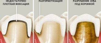
A dentist in a clinical setting stops bleeding in several ways, depending on the severity of the condition and the individual characteristics of the patient. It should be noted that each of the methods will be effective only if you seek medical help in a timely manner.
Stitching
The damaged vessel is ligated over the hole or soft tissue is sutured. The boas of the method stop bleeding with equal effectiveness.
Electrocoagulation
The manipulation is performed under local anesthesia. They resort to this procedure if several capillaries are damaged. The tissues are dissected using a special tool, after which they are soldered, facilitating rapid healing of the wound.
Application of fibrin film
A fibrin film is applied to the place where the tooth was removed. In addition to the hemostatic effect, the film also has an anti-inflammatory, disinfecting and healing effect.
Tamponation
In this case, the dentist uses iodoform turunda. It is inserted into the hole in the same way as a gauze swab, but to a greater depth.
Hemostatic tube
The method is relevant for patients suffering from high blood pressure. The tube is carefully inserted into the hole and held until the AT is brought back to normal.
You can also use gelatin and collagen sponges to stop bleeding.
The dentist makes a decision on the need for additional treatment procedures based on the results of examining the condition of the wound after extraction.
Classification
Coagulopathies are divided into hereditary and acquired.
- Hereditary forms are associated with genetically determined changes in the walls of blood vessels, abnormalities of platelets and plasma blood factors. Hereditary coagulopathies include hemophilia A , von Willebrand disease , hemophilia B , and deficiency of various clotting factors.
- Acquired forms are most often associated with vascular damage of various etiologies (immune, toxicoinfectious and dysmetabolic), platelet damage, pathology of coagulation factors and a combination of all these factors.
The following types of acquired coagulopathy are distinguished:
- Disorders of platelet hemostasis . These include thrombocytopenia of various origins (associated with decreased platelet production, associated with increased platelet destruction, caused by non-immune causes and immune ones - idiopathic thrombocytopenic purpura), HELLP and hemolytic-uremic syndrome, thrombotic thrombocytopenic purpura, thrombocytopathies. With thrombocytopenia, the platelet germ may be primarily affected, platelets can be redistributed and accumulate in the spleen, and there will be an insufficient number of them in the blood. Also, platelets can be destroyed in large quantities (with lupus erythematosus and thrombocytopenic purpura). In addition, platelets can be consumed in large quantities during the formation of blood clots (for example, in DIC syndrome). Thrombocytopathies are characterized by the production of abnormal platelets whose function is impaired. An example of thrombocytopathy is von Willebrand disease and Glanzmann thrombasthenia .
- Types of coagulation disorders. This group includes hemostasis disorders associated with an overdose of anticoagulants ( heparin , warfarin ), hemodilution coagulopathy , vitamin K-dependent (with impaired liver function, poor absorption of vitamin K, taking certain medications).
- Coagulation-platelet (mixed) disorders that developed against the background of liver and kidney failure (hepatic and uremic coagulopathy).
- DIC syndrome is distinguished separately.
Fibrinogen plays an important role in the blood coagulation system . Normally its content is 2-4 g/l. Patients often have hypofibrinogenemia —a decrease in fibrinogen levels. This condition can be hereditary, but more often acquired, due to insufficient formation of this liver protein when it is damaged or its increased dissolution (fibrinolysis). In this condition, blood clotting slows down and as a result, a loose, disintegrating clot is formed. Hypofibrinogenemia manifests itself through the formation of bruises with minor trauma and various bleeding.
However, in most patients, decreased fibrinogen levels do not manifest themselves in any way. A decrease in the level of this protein is observed in cirrhosis , liver necrosis , bone marrow metastases leukemia , shock , anemia , eclampsia , premature placental abruption, complicated childbirth, and sepsis. The cause of acute hypofibrinogenemia is intravascular coagulation when fibrinogen is intensively consumed. Many patients with low fibrinogen levels do not require treatment. Heavy menstruation in women is prevented by hormonal agents and antifibrinolytic drugs.
Afibrinogenemia is a complete absence of fibrinogen in the blood with a normal platelet level. This is a genetically determined disease, but is very rare. In this condition, any injury leads to bleeding, hematomas, and hemorrhages in the joints. Dental manipulations and operations are accompanied by significant blood loss. Children rarely survive to adulthood. In the treatment of this pathology, replacement therapy is used: administration of fibrinogen , cryoprecipitate and fresh frozen plasma .
Use of the drug Sinkumar for blood thinning
Sinkumar is an indirect anticoagulant. This suggests that the drug has a cumulative effect. The peak of its activity occurs 2-4 days after the start of administration. When treated with Sinkumar, you should constantly monitor your doctor and monitor the level of blood clotting. It is also worth stopping treatment by gradually reducing the dose and increasing the interval between doses of the drug.
Contraindications for taking the medication are pregnancy, malignant tumors, liver and kidney diseases, gastrointestinal ulcers, and an allergic reaction to the components of the drug. You can buy Sintrom in Moscow by placing an order at an online pharmacy.
Causes of poor blood clotting
Based on the above, we can name the main causes of coagulation disorders:
- Vascular wall defects are hereditary (associated with collagen abnormalities) and acquired (immune or inflammatory vascular lesions).
- Pathology of platelets. Platelet dysfunction sometimes causes significant bleeding. It includes thrombocytopenia (quantitative changes in platelets) and thrombocytopathies (changes in platelet quality). Acquired thrombocytopathies are caused by taking non-steroidal anti-inflammatory drugs, dipyridamole , antibiotics , uremia , heart valve pathology, and the use of extracorporeal circulation.
- The cause of a bleeding disorder may be a lack of factors (there are thirteen of them) of the blood coagulation system or a reduced synthesis of one or more factors. Prothrombin (factor II) is the main component of blood clotting. It is a precursor of thrombin and is involved in the formation of a clot (thrombus). Fibrinogen (factor I) is produced in the liver. In the coagulation cascade, it is converted into fibrin, which participates in clot formation. The deficiency of this factor was mentioned above.
- Deficiency of factor XI is associated with hemophilia C, factor VIII with hemophilia A , and factor IX with hemophilia B. Among hereditary coagulopathies, the most common (in 95% of cases) deficiency of factors VIII and IX. Deficiency of factors VII, X, V, XI is only 1.5%. Acquired deficiency of prothrombin complex factors (II, VII, X, V) occurs in liver diseases, jaundice , dysbacteriosis , as well as in overdose of vitamin K .
- The cause of bleeding can also be increased fibrinolysis , that is, excessive fibrinolytic activity. This may be a hereditary deficiency of alpha2-antiplasmin or increased formation of plasminogen activators and impaired excretion of these activators in liver diseases.
Hemostasis is the process that stops bleeding
Why does blood clot? Few people think about this crucial mechanism. It’s only after cutting yourself that the thought sometimes slips into your mind: “Why doesn’t the bleeding stop for so long?” The basics of this process are quite a complex topic. But you still need to know the minimum.
In many developed countries, starting from school, classes are held that teach children how to act in emergency situations. Such situations include sudden cardiac arrest, drowning, and trauma, which are often accompanied by external bleeding. For an unprepared person, the sight of human blood can prevent him from taking the right actions. Moreover, it can put a person into panic and stupor. In this case, wasting time is an unaffordable luxury. In the case of bleeding from large arteries, the outcome can be determined in seconds.
What happens in the body when there is bleeding?
The physiology of hemostasis, the process aimed at protecting life during bleeding, is complex. One can only marvel at how thoughtfully nature created man. Hemostasis mechanisms are activated instantly as soon as a blood vessel is damaged. When the inner layer of the vessel or endothelium is damaged, substances are released that signal bleeding. These are serotonin and thromboxane. Pain and these substances cause spasm of the smooth muscles of the damaged vessel, which leads to a decrease in blood flow and blood loss. Thromboplastin, a substance also released during injury, causes activation of platelets and a cascade of reactions leading to the formation of a blood clot. Thrombus is a blood clot that acts as a “plug”. It prevents bleeding.
How does a blood clot form?
Primary, or vascular-platelet, hemostasis is caused by vascular spasm and platelet aggregation. Platelets are small blood cells that have the ability to form a lump when activated. They stick to the edges of the damaged area and to each other. In this case, platelets are a source of biologically active substances that trigger the cascade of the coagulation system or coagulation hemostasis. Under the influence of these substances, as a result of a complex reaction at the site of injury, long threads of protein - fibrin - are formed in the blood. Fibrin strengthens the platelet clot and helps to reliably stop bleeding. Fibrin is constantly present in blood plasma in the form of inactive fibrinogen. Special proteins that regulate the process of fibrin formation, or coagulation factors, are also found in the blood plasma. This is why coagulation hemostasis is also known as plasma hemostasis.
A detailed diagram of hemostasis in the specialized literature takes up more than one page. It is important to conclude: to quickly stop bleeding, it is necessary to create conditions that facilitate the work of the hemostatic system. For example:
- if the wound is on a limb, use a tourniquet, this will slow down or stop blood circulation and bleeding;
- the wound must be squeezed or clamped tightly to reduce the lumen of the vessel;
- if possible, put cold on the wound (snow, ice, cold water in a bottle) to cause vasospasm;
- if a limb is injured, raise it high to slow down the blood flow;
- When applying a pressure bandage or covering the wound with a bandage or other dressing material, the bandage must not be removed or changed, even if bleeding continues. This can destroy an already formed but still weak blood clot, and the bleeding may increase. You should increase the pressure on the wound and use more bandages to get the person to the hospital faster.
Pathologies of the hemostatic system
Impaired hemostasis is a serious pathology. There are diseases in which the formation of a blood clot is impaired and (or) there is a risk of uncontrolled bleeding. Diseases of the hemostatic system:
1. Plasma hemostasis:
- violations of the synthesis of coagulation factors VIII and IX (hemophilia);
- von Willebrand disease;
- disseminated intravascular coagulation syndrome;
2. Platelet hemostasis:
- thrombocytopenia (decreased platelet count);
- Werlhof's disease;
- von Willebrand disease (platelet form);
- thrombasthenia and other diseases.
If a patient with these diseases bleeds, hemostasis will not be initiated properly. The extent of the damage is of great importance. Contrary to popular and erroneous belief, minor cuts and scratches usually do not pose a danger, but any pathology of hemostasis can lead to serious complications during surgical interventions and injuries.
Vascular-platelet hemostasis in people with hemophilia is the only mechanism that can stop bleeding.
Assessment of the state of the hemostasis system
The study of hemostasis is a mandatory item when preparing for surgery, during pregnancy and childbirth. Various analyzes and tests are carried out. Hemostasis and its condition can be assessed using a special analysis - a coagulogram or hemostasiogram. To carry out the analysis, blood must be taken from a vein. The study is carried out in vitro. Hemostasis can be examined by doing the following tests:
- blood clotting time or UCT;
- thromboelastography;
- prothrombin time;
- prothrombin test;
- activated partial thromboplastin time;
- level of fibrin degradation products (d-dimer, RFMK);
- thrombodynamic test, etc.
Based on the test results, it is possible to assess the state of the entire hemostatic system as a whole, or separately plasma and platelet hemostasis. An analysis for complete hemostasis is carried out in case of suspected coagulopathy (for example, hemophilia), to diagnose pathologies of the hemostatic system, in many diseases accompanied by disorders of the hemostatic system (oncological diseases, coronary heart disease, emergency conditions). The hemostasis laboratory is a special department in hospitals that performs these complex tests.
Molecular medicine methods have recently begun to be used to diagnose hereditary pathologies. Modern science allows us to look into a person’s genes and find out what features of the hemostatic system he has, what mutations have occurred, whether there is a disease or a risk of developing coagulation system disorders. The analysis is called polymorphism of hemostasis genes.
The Hemostasis Pathology Center is a leading medical institution that performs all types of necessary tests and provides medical care to patients with hemostasis pathologies. In the regions, this function is performed by the hemostasis center. Chelyabinsk, for example, on the basis of city clinical hospital No. 11, created such a center that serves the entire Southern Urals.
Hereditary diseases of the hemostatic system are a serious problem. Hemophilia is diagnosed in thousands of newborns around the world every year. Treatment of these pathologies is expensive and requires huge expenses. Most patients do not receive proper care.
Hemostasis disorders that occur in other diseases are of great importance in treatment. For example, with atherosclerosis, myocardial infarction, stroke, cancer, varicose veins of the lower extremities.
coagulogram, hemostasis
Symptoms
Whatever the cause of coagulopathy, the main symptom is bleeding of varying severity - from small bruises to severe bleeding during injuries (including minor ones). On the skin side, patients experience small petechiae , hematomas , bruises at injection sites, nasal and gingival bruises, heavy uterine bleeding in women, and often gastrointestinal bleeding.
Thrombocytopathies , both congenital and acquired, are not accompanied by severe hemorrhages . Bleeding in such patients can develop only during operations, injuries and tooth extraction. The most common manifestations are bruising and periodic nosebleeds and gum bleeding.
Thrombocytopenic purpura is a very common disease, especially among women 20-30 years old. Patients develop petechiae , bloody blisters that rise above the skin, bleeding gums and heavy uterine bleeding. The disease begins either gradually or acutely with hemorrhagic syndrome. According to its manifestations, there are two types of purpura: “wet”, when hemorrhages are combined with bleeding, and “dry”, if the patient has only skin hemorrhages. Hemorrhagic syndrome on the skin is observed in 100% of patients.
The number of hemorrhages can be single or multiple. Cutaneous hemorrhagic syndrome is characterized by:
- Various hemorrhagic rashes - petechiae and large hemorrhages.
- Inconsistency of hemorrhages with the degree of injury.
- Spontaneous appearance at night.
- Different colors of skin hemorrhages depending on the age.
- Painless.
- Asymmetry of elements.
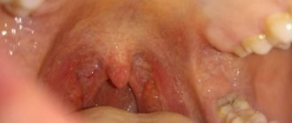
- Hemorrhages in the soft palate and tonsils, sclera, fundus. Scleral hemorrhage sometimes precedes severe cerebral hemorrhage, which occurs quickly and progresses.
- Manifested by dizziness , headache , convulsions .
With von Willebrand disease, there is a tendency to intradermal hemorrhages, effusions of blood into the mucous membranes and severe bleeding after injury.
Duration of bleeding, normal limits
One of the frequently asked questions by patients who are about to have a wisdom tooth removed is how long does the formed socket bleed and what to do if the bleeding does not stop.
As a rule, after 10-15 minutes after surgery, a blood clot forms in the socket; in some patients, approximately 30-40 minutes may pass before the clot appears. Under no circumstances should this clot be removed, because it protects the wound from infection.
Deviations from the norm are situations where bleeding continues after returning home. But some patients worry in vain, mistaking ichor for blood. If the bleeding in most cases stops within 30-60 minutes, then the discharge of ichor from the wound can be observed for up to 12 hours or longer.
Ichor is a yellowish or colorless liquid with a small blood capacity. Each person may have a different intensity of discharge depending on the characteristics of the body, but in any case this is not a cause for concern.
If blood is released from the wound uncontrollably for a long time, you should inform the doctor about this without waiting for complications.
Tests and diagnostics
Coagulological screening includes:
- prothrombin index;
- activated partial thromboplastin time;
- amount of fibrinogen;
- platelet count;
- bleeding time.
In case of isolated prolongation of activated partial thromboplastin time, proceed to the second stage of examination:
- do a correction test;
- activity of factors VIII, IX, XI, XII.
If the activity of factor VIII decreases, they proceed to the third stage of examination:
- lupus anticoagulant;
- specific inhibitor of factor VIII.
In children
Poor blood clotting in a child is often associated with immune thrombocytopenic purpura . Acute purpura develops between the ages of 2 and 9 years. This is an immune-related disease characterized by a constant (or periodic) decrease in platelets of less than 100 thousand. The disorder in children occurs 1-3 weeks after a viral infection. Such pathological reactivity can start not only under the influence of a viral infection, but also after taking medications, vaccination, exposure to temperatures (both low and high), surgical interventions or emotional stress. Against the background of normal health, the child develops a petechial rash (on the mucous membranes and skin), bruises, repeated nosebleeds and bleeding gums. In severe cases, there may be brain hemorrhages and stomach bleeding. Since antigens gradually leave the blood, in most people the disease goes away on its own after 2 months.
Vitamin K deficiency coagulopathy occurs in children in the first months of life and newborns . With Vit-K deficiency, the activity of several factors decreases: prothrombin , proconvertin , Christmas factor and Prower factor . hypocoagulation develops , accompanied by hemorrhagic syndrome.
Causes of Vit-K deficiency in a newborn:
- taking anticoagulants during pregnancy;
- antibiotics;
- anticonvulsants;
- severe damage to the pregnant woman’s liver and intestines;
- the presence of fetoplacental insufficiency;
- gestosis and preeclampsia .
The manifestations of this coagulopathy in newborns are not very specific - skin syndrome, increased bleeding during blood sampling and bleeding from the umbilical wound. With a lack of Vit-K, the duration of bleeding and platelet levels are within normal limits. Many authors recommend prophylactic administration of Vit K to all children immediately after birth - 2-3 administrations for the first 1.5 months, and in some cases weekly administration continues up to 3 months.
In later life, Vit-K deficiency coagulopathy is caused by breastfeeding only. At the same time, 78% of children develop massive intracranial hemorrhages. Much less frequently, the cause of decreased coagulability in children is disseminated intravascular coagulation syndrome in severe sepsis, congenital metabolic changes and hereditary coagulopathies.
Poor blood clotting during pregnancy
A normal pregnancy is always accompanied by important biochemical changes, including in the hemostatic . However, there are pathological conditions that lead to blood clotting disorders. Coagulopathy in pregnancy, what is it? This is a pathological condition that occurs with impaired coagulation and an increased risk of bleeding.
Pathological bleeding in pregnant women can be caused by:
- Congenital disorders in the coagulation system.
- Werlhof's disease.
- Moschkowitz disease.
- Antithrombin deficiency , which often occurs during pregnancy.
- Preeclampsia.
- Eclampsia.
- DIC syndrome associated with preeclampsia. At the first stage, intravascular coagulation occurs, and then the coagulation system is depleted.
- HELLP syndrome, which is also associated with preeclampsia (hemolysis, elevated liver enzymes and thrombocytopenia). Disseminated intravascular coagulation syndrome, a combination of thrombosis and bleeding.
- HELLP syndrome combines a triad of manifestations: hemolysis , decreased platelet levels and increased liver enzymes. During pregnancy with severe preeclampsia, the frequency of this syndrome reaches 20%.
- It develops during full-term pregnancy, premature birth and even after childbirth. HELLP syndrome is considered a subtype of preeclampsia. Diagnostic criteria for this syndrome: platelets less than 100×10 in 9/l, transaminases 2-3 times higher than normal, hemolysis of erythrocytes, bilirubin more than 20.5 µmol/l.
Women experience nausea , swelling and pain in the right hypochondrium. Pregnant women with HELLP syndrome are prescribed magnesium sulfate before and after birth for two days. Platelet transfusion is indicated when platelets are less than 20 × 10 in 9/L if a natural birth is expected and when platelets are less than 50 × 10 in 9/L if a cesarean section . Thromboconcentrate is administered before delivery. Corticosteroids increase platelet levels, so their use is reasonable. In the postpartum period, plasma exchange is used.
In DIC syndrome, a short phase of hypercoagulation is replaced by hypocoagulation. Such changes occur if a woman loses 15-20% of her blood volume. In case of bleeding, a platelet concentrate is infused.
In case of bleeding and if the prothrombin time and activated thromboplastin time are increased, an infusion of fresh frozen plasma is performed. If it is not possible to administer plasma, clotting factor concentrates are administered. Severe hypofibrinogenemia , which is not corrected by plasma transfusion, is then transfused with cryoprecipitate . For hyperfibrinolysis and bleeding, tranexamic acid . Hemolytic-uremic syndrome in women is accompanied by thrombocytopenia , kidney damage and microangiopathy . This condition most often develops after childbirth. Plasma exchange in this pathology is not very effective - the woman requires hemodialysis .
Disturbances in the coagulation system
A decrease in the content or activity of coagulation factors may be accompanied by increased bleeding (for example, hemophilia A, hemophilia B, von Willebrand disease). Excessive activation of coagulation hemostasis (for example, Leiden mutation of factor V) leads to the development of thrombosis (thrombophilia).
Hemostasis and pregnancy
Among all the causes of miscarriage, problems in the hemostatic system are in second place in frequency. Second after obstetric and gynecological reasons. What's the matter?
During pregnancy, the expectant mother's body prepares for childbirth. The hemostasis system is also being prepared to minimize blood loss during childbirth. Hemostasis is activated progressively along with increasing gestational age. If a woman’s hemostasis is initially overactive, then during pregnancy microthrombi may form in the vessels of the uterus or placenta, which leads to miscarriage or frozen pregnancy.
Under what conditions can this happen?
- 1. For hereditary thrombophilia,
more often when there is a violation of the metabolism of folic acid and its compounds (folates), when the amount of homocysteine in the blood increases. The reasons for the increase in homocysteine levels may be a lack of folic acid and vitamin B12 in the diet, thyroid disease, and kidney disease. It can also increase in smokers, coffee drinkers and while taking medications such as theophylline (by the way, a relative of caffeine), nicotinic acid. Homocysteine damages the endothelium (inner layer) of blood vessels, and this damage triggers blood clots.
- 2. For antiphospholipid syndrome (APS)
– this is the name of an autoimmune disease in which antibodies are produced to one’s own clotting factors. As a result, blood clots also spontaneously form in the vessels.
Prevention
It is impossible to influence congenital pathologies of the coagulation system, but people with such pathologies can take a more careful approach to their health, choice of profession and physical activity. Secondary prevention includes the following:
- Avoid physical activity associated with the possibility of getting bruised (sports, football, wrestling, figure skating, etc.).
- Oral hygiene that helps reduce the need for dental procedures and surgeries.
- Normalize weight, which puts stress on the joints and increases the risk of bleeding into the joint cavity.
- Avoid the use of medications that affect clotting. Among such drugs are acetylsalicylic acid , Clopidogrel , Caffeine , Ibuprofen , Naproxen , nitrofurans , barbiturates , carbenicillin .
- Women with congenital factor deficiencies should consult and be examined by a geneticist and find out the risks of having a child with a congenital blood clotting disorder.
Similarly, there is no primary prevention of thrombocytopenic purpura , and secondary prevention is limited to preventing exacerbations. Patients should not be exposed to the sun; they are contraindicated from working in conditions of elevated temperature (hot shop, food shop near the stove). Children are exempt from physical education. After each case of ARVI, it is mandatory to test the blood.
Causes of bleeding
As a rule, hemorrhage (hemorrhage) occurs as a response to damage during soft tissue surgery or as a result of exposure to certain medications. Such problems usually go away within a few hours after the procedure.
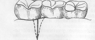
If an ordinary tooth can be pulled out without much difficulty and a cotton swab applied to the wound, then removing third molars is much more difficult. During the manipulation process, it is often necessary to saw the crown of the tooth and pull it out in parts. With deep and curved roots, it is almost impossible to extract them without damaging bone and soft tissue.
In addition, there are also other reasons why the bleeding does not stop after a wisdom tooth has been removed:
- high blood pressure;
- mechanical injuries of a large vessel;
- the patient is taking blood thinning medications, for example, Heparin, Aspirin and others;
- low blood clotting;
- the use of anesthetics, after which the blood vessels dilate. This increases bleeding and slows down the process of clot formation;
- the presence of concomitant diseases can cause heavy bleeding. These include hepatitis, scarlet fever, leukemia, hemophilia and others;
- Overheating of the body or high physical activity immediately after removal increases the risk of hemorrhage.
If immediately in the postoperative period the patient abuses alcoholic beverages or smoking, bleeding can occur as complications.
If wisdom tooth extraction cannot be avoided, then the patient needs to understand that bleeding after such a procedure is a natural and normal process. But even with relative safety, the release of blood requires careful monitoring by both the doctor and the patient himself.
Consequences and complications
With coagulopathies, the following complications may occur with varying probability and severity:
- Anemia.
- Hematuria (blood in the urine).
- Heavy and prolonged periods.
- Brain hemorrhage
- Gastrointestinal bleeding.
- Hemorrhage into the structures of the eye, pleurisy, compression of the larynx and trachea by hematomas.
- Hemorrhage into the joints and, as a consequence, the development of arthritis , arthrosis , osteoporosis and posthemorrhagic bursitis .
- Intravertebral hemorrhages.
- In rare cases, death due to massive bleeding.
Forecast
Acute forms of idiopathic purpura disappear within a few months and it often happens that the disease does not recur. In the chronic form, it is possible to induce remission, but the disease often recurs.
When using modern medications and following all recommendations for lifestyle changes and employment, thrombocytopenic purpura has a favorable prognosis.
Untreated hemophilia Lack of treatment often leads to joint pathology ( hemophilic arthropathy ), which then requires the use of crutches and wheelchairs or special orthopedic treatment. During treatment, life expectancy is practically no different from healthy individuals.
List of sources
- Fatkullin I.F., Zubairov D.M. Hereditary and acquired defects of the hemostatic system in obstetric and gynecological practice. M., 2002. P. 64.
- Galstyan G.M., Sukhanova G.A. Introduction to hemostasis, modern blood products and their effect on coagulation // Medical Council. 2013. from 11-13.
- Barkagan Z.S. Diagnosis and controlled therapy of hemostasis disorders / Z.S. Barkagan, A.P. Momot. – M.: Newdiamed, 2001. – 296 p.
- Barinov S.V., Dolgikh V.T., Medyannikova I.V. Hemocoagulation disorders in pregnant women with gestosis. Journal of Obstetrics and Women's Diseases. – 2013; 62 (6): 5–12.
- Degtyarev D.N., Karpova A.L., Mebelova I.I. and others. Draft clinical recommendations for the diagnosis and treatment of hemorrhagic disease of newborns // Neonatology. 2015. No. 2. P. 75–86.