The eruption of wisdom teeth is often accompanied by various complications. This is due to the lack of space in the dentition for the number eight, which erupts last. In dental clinical practice, third molar retention is often encountered - incomplete eruption of the crown above the gum. Removing a wisdom tooth in the gum is a small operation that requires not only extraction of the tooth, but also the creation of access and an incision in the gum. Our dental clinic performs extractions even in the most complex clinical cases. Dentists have extensive experience and high qualifications, and perform procedures painlessly and safely.
Hematoma
Description
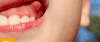
If there is a swollen lump on the gum filled with blood, most likely it is a hematoma. This tumor occurs as a result of an incorrectly removed tooth or injury. Such a lump on the gum above the tooth, as a rule, does not hurt.
Treatment
Dissolves on its own. But if the lump hurts, consult your dentist for advice. The doctor will prescribe painkillers.
Epulis
Treatment
The lump on the root of the tooth is attached to a stalk and is either red or gum-colored. It affects the lower jaw and occurs as a result of malocclusion, permanent mechanical damage, and poor quality dentures. Often observed in women with hormonal imbalance.
Treatment
It is removed by one of three methods: scalpel, diathermocoagulation or cryodestruction. The manipulation is performed under local anesthesia.
Cartilaginous choristoma of the gums: a rare nosological form
Choristoma is one of the types of congenital developmental anomalies, which, in fact, is a heterotopic change in cells. The term "choristoma" is more common. For the first time, this term was proposed to be used by Krolls and colleagues to describe the tumor-like growth of microscopically unchanged tissues in a place unusual for them.
In the oral cavity, this process is considered as a tumor-like process, which develops from a pool of primary cells located far from the original and characteristic tissue or organ. The formation may consist of a large amount of cartilage, bone, glial tissue, fat cells, nerve elements, lung tissue, thyroid tissue and intestinal mucosa.
Cartilaginous choristomas are quite rare in the oral cavity and consist of mature hyaline cartilaginous tissue. This article describes a rare case of cartilaginous gingival choristoma.
Clinical case
A 54-year-old man presented to the Department of Oral and Maxillofacial Surgery with a complaint of gingival swelling in the right lower posterior region of the mouth for the past 5 years (Figure 1). The swelling gradually increased in size, resulting in increased throbbing pain. A single fistula was present on the right side of the face (Figure 2). After an incisional biopsy, the sample was sent to the Department of Maxillary Pathology and Microbiology with a preliminary diagnosis of epulis.
Photo 1: Photograph of a choristoma in the oral cavity during the preoperative period.
Photo 2: Fistula on the right side of the face.
The sample measured approximately 1.7 cm × 0.5 cm × 0.3 cm, was soft in consistency, white in color, had a cylindrical shape and a lobed surface (photo 3).
Photo 3: Photo of a gross specimen of an incisional biopsy.
Histopathological examination identified the presence of peripheral parakeretinized stratified squamous epithelium. The underlying connective tissue stroma revealed a well-defined basophilic structure of mature cartilage.
Excisional biopsy was performed under general anesthesia. The resulting sample, greyish-black in color with creamy white flecks, measured approximately 4 cm × 1.5 cm × 0.6 cm, had a hard consistency, and was approximately triangular in shape with an uneven surface. Subsequently, the sample was divided into two equal parts (photo 4). Histopathological examination showed basophilic foci of mature cartilage in the stroma of dense fibrous connective tissue, together with groups of chronic inflammatory cells - plasmacytes and lymphocytes. Peripheral stratified squamous epithelium was also detected (Figures 7 and 8).
Based on the results of these studies, the final diagnosis was made: cartilaginous choristoma. There were no phenomena of receding education observed over the course of 3 years (photos 5 and 6).
Photo 4: Photo of a gross excisional biopsy specimen.
Figure 5: Intraoral photograph of choristoma after surgery.
Figure 6: Extraoral photograph of choristoma after removal.
Photo 7: Tissue section (parakeretinized stratified squamous epithelium with foci of cartilaginous tissue and inflammation in the connective tissue stroma) (H&E 20x).
Photo 8: Tissue section (basophilic lesions of mature cartilage tissue) (H&E 40x).
Discussion
Choristomas are the result of a dysmorphic proliferation of cells that are not characteristic of the corresponding organ or tissue in which they arise. Like hamartomas, they grow to a certain size, after which their growth stops. The term "choristoma" was first introduced by Krolls and colleagues. Some researchers characterize choristoma as a tumor-like formation that arises from a group of primitive cells located far from their characteristic tissue or organ. A heterotopic gastrointestinal cyst, which contains specific glandular structures, can be found in the tissues of the tongue or floor of the mouth in infants, and is considered a choristoma.
Atypical findings of bone or cartilage tissue in the tongue or thyroid tissue in the posterior third of the tongue are also identified as choristoma.
It can also be assumed that the commonly found Fordyce granules are, in fact, choristomas in the form of nonfunctional submucosal sebaceous glands. Uncharacteristic intestinal replication cysts of the floor of the mouth and osteomas of the tongue are also examples of choristomas. The tissues of the salivary glands in the structure of the lymph nodes can also be considered as choristomas.
Discussions are still ongoing whether choristoma is a developmental defect, a neoplasm, or has a reparative nature.
Some researchers support the theory that choristas develop gums from embryonic remains. This theory also explains the fact that pluripotent mesenchymal cells can differentiate into osteocytes or chondrocytes.
Cartilaginous choristoma was first described by Berry in 1890. The occurrence of this pathology varies within the age group from 10 to 80 years, and is more common in women. However, in this clinical case, the patient was a 54-year-old man. The most common location of diagnosed cartilaginous choristomas in the head and neck region is the oral cavity.
Cartilaginous choristomas are diagnosed as growing edematous formations without accompanying symptoms. This phenomenon was identified in our case, because the patient visited the dentist 5 years ago during the period of choristoma occurrence without any complaints about the tumor.
The tongue is the most common site of choristoma occurrence in the oral cavity. But in our case, the process arose in the gum tissue of the lower jaw. This topography is rare for this pathology.
Perrotti et al described a case of cartilaginous gingival choristoma.
The cartilaginous proliferations of the orofacial soft tissues appear to reflect the multipotent nature of the primitive mesenchymal cells of that part of the body in which their growth may be caused by trauma, irritation or inflammation.
In this case, the edema proliferated from the orofacial soft tissues, stimulated by local factors (tartar, poor oral hygiene).
Cartilaginous choristomas should be distinguished from cartilaginous metaplasia, which often occurs in soft tissues under the bases of poorly made dentures. In this clinical case, the patient had no dental defects.
Histopathologically, cartilaginous metaplasia is characterized by multiple distributions of cartilage cells and calcium at various stages of development in single or clustered cartilaginous lesions.
Histopathological examination in this case revealed basophilic foci of mature hyaline cartilage in a dense fibrous connective tissue stroma with focal infiltration of chronic inflammatory cells—plasmocytes and lymphocytes.
In the overlying peripheral stratified squamous epithelium, a process of parakeratosis was detected.
Conclusion
This clinical case describes a rare example of the occurrence of cartilaginous choristoma in the gum tissue of a male patient. The article focuses on the fact that cartilaginous choristoma is a harmarthroma in nature, and therefore its treatment protocol must be appropriate. Early and accurate diagnosis is possible only with an understanding of the characteristics of this pathology. Intraoral choristoma in most cases requires surgical excision. No relapses of the tumor after adequate treatment have been recorded so far.
Authors: Ramalingam Suganya, Narasimhan Malathi, Subramani Vijaya Nirmala, Chinnaswami Ravindran, Harikrishnan Thamizhchelvan
Periodontitis
Description
A hard lump on the gum, an abscess formed at the base of the tooth root. In the absence of therapy, it transforms into a benign tumor.
Treatment
If such compaction of the gums under the tooth appears, contact your dentist. The teeth are unfilled and root canals are cleaned. After removing the exudate, oral baths with medicinal herbs or soda solution are prescribed. Treatment for periodontitis may require multiple visits to the doctor.
A fragment remains after tooth extraction: why does this happen?
A tooth is an integral organ that consists of a crown and root parts. If a root or splinter remains after tooth extraction, it means the tooth has lost its integrity. This occurs due to mechanical stress during removal. Tooth destruction during extraction occurs for a number of reasons.
- Doctor's mistake.
Lack of doctor qualifications is a fairly common cause of complications. Incorrect tooth extraction technique can lead to the destruction of even a relatively strong tooth. - Poor condition of the tooth root.
If the root of the tooth remains in the gum, this is often due to the fact that the root part was in poor condition, so when you try to remove it, the tooth literally breaks into two parts. - When removing a tooth,
the neighboring one was hit, and a fragment from it fell into the socket. This happens quite rarely, but it still happens. This situation is also a consequence of incorrect extraction technique.
Flux (periostitis)
Description
An inflammatory process of bone tissue, which is accompanied by pain, elevated body temperature, enlarged lymph nodes, and swelling of the oral mucosa. The seals have purulent contents. The cause is an infection of the oral cavity resulting from untreated caries.
Treatment
A visit to the doctor is required to treat the lump. The dentist opens the tooth, places special medications in the cavity, and closes it with a temporary filling. If treatment does not bring positive results, the tooth is removed.
Fibroma
Description
Benign tumor of epithelial cells. Initially, the bumps on the gums above the tooth are small in size and do not hurt. However, mechanical stress and other negative factors can lead to the transformation of fibroma into a malignant tumor.
Treatment
Treatment is carried out surgically. The doctor excises the tumor, applies stitches, and prescribes rinses. Further follow-up visits may be required to monitor the recovery process.
Cyst
Description
A hard, bone-like compaction of gums under a tooth, up to 1 cm in diameter. Occurs in people with weakened immunity, genetic predisposition, as well as due to acute infectious diseases, mechanical damage to the soft tissue of the oral cavity.
Treatment
Treatment is carried out in the same way as for fibroma - by excision. After surgery, mouth rinses and baths are necessary, as well as repeated visits to the doctor to monitor recovery.
A wisdom tooth remains in the gum - is this critical?
Retention or partial eruption is characterized by incomplete emergence of the crown above the gum. Only 1-2 bumps or part of the tooth crown may appear. Retention can be independent or together with dystopia - incorrect positioning of the molar. Symptoms of the disease will depend on the individual clinical situation.
Depending on the location, retention can be vertical, horizontal or angular. With a vertical crown, the tooth crown is located normally in the bone, in accordance with other teeth. Horizontal is characterized by the location of the tooth perpendicular to the others, horizontally to the arch of the jaw. With angular retention, the crown is tilted to the side. According to the depth of its location, the tooth can be covered only by gum, but by a bone plate.
Retention is accompanied by the ingress of food debris and plaque under the gum. This causes inflammation, redness, tissue swelling, and pain. Pericoronaritis may occur - acute inflammation of the gums and mucous hood above the crown of the impacted tooth. The disease can be serous and purulent, causes acute pain, hyperemia, tissue swelling, and makes it difficult to open the mouth.
In most cases, a tooth in the gum is a source of various complications: pericoronitis, the formation of caries and its complications, cysts, stomatitis, periostitis, abscess. Therefore, in case of pathology, dentists recommend removal.
To date, exact methods for preventing retention are unknown. General measures include proper hygienic care of the oral cavity and monitoring the correct development of the child’s jaws and bite. As well as timely and correct orthodontic treatment of malocclusions.
Gum cancer
Description
A rare dangerous disease. It is characterized by the formation of a dense lump on the gum at the root of the tooth; it can develop asymptomatically for several months.
Treatment
Requires serious medical intervention, chemotherapy, radiation therapy, surgery, long-term recovery and follow-up.
Gum cancer, if detected early, is curable, otherwise it ends in death. Red and white bumps on the gums appear in both adults and children. Only a doctor can correctly identify the disease and prescribe appropriate treatment. The sooner you see a dentist, the greater the chances of a quick recovery.
Don't self-medicate! If a ball is inflated on your gum and you don’t know what to do, contact a dentist in Odintsovo. The doctor will conduct a full examination of the oral cavity, accurately diagnose the disease, and prescribe effective treatment.
You can make an appointment for a consultation, as well as find out the cost of admission and treatment by calling the clinic in Odintsovo.
Specialist actions
It doesn’t matter whether you turned for help to a professional dentist in a clinic or to an emergency room, the doctor’s algorithm of action is always the same. First of all, an external examination of the gums and teeth is required, and an x-ray examination is prescribed. X-ray helps determine the number of fragments, their location and depth.
Only after a preliminary examination can the doctor choose a specific technique for extracting fragments. After their removal, the hole must be washed with antiseptic solutions; if necessary, a drug with a powerful antibacterial effect is added to it. If fragments were found very close to the surface, they can be removed using a tool. If they are deep enough, then dissection of the gum tissue will be required.
Today, in all these cases, anesthesia is used, so the patient does not experience any discomfort. In rare cases, general anesthesia may be prescribed. It is very important to seek help immediately after identifying the first symptoms, this will allow you to remove the particles as quickly as possible and avoid serious complications in the future. Unfortunately, even the most highly qualified specialist is not immune from such situations, because everything depends on the complexity of the specific clinical picture and the individual characteristics of the human body.
previous post
What to do if a tooth grows from the roof of your mouth?
next entry