After a tooth was removed, did a lump appear on the roof of your mouth? Most often this is caused by tissue damage and infection entering the wound. Additional risk factors:
- non-compliance with specialist recommendations,
- improper oral hygiene,
- smoking and drinking alcoholic beverages,
- abuse of hot, very spicy and salty foods,
- immunodeficiency.
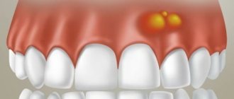
The formation on the palate may not appear immediately after surgery. In order not to miss the growth of the tumor, you need to regularly examine the oral cavity, and if there is swelling, immediately consult a doctor.
A lump appeared on the gum: what could be the reason?
A lump on the gum, by and large, is a compaction that is caused by damage to periodontal tissue in a certain area. The cause of the lesion is another point on which the treatment plan and possible complications in the absence of timely medical care depend. First of all, the doctor determines the nature of the appearance of the lump on the gum, which comes in two types.
- Infectious nature.
Infection caused by pathogenic bacteria is the most common cause of lumps and lumps in the gum area. - Non-infectious nature.
This includes injuries, mechanical, chemical damage, as well as other factors not related to the activity of microorganisms.
A lump on the gum above a tooth or under a tooth (in the case of the lower jaw) appears much more often as a result of infections, however, the treatment regimen is individual for each case, depends on the specific symptoms and is drawn up after collecting an anamnesis. Next, it makes sense to describe in more detail the diseases and pathologies that provoke the appearance of seals on the gums.
Diagnostic methods
To determine the method by which a lump on the roof of the mouth will be treated, the causes of its occurrence are studied. The diagnostic procedure is carried out by a dentist.
Regular visits to the dentist are important to prevent the disease and determine whether a lump has appeared on the roof of your mouth. It needs to be treated at an early stage.
For diagnosis use:
- Radiography;
- Biopsy;
- Blood analysis;
- Radioisotope study;
- Ultrasound.
If you comprehensively examine the ball on the roof of your mouth, this will confirm or deny the presence of a malignant formation, and if it is absent, it will determine the exact diagnosis of a benign one.
Infectious diseases that can cause a lump on the gum
- Periostitis (flux).
Periostitis, or flux, usually occurs against the background of complications of caries, pulpitis and periodontitis, when infection and pus extend beyond the periodontal tissues and penetrate into the soft tissue area. You can notice with the naked eye that there is a swollen lump on the gum. - Complications of periodontitis (granuloma, cyst).
When infections occur at the root apex, a periodontal abscess occurs. Gradually, pus penetrates into the periosteum area, and then directly under the mucous membrane. Thanks to its elasticity, the patient often does not feel any unpleasant symptoms from the lump that appears, so he is in no hurry to see a doctor. Quite dangerous are cases when the pathology takes the form of a fistula, which looks like a small white lump near the tooth. The fistulous tract allows pus to come out, but this does not mean that the inflammatory process will disappear on its own; - Gingivitis and periodontitis.
With periodontal inflammation, swelling and hardening of the gums are almost always observed. Gingivitis is considered the initial stage of the disease, therefore it is characterized by the so-called red ball (gum tumor), while with periodontitis, purulent discharge is observed, and the tumor acquires a beige or grayish tint. In some particularly advanced cases, against the background of periodontal problems, gumboil may appear, which visually looks much more noticeable.
Non-infectious diseases that can cause a lump on the gum
- Epulis.
A small growth that has some kind of stalk, which occurs mainly due to injuries, malocclusions, improper prosthetics, during teething (in children), and also due to hormonal disorders (less often). In appearance, epulis may resemble a tumor due to gingivitis, so diagnosing the disease may require x-rays and histological examination. - Ecostosis.
This pathology is a bone growth that is formed due to a jaw anomaly. The main cause of ecostosis is considered to be a genetic predisposition, but pathology can develop after injuries and traumatic tooth extraction. In this case, the bone extends beyond its usual boundaries, resulting in the formation of a noticeable lump. The disease is not accompanied by an inflammatory process, but it can occur due to mechanical damage to the growth. - Hematoma.
It is formed as a result of a strong blow and may well go away on its own or with the use of special means. If the tissue structure is disrupted as a result of injury, the help of a surgeon is needed.
It hurts/doesn’t hurt: in what cases does pain occur?
Periostitis is considered the most painful. With this complication, the main inflammatory process is localized in the area of the bone and periosteum, which causes severe pain. Seals caused by the inflammatory process in the periodontium (gingivitis, periodontitis) are disturbing when pressed, thermal and mechanical influences are applied. Epulis and ecostosis, as a rule, are not accompanied by pain, but when damaged and inflamed, they also disturb the patient.
A lump near a tooth with periodontitis may not cause noticeable pain, especially if it has a fistulous tract. It is important to understand that the absence of pain does not mean that treatment can not be carried out. In most situations, the tooth can be saved, but some patients are very negligent about their health and consult a doctor in extreme cases. A lump above (or under) a tooth is quite an alarming signal, which often indicates the development of quite serious diseases.
Content:
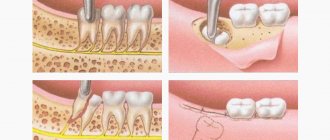
- In what cases are molars removed?
- Reasons for the formation of a lump after wisdom tooth removal
- Dangerous symptoms
- How to treat a lump after tooth extraction
- What happens if you don’t treat a lump after a tooth extraction?
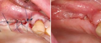
Most patients at dental clinics believe that pulling out a diseased tooth will relieve them of painful symptoms.
They treat the extraction procedure as a life-saving one. But, unfortunately, it is not always possible to solve the problem completely surgically. It happens that a small tumor soon forms in the vacant space. A seal on the gum after tooth extraction indicates the need to urgently visit a doctor. Below we will tell you why it occurs and how doctors fight it.
Lump after crown installation
Improper denture fitting is a very common cause of lumps on the gums. Most often, errors occur at the tooth preparation stage, when the depulpation procedure is carried out. Poor canal filling provokes the development of infection, which, in turn, leads to periodontitis. Another common reason is a poorly made prosthesis or defects during its installation. In this case, the prosthesis injures the mucous membrane and causes swelling, abscesses and growths. Treatment almost always involves removal of the prosthesis and subsequent replacement of the prosthesis (if possible). The sooner a complication is detected, the higher the chance that it can be successfully treated. A well-installed prosthesis should not cause pain, discomfort or injure the mucous membrane. The tooth under the crown should also not hurt, since the dental nerve is killed before permanent prosthetics.
Treatment of lumps on the gum
The treatment plan depends entirely on the results of the diagnosis, which is designed to determine the root cause of the lump or ball on the gum, which is why the patient should not self-medicate, but go to the doctor as soon as possible. If the cause of the lump is an infection, then you need to eliminate the cause of its appearance. For complications of pulpitis and periodontitis, treatment can take a long time. The lump is opened, a drainage is installed to pump out the pus, after which the doctor prescribes conservative or surgical treatment to the patient, depending on the severity of the disease. In case of periodontal inflammation, professional cleaning and sanitation of the oral cavity are carried out, special ointments and antiseptics are prescribed. In more complex cases, curettage, physiotherapy, and so on are used. With epulis and ecostosis, pain and inflammation are often absent, so the decision about intervention is made together with the patient. Most often, the pathology will be removed after surgery: even a small bump on the gum (especially in the smile area) spoils the aesthetics, so it is understandable that a person wants to get rid of it.
Preliminary determination of lump symptoms
A lump on the palate is the cause of a dental disease or inflammatory process. Each type of growth has its own causes and subtleties of symptoms, and therefore requires separate consideration.
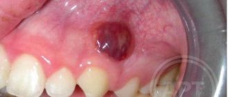
Angioma
Angioma is a tumor in the palate caused by disruption of processes in the tissues of the blood vessels of the soft palate. The shape of the cone resembles a rolled corkscrew. The tubercle has a blue or purplish-black characteristic color. The ball is a product of a disorder in the development of blood vessels, so the color is caused by the excess amount of blood in the formation.
Pressure causes bleeding, which should not be experimented with. There is a pulsating response to pressure.
Such a lump on the upper palate is life-threatening for the patient. Bleeding is difficult to stop, and large blood loss is fatal.
At the first signs of such compaction, you must seek qualified help.
Cyst
A lump in the mouth on the palate is classified as a cyst when a hard ball appears on the palate and its size does not exceed 12 cm. The cyst appears due to a disorder of the sebaceous glands. It won’t hurt, but it will make it significantly more difficult to eat, and then completely disrupt the correct functioning of these secretions.
A cyst on the palate requires surgery and can only be treated with it.
Pemphigus
Children are most susceptible to developing pemphigus. It pops up in the shape of a white ball at the top of the mouth. The bumps are a consequence of erosion and later develop into ulcers. When pressed it bleeds and hurts. Diagnosed by visual examination and using the Nikolsky method.
If the tumor is not removed, erosion will develop into massive exfoliation of the oral epithelium, disruption of digestive processes, and general weakness. If there is no treatment, the bumps become widespread. Pressure causes rupture of tumors, the contents of which cause intoxication of the body.
Myxoma
A myxioma is a hard, white growth at the top of the mouth. The disease affects the hard part of the palate, and upon visual examination it is almost invisible; pressure on the tumor is not felt. This significantly complicates diagnosis and delays the patient’s visit to the doctor.
The diagnosis can be confirmed or refuted by a biopsy of the palate.
Oncological disease
Cancerous lump is classified into two diseases:
- Hard palate cancer - starting from the bone tissue between the nasopharynx and the palate, the disease spreads to all mucosal tissues.
- Cancer of the soft palate - a lump appears due to an oncological process in the muscle and mucous tissues of the mouth.
Additionally, the oncological tubercle on the roof of the mouth is divided according to the tissue from which the spread of the disease began:
- Cylinder - the maternal tissue is the tissue of the glands, the cancer spreads quickly and invades the oral cavity;
- Adenocarcinoma - begins expansion of the mouth from the soft tissues of the cavity;
- Squamous cell carcinoma is a malignant formation that begins to develop from the tissues of the mucous membrane.