Tooth extraction is one of the most common operations in dentistry. It is indicated for severe destruction of the coronal part, periodontitis, inflammation with suppuration, a cyst against the background of advanced caries. After extraction of a dental unit, a hole filled with blood remains in the jaw. You need to take proper care of it to avoid complications. On average, wound healing takes 14-20 days. We tell you what you should not do after tooth extraction and what symptoms are a reason to contact your doctor.
Content:
- When white plaque is the norm
- When white plaque is a harbinger of disease
- Why do complications occur after tooth extraction?
- How to recognize the problem
- Diagnostic measures
- Treatment of alveolitis
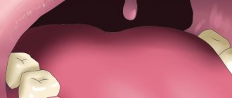
After tooth extraction, it is important to monitor the condition of the mucous membrane.
Very often a white coating appears in the extraction area. It frightens patients because it seems something strange to them, and to some it resembles an accumulation of purulent masses. It should be noted that white plaque in the socket after tooth extraction can be either normal or a complication.
In the first case we are talking about natural regeneration, in the second - about alveolitis.
Wisdom tooth transplant as a way to replace a defect
Of course, Kim was very surprised, but we showed her photographs of successful operations (and we have all of them successful), apparently our persuasiveness and the patient’s desire played a positive role in making the decision to transplant the eighth tooth. She was happy and agreed to the operation.
Moreover, in this case we did not even have to make a stereolithographic model of the tooth, since all surgical procedures (extraction and transplantation) were performed on the very first day of her visit to our clinic.
Dental transplant surgeries typically require several days of preparation, as we describe in the following clinical case study. But in this case, taking into account the current clinical situation and relying on our many years of successful experience in tooth transplantation, we were able to transplant tooth 8 into the place of tooth 6 as quickly as possible. We had all the indications for current dental transplantation.
The donor was an impacted (completely hidden under the gum) wisdom tooth 48. In fact, as we joked, it was a neighbor tooth to the problematic six. That is, the tooth grew and grew for 20 years, waited in the wings, and that hour struck! Now this donor tooth is for another 30 years
at least he will live in a new place.
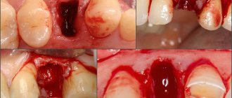
If you look at the sixth tooth from the side, you can see bluish, literally purple gums
:
This color of the gums is due to the developed inflammatory process. The patient developed destruction of bone tissue against the background of advanced periodontitis. There was a crack in the root of the sixth tooth.
When white plaque is the norm
Nature intended it so that damaged tissues should be protected from the penetration of infectious agents. This is why, after a tooth is removed, a clot and white plaque forms in the socket. The latter is nothing more than fibrin, a protein needed for blood clotting. It protects the wound surface from infection and does not allow the inflammatory process to spread.
At first, the white film seems very thin, but gradually it turns into bone tissue. Some even mistake plaque for food debris and try to remove it with their tongue or using foreign objects. Under no circumstances should this be done. If the wound heals according to plan, there are no severe pain symptoms, there is no need to worry or touch it. This can lead to complications.
So, fibrin in the form of a whitish coating is needed to:
- the wound healed quickly;
- no infection penetrated into the socket;
- soft tissues were not inflamed.
If the patient has questions regarding the white clot on the surface of the damaged gum, he should consult with his dentist. You don't need to do anything yourself.
Prevention
Regular cleaning of the oral cavity in an office setting has a positive effect on dental health. When the gum mucosa is affected, you should not touch it with your hands, burn it with alcohol, or eat hot and spicy food. You should carefully observe the rules of hygiene and systematically care for your oral cavity. It is good if the patient can quit smoking and drinking alcoholic beverages. The intake of salty, spicy foods and spices should be reduced to a minimum. You need to use a separate towel and dishes. It is important not to miss visits to the dentist, visit the dentist 2 times every six months.
It is better to rinse after breakfast, lunch, and dinner. This helps maintain the oral microflora in order. The risk of pathology is reduced if you perform a special massage of the gums in a circular motion. This improves blood circulation. It is useful to walk in the fresh air, take walks in a park or square. Avoid stress, alternate rest with feasible physical activity.
When white plaque is a harbinger of disease
It also happens that a whitish coating indicates the development of alveolitis after tooth extraction. This means that the tooth socket has become infected. In this condition, the patient is concerned about:
- severe pain in the area of the removed unit;
- increased body temperature;
- weakness, decreased performance;
- enlargement of the submandibular lymph nodes;
- headache;
- unpleasant putrid odor from the mouth and the same taste in the mouth.
If you experience these symptoms, you should make an appointment at the dental clinic as soon as possible.
Symptoms and treatment of leukoplakia -
Leukoplakia is characterized by the fact that under the influence of various irritants, keratinization of the epithelium of the oral mucosa begins to occur. This process occurs due to a violation of the desquamation of the epithelium in those areas of the mucous membrane that is exposed to pathological irritants. These irritants can be:
- smoking,
- sharp edges of carious teeth,
- overhanging edges of fillings,
- malocclusion,
- excessive consumption of spices, very hot foods,
- poorly manufactured removable dentures,
- the presence of artificial crowns made of different metals on the teeth, which gives rise to galvanic currents in the oral cavity.
White spots on the gums with leukoplakia: photo
Symptoms of leukoplakia:
The main element of leukoplakia is a white plaque, which may rise above the level of the mucous membrane, but may not rise (as in Fig. 4). In addition, the plaque can have both clear and blurred boundaries. Patients most often call such plaques white spots. As a rule, white spots do not bother their owners and are discovered completely by accident.
In some cases, patients may be concerned about the roughness of the mucous membrane at the site of the white spot, as well as the fact that the mucosa in this place may rise above the level of the mucosa. In some cases, cracks and ulcers may appear on the surface of the stain; in this case, the patient may also be bothered by burning and pain. If leukoplakia develops as a result of smoking, then, as a rule, patients first of all complain of dryness and burning of the oral mucosa (24stoma.ru).
Treatment of leukoplakia will primarily involve eliminating exposure to the irritating factor. Those. it will be necessary to eliminate the impact of the overhanging edge of a filling or crown, replace old low-quality dentures, normalize oral hygiene, and of course, solve something with the effects of nicotine and hot dry air on the mucous membrane (which occurs when smoking).
Only after this are prescribed keratolytic agents for topical use (3-5-10% salicylic acid, celandine infusion), and vitamin therapy. Applying any treatment procedures without eliminating the causative factor will be useless.
The danger of leukoplakia –
Leukoplakia belongs to the category of precancers. Malignancy of leukoplakia, i.e. its transition to cancer occurs in 15-75% of cases (depending on its form). Therefore, if you do not want to develop cancer of the oral mucosa, you need to urgently go to the dentist and look for the cause of its appearance. But in the photo below you can see photos of two clinical cases when leukoplakia transformed into a malignant tumor of the oral mucosa and the red border of the lips.
Cases of cancer associated with leukoplakia –
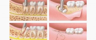
Why do complications occur after tooth extraction?
The most common reason for the development of complications is the patient’s failure to comply with medical prescriptions. Normally, the hole should heal within a few days. She shouldn't be sick for a long time. As a result of the fact that the blood clot closes it, it is reliably protected from viruses and food debris.
It happens that a person actively rinses the wound or constantly touches it with his tongue, fingers, and tries to remove the light coating. Then the clot displaces or does not form completely, the tissue becomes infected and alveolitis develops.
Untreated neighboring teeth can also lead to disease (which is why all carious “cavities” need to be treated before removal), and failure to comply with the rules of oral hygiene. If you properly prepare for extraction and follow all the doctor’s instructions, the risk of developing alveolitis after removal will be minimal.
Using antiseptic baths
To prevent the development of an infectious process, it is recommended to use baths with an antiseptic. They may contain the following medications:
- Chlorhexidine;
- Miramistin;
- healing herbs.
It is recommended to rinse in the following steps:
- take a small amount of liquid into the mouth;
- hold for 2-3 minutes;
- spit.
After rinsing, do not eat food for 1 hour. Baths can be repeated 2-3 times a day.
Baths are not recommended for all patients. They are prescribed only in case of active inflammatory process or risk of infection. They are often prescribed after opening the flux or if there is a tooth with caries next to the hole. Baths are useful for patients with periodontitis or periodontal disease.
How to recognize the problem
How to understand that white plaque signals a problem?
If the gums heal well, then within 2-3 days after surgery the pain becomes very weak and barely noticeable. The wound closes with a dense clot and practically does not bother the person. With the development of alveolitis on days 3-5, the pain, on the contrary, intensifies. It's pulsating. It can become unbearable. Sometimes it radiates to the entire half of the face. Body temperature rises, lymph nodes enlarge, appetite decreases, salivation becomes stronger. The face may also become swollen.
In such a situation, staying at home is dangerous - inflammation can spread to the deep tissues of the jaw. You need to immediately go to the dental clinic.
Treatment of white spots on gums
Depending on the diagnosis, the problem is eliminated by a therapist, dentist, endocrinologist, gastroenterologist, infectious disease specialist and other specialists. Therapeutic methods:
- Removal of soft deposits and hard stones.
- Treatment of pathological masticatory organs.
- Using baths and applications.
- Carrying out rinsing with antiseptic substances.
Cysts and granulomas most often require surgical intervention. Classical therapy can be carried out, which involves opening the canals, thoroughly cleaning the passages, and mandatory rinsing with special solutions. New technologies require modern equipment, as well as medicines. For leukoplakia, the doctor eliminates the irritating factors of the disease. Vitamin preparations are prescribed. If fungal infections are detected, complex therapy is carried out, which includes sanitation of the oral cavity, taking medications, and treating the affected areas with antiseptics. The patient must adhere to a diet.
Stomatitis is treated with anti-viral drugs. In case of an allergic reaction, it is recommended to take antihistamines. If you need to remove a fistula, pulpitis and caries are treated first. Fat deposits are removed surgically. A modern method of removal is the use of laser beams. Herpes is easier to prevent. Most often, Acyclovir is prescribed for this disease.
Treatment of alveolitis
If alveolitis occurs after tooth extraction, you need to undergo dental treatment aimed at:
- elimination of the infectious focus;
- preventing possible complications;
- maintaining the integrity of the rest of the dentition.
First, the dentist cleans the socket and rinses it. It is important to wash away all purulent masses and dead tissue from it. Antiseptics and hydrogen peroxide work well for this purpose.
To reduce painful symptoms, which deprive the patient of the opportunity to fully rest and work, analgesics are used. It is better to use them by applying applications. Compresses are changed every half hour. It is also possible to take drugs orally, that is, in the form of painkillers and anti-inflammatory tablets.
If the patient has concomitant diseases or reduced immunity, the doctor may decide to prescribe antibiotics. They quickly relieve inflammation and speed up the healing of damaged tissue.
If you seek qualified dental care in a timely manner, the prognosis for alveolitis is favorable. After 3-5 days, the symptoms begin to subside, and the person’s well-being improves. Residual socket pain may persist for another 2-3 weeks, but it does not pose a threat.
Should wisdom teeth be preserved?
We think yes.
Although wisdom teeth are not involved in the process of chewing food, they can still benefit a person. There must be clear medical indications for their removal. And now we will provide you with another proof of the benefits of your own teeth-eights. Our patient Kim is about 20 years old, she is from Korea and lives in Moscow, as her parents work at the Korean embassy. I contacted the German Implantology Center due to inflammation of tooth 4.6 – the lower right six. The tooth was destroyed
, an abscess developed. There was a temporary filling on the tooth (placed in another clinic):
Where a temporary filling was placed, she was told to remove the tooth. That's why Kim came to us with one simple goal - to atraumatically remove the six, which began to greatly bother and hurt her. After doing a CT scan and looking at the tooth, we offered her an option, which, upon hearing, Kim at first did not believe her ears. Our proposal was to remove the six and immediately in its place... transplant its lower right wisdom tooth! This is called autotransplantation surgery.
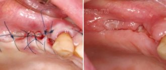
After autotransplantation 2 weeks
Patient Kim came to us after 2 weeks
for medical examination and suture removal:
Nothing bothered her anymore. The tooth had acceptable physiological mobility. The gums have become lighter. Of course, all the purple color is not gone yet, but she has already become more pink.
Pay attention to the white area - this is not pus, but fibrin plaque, which we will talk about in more detail below.
The picture shows that the donor tooth has clearly positioned itself in the position of the extracted tooth 46:
Our Kim has a new six molar. Both the patient and the medical staff of the Research Center are very pleased with the outcome of the tooth transplantation operation.
Occurrence of complications
Operations to pull out figure eights are considered complex dental procedures, which are due to:
- difficult access;
- frequent retention;
- unpredictable structure;
- features of the location of the mandibular alveolar nerve.
Such procedures are very traumatic, which creates the preconditions for the occurrence of general and specific types of complications.
Are common
The nature of complications largely depends on the location of the tooth: on the upper or lower jaw. But there are also general complications that arise in almost everyone, regardless of the location of the figure eight and its initial condition. The most common overall effects are listed below.
Painful sensations
Approximately 2-3 hours after molar extraction, you will experience noticeable pain in the gums. This is a normal reaction of the body to trauma, which is dental surgery. During it, soft tissues are torn or cut, the bone is injured (if the tooth is located under it), blood vessels and nerves are damaged. Painful sensations should completely disappear after 2-4 days, and for some people disappear within a few hours. They can be reduced with the help of painkillers prescribed by the dentist. If the pain does not go away, and the cheek is very swollen, it means that the gum healing process is proceeding with complications.
Swelling of the tissues of the face and neck
Often after figure eight extraction, especially impacted ones, swelling of the soft tissues of the gums or cheeks is observed. This is also a reaction to injury, which can be called a normal consequence of a dental procedure. In addition to swelling, the following symptoms may occur:
- swelling of the lymph nodes;
- discomfort when swallowing;
- painful sensations during mouth movements, radiating to the ear.
Normally, severe swelling should go away completely in 2-3 days, and if it doesn’t go away, then we can talk about more dangerous consequences. If the condition worsens every day, difficulties arise when breathing, fainting, the temperature rises and the skin becomes covered with a rash, then such swelling is provoked by an allergy and it can lead to anaphylactic shock.
Hematomas
Hematoma due to extraction is usually expressed by minimal bluishness of the cheek, which goes away after a few days. But there are situations when the appearance of a bruise is accompanied by severe pain, swelling, and an increase in temperature. In such a situation, medical attention is needed. Hematomas form after vascular damage in people with increased capillary fragility, as well as when the patient has hypertension.
Alveolitis
complications after wisdom teeth removal
Alveolitis is often provoked by non-compliance with doctor’s recommendations after treatment procedures. It is a local inflammation of the gums with the following additional symptoms:
- the gums swell and turn red;
- local pain and headache are observed;
- sore throat;
- the temperature rises, muscle aches appear;
- lymph nodes become inflamed, most often the submandibular ones.
If inflammation occurs, the cause is often the loss of a blood clot from the socket and infection. Various infections that enter the wound due to poor hygiene can provoke extensive inflammation. In advanced cases, the above complication develops into osteomyelitis, which is expressed by:
- increased persistent temperature;
- poor general health;
- severe migraine-like pain;
- nausea;
- other signs of intoxication of the body.
Increased body temperature
A slight increase in body temperature to 37.5-38 °C also often occurs in the postoperative period. This complication occurs due to a reaction to inflammation. The temperature should completely return to normal within the first day, and if it continues to rise and rise, it means that more serious pathologies have arisen and you need to go to the hospital again.
Bleeding
The dentist will never dismiss a patient with severe bleeding, especially if it is complicated. After removing the figure eight, the bleeding is stopped in the hospital, and then sent home with a gauze pad on the hole. If blood clotting is normal, then the bleeding will stop within 10-15 minutes, after which the tampon must be removed. Severe prolonged bleeding is provoked by:
- rupture of large vessels;
- capillary fragility;
- hypertension.
Damage to the roots of adjacent teeth
Such a complication is observed extremely rarely and only if the patient did not undergo X-ray diagnostics before the procedure. This procedure is mandatory for all people with indications for the removal of figure eights and allows you to fully assess the condition, location, as well as other features of the tooth and its roots.
Flux
Flux in the postoperative period develops in cases where the gums become infected due to the fault of the dentist or patient, after which the infection quickly reaches the periosteum and provokes its inflammation. This complication is not considered normal and acceptable and must be treated. Main signs of flux:
- redness, suppuration and swelling of the gums;
- severe shooting pain;
- temperature increase;
- weakness.
Others
Among other common complications, the most commonly observed are:
- displacement of the seventh tooth (2 molars);
- mouth rupture; cut gums or cheeks;
- jaw injuries.
In addition, complex extraction of the 8th tooth can cause the formation of a cyst. This is a small tumor located at the root of the tooth and filled with fluid. The cyst often serves as an insulator for infected cells from healthy ones. To prevent its occurrence, the dentist prescribes antibiotics, and the treatment in this case (if the cyst is located at the roots of the tooth) will be resection.
Diagnostics
The main stages of diagnosing reimplantitis are visual examination and targeted x-ray. Additionally, a panoramic image, tomography, and blood tests may be prescribed.
A comprehensive examination allows you to exclude other diseases of the oral cavity in which the mucous membranes become inflamed. Thus, the appearance of white plaque on the gums after tooth implantation may be accompanied by:
- stomatitis,
- thrush,
- periodontal inflammation,
- cyst in the area of the tooth root,
- leukoplakia.