- What kind of disease is candidiasis?
- Symptoms
- Causes
- Treatment
- Therapy methods
- Preventive measures
Many parents are aware of such a common problem in children as thrush, which is characterized by the appearance of a white coating on the tissues of the oral cavity. In medicine, this pathological condition is called “candidiasis” and refers to fungal diseases. Candidiasis most often develops in a child’s mouth in the first year of life. The symptoms of thrush are very disturbing for the baby, but timely treatment allows you to get rid of the fungus quickly and without consequences.
What kind of disease is candidiasis?
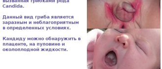
According to statistics, about 30% of infants experience candidiasis. The causative agent of the disease is Candida fungus. These specific microorganisms are normally present in the body of every person, even in the absence of health problems, but only in small quantities.
Oral candidiasis in children is much more common, since the immune system of children is not yet formed and is not able to resist pathogenic microorganisms and the effects of negative external factors.
With weakened immunity and the concomitant influence of provoking factors, the fungus begins to actively multiply, affecting the mucous membranes. Without therapeutic treatment, candidiasis is eliminated in exceptional cases. Much more often, if thrush is not treated, complications arise, and the infection itself spreads throughout the body.
Candidiasis of the intestines and other digestive organs
After Candida damage to the skin and visible mucous membranes, candidiasis of the gastrointestinal tract in infants ranks third. Carriage of microorganisms is quite common when the pathogens do not cause disease. Good immunity inhibits the development of the infectious process. But under certain circumstances, weakened children may develop candidal esophagitis (inflammation of the esophagus), gastroduodenitis (inflammation of the stomach and duodenum), enteritis (inflammation of the small intestine), enterocolitis (inflammation of the small and large intestines).
Factors contributing to the development of gastrointestinal candidiasis
The following factors contribute to the development of intestinal candidiasis:
- An imbalance between Candida colonization in the intestines and defense mechanisms that prevent excessive growth of microorganisms.
- The presence of physiological intestinal dysbiosis.
- Features of the structure of the mucous membrane in newborns. Failure of its protective and secretory functions.
- Long-term antibiotic therapy leads to the death of “good” bacteria and excessive growth of opportunistic flora, including fungi.
Signs and symptoms of candidal esophagitis
The disease may be asymptomatic or have nonspecific manifestations. Classic typical manifestations of candidal esophagitis are recorded extremely rarely. The infectious process develops in children born to mothers with vaginal candidiasis on the second day of life. Lack of appetite, refusal to breastfeed, excessive regurgitation containing whitish lumps, vomiting and rapid increase in dehydration are the main symptoms of the disease. Toxicosis is slightly expressed.
The basis of diagnosis is fibroesophagogastroscopy and culture of vomit on nutritional media in order to identify fungi of the genus Candida.
Rice. 10. Microscopy reveals pseudomycelium of Candida b and budding cells (photo on the left); when inoculating the vomit, an increase in colonies of pathogens is noted (photo on the right).
Signs and symptoms of intestinal candidiasis
Intestinal candidiasis in children occurs with symptoms such as lack of appetite (sometimes infants completely refuse to eat), regurgitation and vomiting, pain, bloating and rumbling in the abdomen, and loose stools. The vomit contains greenish or whitish mucus, and the stool contains yellowish-greenish mucus and white lumps. The smell of stool is normal. Symptoms of toxicosis are mild or absent. Microscopy of vomit and feces reveals fungal mycelium and budding pathogen cells. Candida is isolated from feces in crops in quantities of more than 10 4–5 CFU/ml.
In more than half of infants, intestinal candidiasis is combined with lesions of the skin around the anus, buttocks and visible mucous membranes.
Rice. 11. Often in infants, intestinal candidiasis affects the skin around the anus and buttocks.
Treatment of candidiasis of the gastrointestinal tract
In the non-invasive form of candidiasis, fungal cells do not form pseudomycelium and do not germinate the intestinal mucous layer. They multiply intensively in the lumen of the organ, disrupting cavity and parietal digestion. Treatment in this case is carried out with antifungal drugs that are not absorbed from the intestinal lumen - Nystatin.
In the invasive form of intestinal candidiasis, candida germinates the epithelial layer and penetrates beyond the basement membrane. The pathogens then enter the lymphatic system and bloodstream and spread throughout the body. In this case, Flucanazole, Ketoconazole and Intraconazole are effective.
The drugs are completely absorbed in the upper gastrointestinal tract.
If treatment is ineffective in children in intensive care units, Amphotericin B is used intravenously to treat candidiasis. For intestinal candidiasis in newborns, the drug is rarely used due to its high toxicity.
To correct the composition of intestinal microflora, probiotics are used. Immunocorrectors are used according to indications.
Symptoms
Oral candidiasis in children can have different forms of manifestation and severity of symptoms: mild, moderate and severe. As a rule, each form corresponds to the stage of development of the disease. If therapy is not carried out in a timely manner, the signs of fungal infection become more intense and the number of symptoms increases.
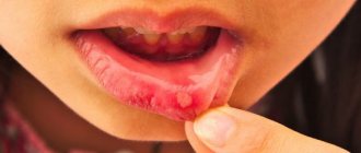
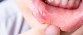
In mild forms of the disease, a red rash appears on the oral mucosa, which is covered with a white coating on top. At the next stage, the child experiences swelling of the tissues and the formation of localized white spots with a coating of curd consistency. Gradually these spots merge into a larger affected area. When plaque is removed, bleeding ulcers open. If thrush starts, the fungus spreads to the entire oral cavity, including the lips, tongue and throat. All fabrics are completely covered with a curd coating.
Common symptoms of candidiasis in children include:
- burning and itching sensation in the mouth;
- discomfort and pain when eating;
- frequent regurgitation in babies;
- formation of cracks in the corners of the lips;
- temperature increase.
Children in the first two years of life report their condition by refusing to eat, constant whims and crying for no reason. It is not difficult to see the signs of candidiasis, so if a child has a sharp rise in temperature or refuses to eat, pediatricians and dentists recommend checking the oral cavity for the presence of white plaque. If you suspect thrush, it is not advisable to delay a visit to a specialist, since the fungal infection progresses quickly.
What does thrush look like in a child?
Any part of the oropharynx can be affected: tongue, lips, corners of the mouth, gums, mucous membranes of the cheeks, palate, pharynx, tonsils. White grains (spot coating) appear, reminiscent of curdled milk, or a film (solid coating) of a cheesy appearance. These elements are easily removed by scraping with a spatula, revealing a bright red base, sometimes with a bleeding surface. As a rule, the course of the infection is painless and has only local manifestations. General health is not affected.
Causes
The main reason for the development of candidiasis of the oral mucosa in children is weakened immunity. If a baby is born premature, the likelihood of developing thrush is very high. Children who are breastfed or have congenital pathologies are also often exposed to fungal infections.
Provoking factors include:
- the presence of vaginal candidiasis in the mother during pregnancy (the child can become infected when passing through the birth canal);
- insufficient hygiene of the female breast during breastfeeding (the fungus is often localized specifically on the nipples due to the environment favorable for it);
- poor handling of the child’s initial things (bottles, pacifiers, etc.);
- parents’ habit of licking nipples (even if the adult has no signs of thrush, there may be a fungus in the mouth that can be passed on to the child);
- long-term use of drugs from the “antibiotics” group (medicines help reduce one’s own immunity);
- frequent regurgitation in infants (after regurgitation, an increased acidic environment will remain inside the oral cavity, favorable for fungus);
- excessive and frequent dry mouth (lack of saliva as a protective agent against the activity of pathological microorganisms).
Infection with Candida fungus in children over 2 years of age can occur as a result of consuming unwashed foods, raw milk or running water. If a child over 3 years of age suddenly shows signs of thrush, they should be examined not only for a fungal infection, but also for other possible diseases that may be accompanied by a “decay” of the immune system.
Skin candidiasis
The disease in infants is often mistaken for allergic dermatitis. The disease occurs with a frequency of 10 to 24% and usually manifests itself at 2 to 3 weeks of a child’s life. Its distinctive feature is the predominance of the erythematous form of inflammation.
Skin candidiasis in children can be congenital or acquired. It occurs in erythematous or vesicular forms. It has several variants of the course - damage to large folds and candidiasis diaper dermatitis.
Congenital candidal dermatitis in children appears more often on the second day of life. With cephalic presentation, the skin of the upper part of the body is affected: face, head, upper back. The mucous membranes are not affected.
Acquired candidal dermatitis in children begins with large folds and always occurs with thrush in the mouth. In the absence of adequate treatment, the infectious process spreads to the smooth skin of the thighs, abdomen and back. In advanced cases, the entire skin is affected. The disease should be distinguished from bacterial diaper rash, which is characterized by weeping, which is not characteristic of skin candidiasis and the absence of peripheral foci of screening and other dermatomycosis in newborns.
Diagnosis of mycosis is based on the detection of pseudomycelium and budding candida cells by microscopy and/or isolation of pathogen cultures.
The vesicular form is characterized by the appearance of a significant number of flabby and small bubbles against the background of hyperemia.
The erythematous variant of inflammation is characterized by the appearance on the skin of smooth, shiny, moist, tense areas of a dark red color. Dark red color of the lesions, distinct borders, scalloped edges, varnish shine, a moist rather than weeping surface, a narrow border of desquamated epithelium along the periphery, the presence of dropout foci (erosions and fresh blisters) along the periphery, absence of dryness and itching even in cases of widespread lesions are the distinctive features of skin candidiasis in children.
Rice. 12. Erythematous form of skin candidiasis in infants (diaper dermatitis).
Rice. 13. Vesicular form of skin candidiasis in a child.
Candidal dermatitis of large folds
Damage to the skin of large folds begins with redness and swelling, against which small blisters and pockets of screening appear along the periphery. Candidal dermatitis is often mistaken for diaper rash. Clear lesions with scalloped edges, a tense, smooth and shiny surface and the presence of desquamated epithelium (epidermal corolla) along the edge of the white border are its distinctive features.
Rice. 14. Erythematous form of candidal dermatitis.
Candidiasis diaper dermatitis
The disease often develops in children who carry candidiasis in the intestines. On the skin, as a result of massive fusion of vesicles and lesions, erythematous plaques with a fringed, clearly defined edge appear, along the periphery there are foci of screening.
Typically, the infectious process involves the perianal area, inguinal folds, perineum and lower abdominal wall. In boys, the skin of the penis and scrotum is affected, in girls - the vagina and labia.
Rice. 15. Erythematous form of skin candidiasis in infants.
Treatment of skin candidiasis in children
To treat the disease in newborns, antifungal drugs are used in the form of creams and ointments: Ketoconazole, Isoconazole, Clotrimazole, Econazole, Sertaconazole. At the same time, oral thrush and intestinal candidiasis are treated. For congenital candidiasis and severe condition of the newborn, Fluconazole is used orally or intravenously. If there is no effect, Amphotericin is prescribed.
Rice. 16. Skin candidiasis in a child (inflammation of the periungual ridges).
Treatment
Fungal diseases in children can be treated by a pediatrician, an infectious disease specialist or a dermatologist. If we are talking about the treatment of oral thrush, then therapy can also be carried out by a dentist.
The diagnosis of candidiasis is determined in most cases based on an examination of the oral cavity performed by a specialist. If there is any doubt, the doctor refers the patient for additional examinations. More often, to confirm the disease, a laboratory test is used to test a smear taken from the mouth for the presence of fungus.
Treatment of thrush in children requires an integrated approach. If the disease is not advanced, then local therapy is carried out in combination with measures to strengthen the immune system. In severe cases, oral candidiasis in children is treated with systemic drugs, local agents and compliance with preventive recommendations.
Types of fungal infections
Fungal infections can appear at any age. Infants most often encounter this problem - redness and peeling appear. The symptoms are similar to those of an allergic reaction, so therapy is often carried out, which is ineffective.
The clinical picture depends on the type of disease:
- Because Candida thrives in moist and warm environments, it multiplies in the diaper area of babies. This manifests as intense, large-scale redness with scaly edges.
- Oral thrush is characterized by the appearance of a tight, gray-whitish film in the cheeks and throat. This type of disease is also caused by Candida and is most common among infants.
- Ringworm is accompanied by severe itching, the appearance of round bald spots and blackheads. There is excessive hair fragility, which results in bald spots.
- Vaginal (genital) candidiasis also occurs in childhood, although much less frequently. The victims may be babies in the first weeks of life, whose mother gave them Candida at birth. Babies still have their mother's female sex hormones (estrogens) in their bodies. They appear to inhibit the regenerative ability of the mucous membranes, which in turn contributes to the progression of the disease.
Pet-borne dermatophytes should not be underestimated. The head and torso are affected. The main carriers are cats, as well as guinea pigs and rabbits. Accompanied by severe itching, redness and excessive dryness.
Table - Classification of mycosis pathogens
| Pathogen name | Localization area | How it manifests itself |
| Tinea capitis or Microsporum (microsporia or “ringworm”) | Scalp | Ring-shaped, sharply limited red spots appear, in which the hair either breaks or falls out. Sometimes the scalp starts to ooze due to weeping. Small red pustules may appear. |
| Tinea corporis (dermatophytosis) | Torso and upper limbs | It appears as pale red areas with characteristic peeling. In addition, there is unbearable itching. Therefore, it is important to start treatment as soon as possible to avoid spreading the fungus to other parts of the child’s body through scratching. |
| Tinea pedis (onychomycosis) | Nail plates | The filamentous fungus damages the toes or nails and gives them a characteristic yellowish coloration. The plates with onychomycosis become thick and brittle, with a “loose” structure. With the exception of visual changes, symptoms such as itching do not appear, so onychomycosis is usually diagnosed in the later stages of progression. |
Therapy methods
Treatment of candidiasis begins with treatment of the oral cavity. The first procedure is performed by a specialist at the appointment. Antiseptic agents are used to remove plaque. The doctor may then apply an antifungal agent.
The parent should closely monitor the dentist’s actions, since in the future they will have to treat the oral cavity themselves at home. The necessary medications will be prescribed by a specialist. Local antifungal agents are dangerous in case of overdose, especially for children, so you must strictly follow your doctor’s recommendations.
In advanced cases, medications are prescribed in the form of solutions, drops or tablets. There are few drugs available to treat candidiasis in children. Replacing them with adult counterparts is also dangerous. The dosage must be strictly observed.
A mandatory addition to the main therapy is maintaining good hygiene, good sleep, proper nutrition and taking measures to strengthen the immune system (eating healthy foods, taking pharmaceutical vitamins if necessary, walking, etc.).
Diagnosis of mycosis
To make a diagnosis, an examination is carried out by a dermatologist or mycologist. The first step is a microscopic examination of the scales of the epidermis, hair and nails.
Laboratory testing allows you to determine the presence of spores or hyphae, which is necessary to clarify the type of pathogen and make an accurate diagnosis. The dermatologist also performs an examination under a Wood's lamp. This approach allows us to determine the type of disease and differentiate it from dermatitis.