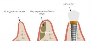
Ridge splitting is a type of bone grafting - a procedure that increases the volume of bone tissue. This operation is often required before implantation if there is a lack of bone tissue. There are various bone grafting techniques, but we will look at one very common one – bone splitting.
Why do some patients need bone grafting?
Before installing dental implants, the implant surgeon must make sure that the volume of bone tissue is sufficient for their reliable fixation. After all, artificial roots must withstand daily chewing loads without the threat of loosening the structure. To do this, a computed tomography (CT) scan is performed, which shows the height and width of the bone.
If the patient’s CT results show significant bone tissue atrophy, the dentist-implantologist will prescribe osteoplasty surgery to increase the bone volume to the required levels. This is a guarantee of a long-term result - that the implant will be firmly fixed in the bone and will not be rejected over time.
Bone grafting is the same as osteoplasty and augmentation.
Why is the procedure needed?
The alveolar process is the basis for holding the implant in the jaw bone. Due to the lack of chewing load, the bones gradually thin out and become looser, decreasing in size. This makes it difficult to securely fix the implant.
The longer you put off going to the dentist after losing teeth, the more difficult it is to restore your dentition by installing implants.
Implantation is also difficult to do if the jaws are deformed. It is provoked by neoplasms in the mouth, severe infectious diseases, and injuries of the maxillofacial apparatus.
Therefore, in case of atrophy and deformation of bone structures, bone grafting is prescribed before implantation, in particular, expansion of the alveolar ridge using the splitting method.
Alveolar ridge splitting
If it is necessary to increase the thickness of the alveolar process (the implant is installed into it) in the lower or upper jaw, the Split-Control technique is used. It allows the alveolar process to be split simultaneously with the installation of the implant.
The implant surgeon carefully makes an incision in the center of the alveolar ridge, fixes a titanium pin, fills the area around it with bone chips, covers it with a resorbable membrane and sutures the wound. After 3-6 months, when the implant has taken root, it can be loaded with a permanent crown.
Did you know that a large number of blood vessels pass through the cancellous bone of the alveolar process. There are also a lot of osteoblasts - cells that produce intercellular bone substance. Osteogenesis, i.e. formation of new bone occurs quickly.
Patient reviews
In general, on the Internet on forums, patients speak positively about this operation, especially if it is performed on the upper jaw. After it, recovery is faster, especially when compared with grafting a bone block. A problem such as shifting of the installed bone material is much less common, that is, complications arise less often. Swelling and pain also go away very quickly. Patients are especially pleased that implants can be placed immediately during surgery, and not some time after bone grafting.
Guided bone regeneration
It is performed to increase both the height and width of the alveolar process. The doctor fills the atrophic defect with osteoplastic material and screws it into the implant. Then the surgical site is covered with a PRF membrane (made from blood plasma) and a barrier membrane. Installation of membranes is mandatory; it is necessary to protect the atrophic defect from negative factors.
If it is planned to carry out directed tissue regeneration simultaneously with simultaneous implantation, then the implant surgeon must grow bone tissue several millimeters more than the required values. The fact is that the bone that appears after the implantation of osteoplastic material is subject to partial atrophy. If the patient has a small gum thickness, then the “reserve” should be even greater.
Guided bone regeneration The process of bone regeneration surgery
Advantages and disadvantages
The main factor that motivates the choice of technology is the absence of the need for surgery to increase bone tissue. The results of implantation are not inferior to other methods, and the procedure itself guarantees a number of advantages, such as:
- Elimination of the consequences of atrophy;
- Formation of a dense bone structure;
- Reducing the postoperative period;
- Possibility of installing a prosthesis in 3-4 months;
- Accelerated tissue regeneration;
- Reducing costs for dental restoration.
It is also worth noting that, in comparison with traditional two-stage prosthetics, the procedure involves less surgical intervention and, as a result, reduces the traumatic nature of the restoration.
Among the disadvantages characteristic of the technique are:
- The limitation is determined by the extent of edentia - only areas where no more than four consecutive units are missing are subject to restoration;
- The need for strict adherence to medical instructions to ensure the achievement of the desired result.
In addition, a limited number of specialized specialists know the technology of alveolar splitting, which makes it difficult to choose a suitable dental clinic.
Systems used
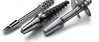
The design features of implants used for longitudinal division of the ridge structure include the use of a cone-shaped shape with a wide thread applied to the apex area. The processing technology eliminates damage to surrounding tissues and guarantees tight contact. Tapered apical edges provide ease of implementation and a tight fit, while the use of a cylindrical end allows for placement of the structure in narrow areas. The technology for manufacturing implants takes into account the peculiarities of the anatomical structure and promotes accelerated integration of artificial foundations, allowing you to secure a permanent crown within 3-4 months after surgery.
Bone block transplantation
This method of osteoplasty involves transplanting a piece of bone taken from the patient himself (for example, from the area of the zygomatic-alveolar ridge of the upper jaw). It is possible to transplant a bone block of xenogeneic origin (for example, a block of bovine bone), but it takes less well.
The doctor fixes a prepared block of allogeneic or xenogeneic origin in the area of the defect using titanium screws. After which it is covered with a protective membrane and the surgical site is sutured.
The NCR method gives a highly predictable result, but has several disadvantages. Firstly,
Additional surgery to remove the bone block may be required.
Secondly,
in some clinical cases, performing NCR with simultaneous implant installation is contraindicated. You have to wait more than six months, and only then can the implant be placed.
Autotransplantation The process of autotransplantation surgery
Why is the operation performed?
In the presence of a narrow ridge, it is impossible to carry out high-quality dental restoration, especially on toothless jaws. The jaw may have this shape naturally or acquire it as a result of atrophy, injury, removal of a tumor, or a previous disease. Indications for surgical intervention:
- Insufficient tissue volume.
- Thin alveolar process.
- Atrophy or other defects of the jaw.
Sinus lift
If the results of a computed tomography scan reveal a lack of bone tissue in the area of the upper lateral teeth, implantation is not possible due to the risk of damage to the maxillary sinuses. It is necessary to first increase the volume of the bone using sinus lift surgery.
The open version is indicated for severe atrophic defects, as it allows to significantly increase its volume.
A closed sinus lift is indicated for patients whose natural bone volume is at least 5 mm.
Sinus lift surgery is no more dangerous than regular tooth extraction. You should not worry too much, especially if the operation is performed by an experienced implant surgeon. In our clinic, sinus lifting is performed by an Israeli implantologist with more than 20 years of experience.
The essence of technology
Implantation of an implant without replenishing the lack of bone mass has become possible thanks to developments involving splitting the alveolar. At the same time, bone volume increases significantly. This is due to a longitudinal incision and spreading of the gums to the sides.
If such an incision is not enough to implant an artificial rod, the dentist places synthetic shavings into the voids and stitches the edges of the wound. To reduce the risk of rejection, an autoimplant can be installed instead of artificial bone.
This technique allows you to reduce the time from the start of treatment to installation of a permanent prosthesis by 3 times.
Is it possible to avoid bone grafting?
Yes, you can. If there is a deficiency of bone tissue in width, if the clinical situation allows, small-diameter implants can be used. And if there is a lack of bone height, short implants are needed.
All-on-4/All-on-6 technologies allow the patient to return teeth with complete edentia without osteoplasty. This becomes possible thanks to the inclined installation of implants in the lateral sections. When implanting titanium screws at an angle of up to 45°, significantly less bone is required in height.
Expert opinion Alexey Isaevich Yakubov Chief physician, orthopedic dentist, implantologist Experience 21 years
For the majority of patients who contacted the Akademstom clinic for a full jaw implantation service, we were able to insert new teeth without bone grafting
.
If the upper jaw is severely atrophied, zygomatic implantation can be performed without restoring the lost bone volume. Elongated titanium rods Zygoma/Zygomatic are fixed not into the jawbone, but into the zygomatic bone. It does not atrophy even with prolonged absence of teeth, so augmentation is not required.
The Akademstom clinic is one of the few Moscow dentistries that performs zygomatic implantation!
Is implantation always possible without bone tissue augmentation?
Typically, the jawbone is oval in shape. Then, during the process of atrophy, only its upper part on the buccal (front) side loses volume, while its base remains wide and supports the rest of the bone. In this case, we can safely carry out “splitting” and not be afraid that the bone will break. However, if the ridge lacks thickness along its entire length, or is less than two millimeters, bone grafting is necessary. True, such situations are quite rare, and in these cases doctors are forced to follow the classic path - performing plastic surgery using bone blocks.
What types of bone materials are used in implantology?
1
Autograft
The bone block is taken from the patient from another area of the body (for example, from the iliac crest). Autologous bone has a high osteogenic potential and is completely biocompatible. But additional surgery is required.
2
Allograft
A block of bone of the required size is taken from a donor (for example, during hip surgery). It is processed to minimize the risk of transmission of infections (HIV, hepatitis, etc.), as a result of which the osteogenic properties are lost.
3
Xenograft
The donor of the bone block is cattle. Xenobone takes root well and is inexpensive.
4
Alloplastic
Osteoplastic material that stimulates tissue growth. A less traumatic method of increasing bone tissue volume that gives good results.
Stages
Splitting the ridge requires several steps:
- First, the patient undergoes an initial examination and visits a dentist-therapist. Also at this stage, a thorough cleaning of the teeth is carried out before the operation.
- Next, the patient is given local anesthesia so that the surgeon can begin to perform the task.
- The third stage involves making an incision in the gum and then removing the tissue. The process is carried out using a surgical scalpel or laser. The incision is made horizontally, the edges are moved apart.
- The material responsible for the formation of bone tissue is injected into the space that appears as a result of gum recession. If necessary, the surgeon additionally introduces a membrane to protect the affected area.
At the last stage, the gum is brought back to its original place and stitched.
Questions and answers
Does osteoplasty surgery hurt?
The operation is performed under local anesthesia, so the patient does not experience any pain. The only thing he feels is the mechanical movements in the oral cavity performed by the doctor.
Will there be severe swelling after a sinus lift?
Slight swelling may persist for 5-7 days. This is a normal reaction to surgery. After the third day, the swelling begins to subside and completely disappears on the 7th day. If the swelling does not subside after 7-10 days, you should definitely consult a doctor.
Are there any contraindications to augmentation?
Yes, it is impossible to carry out if the patient:
- autoimmune disease;
- blood clotting disorder;
- oncology.
It will be necessary to postpone the operation for a certain time if the patient has an ENT disease.
Possible complications
Of course, after the intervention, swelling and pain appear, but they quickly pass and are easily relieved with pharmaceutical painkillers. Also, if too much stress is placed on the surgical area, the stitches may come apart, but the dentist can quickly fix this problem. But still, we do not recommend putting too much stress on the operated area for several months. It’s better to give up food that needs to be chewed hard and for a long time, don’t crack seeds and don’t try to use your teeth as a bottle opener - then there won’t be any problems. Well, be sure to maintain oral hygiene to prevent infection and inflammation.
What is the cost of gum augmentation?
The price of bone grafting depends on the method of surgery and the type of material used, so the cost of treatment is calculated on an individual basis.
The quality and quantity of bone material plays a huge role in pricing. Clinical experience from previous years shows that only the use of high-quality and purified osteoplast with improved biocompatibility reduces the risk of an inflammatory reaction and increases the efficiency of osseointegration.
Carefully choose a clinic and specialist when planning treatment. A qualified doctor is half the success and a guarantee of quality results.
Atrophy of the alveolar process of the upper jaw and parts is a natural process that occurs as a result of tooth loss.
Most often, bone loss occurs within a few weeks after tooth extraction. The blood clot that forms in the hole after tooth extraction often fills it completely. It may be lost as a result of pushing out by the tongue, as well as infection with subsequent development of suppuration.
Types of alveolar bone defects and treatment options
There are three topographic forms of alveolar process atrophy:
- single (loss of bone tissue within one tooth);
- segmental (within several missing teeth);
- complete (of the alveolar process of the jaw as a whole).
Seibert (1983) divided alveolar bone defects into three classes:
- Class I. Bone deficiency in the buccolingual plane with normal thickness of the bone margin in the apical-coronal plane.
- Class II. Deficiency of bone tissue in the apical-coronal plane with normal thickness of the bone edge in the bucco-lingual plane.
- Class III. A combination of buccolingual and apical-coronal tissue deficiency, leading to loss of normal thickness and width of the bone margin.
Evers et al (2010) proposed a new classification of methods for increasing bone volume, which depends on the degree of its vascularization:
- Class I - free bone grafts with microanastomosis;
- Class II - distraction osteogenesis method;
- Class III - use of a pedicle flap;
- Class IV - morphogenetic induction bone grafts;
- Class V - non-vascularized bone grafts.
Nicolas Caplanis in 2009 proposed a new classification of bone defects, which allows for a qualitative clinical assessment of the defect immediately after tooth root removal and to determine recommendations for further orthopedic rehabilitation using dental implantation.
The Caplanis classification distinguishes 4 types of defects:
- type 1 is characterized as a clean socket of an extracted single-rooted tooth with unaffected socket walls;
- type 2 refers to minor destruction of the alveolar ridge and loss of septal bone tissue of no more than 2 mm;
- type 3 is characterized by loss of soft tissue and bone from 3 to 5 mm, accompanied by the destruction of one or two bony walls of the socket;
- type 4 is accompanied by soft tissue injury with a loss of more than 5 mm.
The success or failure of orthopedic treatment largely depends on the volume of the alveolar process. A lack of width makes it difficult to carry out dental implantation or makes it impossible to carry it out without additional measures.
Adaptation of a patient to orthopedic structures with significant atrophy of the jaws due to their unsatisfactory fixation is associated with serious problems, and sometimes may not occur at all.
Until recently, such patients were repeatedly subjected to orthopedic treatment, and sometimes this entire cycle was unsuccessful. But currently, in such cases, reconstruction of the atrophied alveolar process is recommended.
Indications for surgical interventions to increase bone volume:
- preparing the oral cavity for orthopedic treatment;
- adaptation of the atrophied alveolar ridge to the orthopedic structure;
- planning subsequent dental implantation;
- replacement of bone tissue defects that formed after the removal of benign tumors or impacted teeth;
- raising the floor of the maxillary sinus;
- injuries of the facial bones in patients with osteomyelitis of the lower jaw;
- fractures of the bones of the middle and upper zone of the face;
- post-traumatic deformities of the bones of the facial skeleton.
Methods for increasing bone volume
Horizontal and/or vertical augmentation of the alveolar process can be performed immediately after tooth extraction or after a certain period of time.
For each surgical treatment method, different techniques and materials are used, separately and in combination. The ultimate goal of treatment is to restore the functional and aesthetic dentition, taking into account the characteristics of the clinical case.
The operation of raising the maxillary sinus (sinus lift elevation, sinus lift) makes orthopedic treatment possible using intraosseous implants in the lateral area of the upper jaw.
The sinus lift procedure was first performed in 1975 by Dr. Hilt Tatum, and the first comprehensive scientific description of the method was carried out by Boyne and James in 1980.
Yamurkova N.F. et al. (2015) note in their studies that volumetric reconstruction using the developed methods of surgical treatment of severe atrophy of the alveolar process of the upper jaw ensures the creation of sufficient parameters for the installation of dental implants and orthopedic restoration of chewing function.
Among these methods, the authors highlight plastic surgery using an L-shaped autograft, reconstruction of defects using local bone tissue (sandwich plastic method, sliding bone fragment method, intercortical osteotomy method and splitting bone tissue in the defect area).
These authors have proven the effectiveness of using in clinical practice three developed methods for plastic closure of the Schneiderian membrane, which are used during open sinus lift surgery.
The use of these techniques makes it possible to isolate defects in the mucous membrane of the maxillary sinus and continue vertical augmentation.
Razmyslov A.V. in his study (2011) indicated that the use of osteoplastic materials in dental practice enhances osteogenesis and allows the creation of a bone matrix of new bone tissue of optimal density in a period of 6 to 12 months.
When the size of the alveolar process was increased with an autogenous bone graft, a tendency to recovery was noted in the period from 3 to 6 months.
An increase in the alveolar process of the upper jaw (sinus lift) with the complex use of osteoplastic materials, resorbable membranes and autogenous bone filings is characterized by an early and significant increase in the density of the formed bone tissue.
In other works, Kozlova L. (2015) suggests, for atrophy of the alveolar processes of the jaws with a decrease in bone density, complex anti-osteoporotic therapy with bisphosphonates in combination with calcium and vitamin D3 preparations, which helps to normalize the architecture of trabecular bone.
To predict the effectiveness of orthopedic treatment of patients with partial and complete edentia, a clinical diagnostic algorithm is used, which includes:
- study of the microarchitecture of bone tissue according to cone-beam computed tomography data;
- detection of crystallization of the patient’s saliva to assess possible systemic disorders of bone remodeling;
- densitometric determination of bone mineral density.
Meltzer (1979) first published a clinical report on the use of soft tissue flaps exclusively for aesthetic correction of vertical marginal defects. The method is performed by forming a free gingival flap, with the help of which an increase in the thickness of the soft tissue of the alveolar process is achieved.
Garber and Rosenberg (1981) developed a technique for correcting horizontal bone deficiency by using a connective tissue graft and placing it under the epithelial surface, which in turn stabilizes and increases the volume of the alveolar ridge.
In 1985, Allen et al., analyzing the results of early studies, developed an improved surgical technique for local marginal defects. It consists of creating a mucoperiosteal flap using two vertical incisions, which are connected by a horizontal one.
Next, the flap is separated from the bone tissue, and the resulting cavity is filled with one of the hydroxyapatite or graphite-based allomaterials. After this, the cavity is sutured.
Shcherchkov S.V. (2013) to increase the effectiveness of implant treatment of patients with alveolar process atrophy, proposed a modification of the method of autogenous bone grafting similar to the veneer technique - through osteoperforation of the autograft and the recipient site.
This modification with delayed installation of dental implants allows for orthopedic treatment to be carried out within a period of up to 8 months, and the technique is indicated for bone widths of at least 3 mm and cortical thickness of no more than 1.5 mm.
With the immediate installation of dental implants, orthopedic treatment can be carried out after 5 months, the important condition being that the bone width is more than 3.5 mm and the thickness of the cortical layer is no more than 1 mm.
G.A. Ilizarov developed the method of distraction osteogenesis in the late 1960s. The first attempts to use this principle for vertical bone distraction in maxillofacial surgery were made about 15 years ago.
Distraction osteogenesis does not require obtaining a bone graft, suturing the donor site, or performing additional surgery to correct the mucogingival attachment, therefore this method is associated with fewer postoperative complications compared to other regenerative interventions.
However, when using this method in long-term studies, bone resorption was observed after vertical callus distraction. Another disadvantage is the possibility of exclusively vertical regeneration.
When simultaneously increasing the height and width of the bone crest, additional bone block transplantation or directed bone regeneration is required.
The sandwich osteotomy method has become an alternative to distraction osteogenesis.
With this technique, the mobilized bone fragment is fixed in the desired position with plates for osteosynthesis, and the gaps are filled with autogenous bone chips. This method does not require the installation of extrawell distractors.
However, the relatively dense mucous membrane of the palate limits the capabilities of this technique. This method allows you to increase the height of the alveolar process by a maximum of 5 mm, while minimally increasing the width of the bone.
Gerasimenko A.V. et al. (2013) proposed a technique for augmentation of the alveolar process by subperiosteal injection of osteoplastic materials.
In this technique, blood is drawn from a patient's vein, after which the blood plasma is enriched with platelets, thus obtaining platelet-rich plasma.
The latter, using an injection needle, is injected subperiosteally from the vestibular side in the projection of the tooth, infiltrating the area of deformation of the alveolar process.
To enhance the result, after 10-15 minutes, a suspension of powdered bone material in an isotonic sodium chloride solution is injected subperiosteally into the tissue of the same area.
The literature describes a method of augmentation of the alveolar process in difficult anatomical conditions in the area of the chewing teeth of the upper jaw, proposed by D.S. Avetikov. et al (2014).
It involves taking a bone autoblock of the desired shape and volume from the anterior surface of the body of the upper jaw using a piezo scalpel. Then the bone fragment is fixed with screws to the vestibular surface of the alveolar process of the upper jaw, and the interosseous spaces are filled with autologous and xenogeneic bone chips and stabilized with a collagen membrane.
Researcher Malanchuk V.A. (2002) proposed an alternative treatment method.
After tooth extraction, the mucoperiosteal flap of the alveolar process is detached from the vestibular side with mobilization of the vestibular mucoperiosteal flap. Using a dental bur, the cortical layer of the side walls and top of the tooth socket is additionally removed, revealing the bone marrow spaces, leaving shavings of the cortical layer in the socket and suturing it.
Open and closed sinus lift
The closed sinus lift procedure is carried out by separating the periosteum, after which a pilot canal is formed in the area of the missing tooth without perforating the bottom of the cavity with a cutter. Next, carefully continue preparation using a cutter with depth marks until contact with the bottom of the cavity is felt.
After this, a bed for the dental implant is formed. The last stage consists of perforating the floor of the paranasal sinuses, leaving the mucous membrane of the maxillary sinus intact. Finally, mobilization and elevation of the shell are performed and a resorbable membrane and bone material are introduced through the formed bone bed, the length of the corresponding implant is measured and it is installed.
The open sinus lift procedure is recommended when the residual bone height is up to 3-4 mm. The incision configuration depends on the prosthetic planning.
If you plan to make a fixed bridge structure, the incision must be shifted from the center of the alveolar process by 2 mm to the palatal side. If a removable structure is planned in the future, then the incision is made in the center of the alveolar process.
Next, the mucoperiosteal flap is separated. After exposing the lateral bone wall, corner points are applied, connecting them with a diamond bur to form a bone window. Through the window, the mucous membrane of the maxillary sinus is mobilized and lifted and bone material is introduced, after which the wound is sutured.
The double window technique is an open sinus lift technique.
The indication for the use of this technique is an enlargement of the maxillary sinus, which usually occurs with prolonged absence of teeth. The bone partition created with this method between the two windows corresponds in its location to the zygomaticalveolar ridge.
With the double window technique, the maxillary sinus mucosa can be mobilized from both the medial and distal windows, after which bone material is injected.
Raising the floor of the nasal cavity is used before installing dental implants, in case of insufficiency of bone tissue in the frontal area of the upper jaw, or in order to prevent its perforation.
The mucous membrane of the bottom of the nasal cavity is thicker, in contrast to the mucous membrane of the maxillary sinus, and has a stronger fixation to the bone.
This procedure is carried out through a formed bone canal for installation of a dental implant. The mucous membrane of the bottom of the nasal cavity can be separated, protecting it from perforation during the formation of the implant bed.
Kasiyanchuk M.V. (2009) proposed a method of performing a combined sinus lift, involving trepanation, preparation, lift and peeling.
In this case, trepanation is carried out slit-like along the alveolar ridge. The preparation is carried out separately along the vestibular and palatal walls. The lift is performed in an open way, detachment of the Schneiderian membrane from the bone walls is done in a closed way, after which it is performed with osteotropic material and a membrane.
Sennikov A.N. and co-authors (2009), when performing a sinus lift, propose preparing the anterolateral wall of the maxillary sinus with mutually intersecting lines that connect along the perimeter of the window to form a perforation hole.
The depth of the cuts is determined visually when the mucous membrane of the maxillary sinus appears adjacent to the bone tissue, and the peeling of the mucoperiosteal flap is carried out only along the periphery of this opening.
Research Kekukh E.O. (2013) are focused on the use of endoscopic sinus lift, which can reduce surgical trauma to soft and bone tissues, and reduce the recovery time for patients after surgery
Endoscopic sinus lift eliminates many of the disadvantages inherent in the traditional procedure on the maxillary sinus, prevents scarring of the mucous membrane, reduces blood loss and preserves local blood microcirculation.
This method involves restoring the integrity of the oral mucosa with the immediate elimination of the defect in the bone tissue of the alveolar process and the creation of the necessary volume of bone tissue for the subsequent rehabilitation of patients using delayed dental implantation.
Modern clinical, endoscopic and radiological research methods, as well as analyzes of long-term results of surgical treatment of patients using this method prove the effectiveness of endoscopic sinus lift.
Conclusions on the treatment of alveolar ridge atrophy
The above literature review allows us to draw the following conclusions:
- The data obtained as a result of a systematic retrospective analysis do not provide an adequate evidence base for identifying clear advantages of one method of surgical treatment of atrophy of the alveolar process of the maxilla over alternative methods.
- Each of the above-mentioned techniques should be selected according to the anatomical characteristics of the patient. The scope of surgical interventions depends on the severity of jaw bone atrophy. It is especially important to obtain research results in combination with concomitant diseases.
- To obtain reliable results, prospective clinical studies are needed, especially among postmenopausal patients, who account for a significant portion of the known anatomical and functional disorders.
Taking into account the results of studies in the future may make it possible to adjust the process of bone restoration during surgical treatment of alveolar atrophy and significantly increase its predictability.
Possible consequences
Bone grafting during dental implantation is a surgical procedure that is accompanied by a risk of complications. Some of them are close to normal, but there are also those that pose a greater danger. Common adverse effects of bone grafting include:
- Formation of hematomas of various locations and sizes;
- Dryness and cracks in the corners of the lips, as well as the occurrence of herpetic rashes, especially if there is a predisposition to them;
- The appearance of a slight sore throat and increased temperature (if the fever progresses and does not subside within 3 days, consult a doctor immediately);
- Pain in the ears, neck, head, different parts of the oral cavity (irradiation from the operated area, goes away on its own);
- Increased sensitivity of teeth to temperature changes in food (the phenomenon goes away a few weeks after surgery);
- Increased tooth mobility for several weeks after the procedure;
- Numbness of the jaws that lasts more than 4 hours (immediate consultation with a doctor).
To avoid dangerous complications of bone grafting, the healing and condition of the oral cavity should be periodically monitored by a dentist. The patient is obliged to follow every doctor’s recommendation and turn off amateur activities.