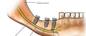
The biomechanics of the lower jaw is considered from the point of view of the functional purpose of the dental system (speech, chewing, swallowing). Movements of the lower jaw are realized as a result of the interaction of the temporomandibular joint (TMJ), teeth and masticatory muscles.
This interaction is coordinated and controlled by the central nervous system. Voluntary and reflex movements are regulated by the neuromuscular system; they are carried out sequentially.
For example, the initial movements—biting food and placing it in the oral cavity—are voluntary. Chewing and swallowing are then carried out unconsciously.
Everything is not as simple as it seems
Vertical movement of the lower jaw.
The initial position of the lower jaw when opening the mouth is the state when the lips are closed. In this case, there is a gap of 2-4 mm between the dentition of the lower jaw and the upper jaw. This state is called a state of physiological rest. The movement of the lower jaw in the vertical plane occurs when opening and closing the mouth, due to the active contraction of the muscles: - depressors (mylohyoid, geniohyoid, anterior belly of the digastric muscle) - levator (proper masticatory muscle, temporalis, medial pterygoid muscle). The amplitude of mouth opening is strictly individual. On average it is 4-5 cm.
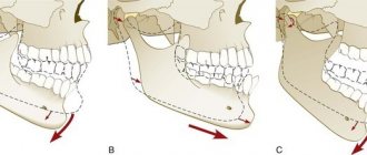
Phases of lowering the lower jaw.
1. With a slight lowering of the lower jaw (quiet speech, drinking), the articular heads in the infero-posterior part of the joint rotate around a horizontal axis passing through their centers. 2. With a significant lowering of the lower jaw (loud speech, biting), the hinge rotation in the infero-posterior part of the joint is joined by sliding of the articular heads together with the discs forward along the circumference of the articular surface. The result is a combined movement of the articular heads, in which the point of contact of two convex articular surfaces moves. 3. With the maximum lowering of the lower jaw, the sliding of the heads is delayed at the apex by the tension of the articular capsules, articular ligaments and muscles, and one hinge movement continues in the joint. The trajectories of movement of the lower teeth are concentric curves with a common center in the head of the lower jaw. They, just like the axis of rotation of the head, can move in space.
Angle of transversal articular path (Benét's angle)
On the side of the contracted muscle, the articular head moves downward, forward and somewhat outward. Its path during this movement is at an angle to the line of the sagittal articular path. This angle was first described by Benet and for this reason is named after him. Otherwise it is called the angle of the lateral articular path. It is on average 17°. On the opposite side, the ascending ramus of the lower jaw moves outward, thus becoming at an angle to its original position (Fig. 34).
Transversal movements are characterized by certain changes in the occlusal contacts of the teeth. Since the lower jaw shifts to the right and left, the teeth describe curves intersecting at an obtuse angle. The farther the tooth is from the articular head, the blunter the angle. The most obtuse angle is obtained from the intersection of the curves formed by the movement of the central incisors. This angle is called the angle of the transversal incisal path or the Gothic angle (Fig. 35). It determines the range of lateral movements of the incisors and is equal to 100-110°.
Of significant interest are changes in the relationships of the chewing teeth during lateral excursions of the jaw (Fig. 36). When lateral movements of the jaw are made, it is customary to distinguish between two sides: working and balancing. On the working side, the teeth are set opposite each other with cusps of the same name, and on the balancing side they are opposite, i.e. e. the buccal lower cusps are set opposite the palatal cusps.
Until now, when studying the movements of the lower jaw, the latter were artificially decomposed into their component elements (lowering, moving forward, to the sides). This was done for methodological reasons. In reality, mandibular excursions are very complex because they are a combination of different movements. Chewing movements are of greatest practical interest for orthopedic dentistry. Knowing them can facilitate the manufacture of dentures and artificial teeth. When chewing food, the lower jaw undergoes a cycle of movements. Gisi presented the cyclical movements of the lower jaw in the form of a diagram (Fig. 37). The initial moment of movement is the position of the central occlusion. Then four phases follow continuously one after another. In the first phase, the jaw lowers and moves forward. In the second phase, the jaw moves to the side (lateral movement). In the third phase, the teeth close on the working side with cusps of the same name, and on the balancing side with opposite cusps. In the fourth phase, the teeth return to the position of central occlusion and the chewing cycle repeats. After chewing is completed, the jaw is set to a position of physiological rest.
There is no doubt about the statement that on the working side there is a closure with the same tubercles. A different relationship of the lateral teeth would not ensure grinding of food. As for the balancing side, here it is possible either the formation of contact between opposite tubercles, or their absence. The latter is confirmed by research by A. Ya. Katz and A. K. Nedergin. This appears to depend on the severity of the transversal occlusal curves.
Sagittal movements of the lower jaw.
The forward movement of the lower jaw is carried out by bilateral contraction of the lateral pterygoid muscles. The movement of the head of the lower jaw is divided into 2 phases: 1- the disc together with the head slides along the surface of the articular tubercle; 2- the sliding of the head is accompanied by its articulated movement around its own transverse axis. The distance that the head of the mandible travels when it moves forward is called the sagittal articular path. This distance is on average 7-10 mm. The angle formed by the intersection of the line of the sagittal articular path with the occlusal plane is called the angle of the sagittal articular path. According to Gisi, it averages 33º. With an orthognathic bite, the protrusion of the lower jaw is accompanied by sliding of the lower incisors along the palatal surface of the upper ones. The path taken by the lower incisors when moving the lower jaw forward is called the sagittal incisal path. The angle formed by the intersection of the line of the sagittal incisal path with the occlusal plane is called the angle of the sagittal incisal path. On average, its value is 40-50°. Bonneville's three-point contact. When the lower jaw is advanced to the position of anterior occlusion, contact of the dentition is possible only at three points. Two of them are located on the distal cusps of the second and third molars, and one on the anterior teeth.
U
Carbon steel
(see Carbon steel).
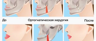
Deepening of the vestibule of the oral cavity
- an operation that allows the movement of soft tissues to create an increase in the volume of space in the vestibule of the oral cavity.
Balkville corner
(FG Balkvill - British dentist) - the angle between the line connecting the head of the lower jaw (upper surface) and the midpoint on the cutting edges of the incisors, on the one hand, and the Camper horizontal line, on the other. Equal to 22-27°. It is important for finding the occlusal plane and installing models in the articulator.
Contact angle
- the angle between the tangent to the surface of a liquid drop and the surface of the solid body on which this drop is located.
Usually the contact angle is denoted by a Greek letter. This indicator is used to measure wetting.
Mandibular angle
- a section of the lower jaw located at the point of transition of the body of the lower jaw into the ramus of the lower jaw. Normally it is 110-120°.
Angle of sagittal incisal path
(from Latin
sagitta
- arrow) - is formed by the intersection of the trajectory of the sagittal incisal path with the occlusal plane. On average it is 40-50°.
Articular sagittal path angle (Gysi angle)
- the angle formed by the intersection of the line of the articular sagittal path with the occlusal plane. Depends on the degree of severity of the articular tubercle of the temporal bone and the tubercles of the lateral teeth - on average it is equal (according to Gysi) 33°.
Angle of transversal incisal path (Gothic angle)
(from Latin
transvertere, transverses
- turn to the side) - is formed by the intersection of the trajectory of movement of the incisors during lateral movements of the lower jaw (transversal incisal path) to the right and left and is equal to an average of 100-110°.
Angle of transversal articular path (Bennett's angle)
(Bennet NG - English dental surgeon) - is formed during transversal (lateral) movements of the lower jaw. The head of the mandible on the working side (offset side) rotates around a vertical axis. On the opposite balancing side (the side of the contracted lateral pterygoid muscle), the head of the mandible slides down, forward and somewhat inward along with the disc along the surface of the slope of the articular tubercle, making a lateral articular path. The angle formed by the lines of the sagittal and transverse articular path is the angle of the transversal articular path and is equal to an average of 17°.
Functional significance of the tubercles
The buccal cusps of the upper and lower molars, as well as the lingual cusp of the lower molar, perform a protective function. The palatal cusp of the upper molar is the supporting one.
In the process of closing the teeth in the central position, contact occurs between the palatal tubercles of the upper teeth and the central fossae or marginal projections of the molars and premolars of the lower jaw. There is also contact between the buccal tubercles of the lower teeth and the central fossae and marginal projections of the molars and premolars located above.
Note! The buccal tubercles of the teeth of the lower jaw and the palatine tubercles of the upper are supporting and holding ones. The lingual cusps of the lower teeth and the buccal cusps of the upper teeth are guides and protective (prevent biting the cheek and tongue).
When performing chewing movements, the lower jaw should slide over the surface of the upper jaw teeth without obstacles. The tubercles slide smoothly along the antagonist slopes without disrupting occlusal relationships.
At the same time, they must maintain tight contact. Sagittal and lateral movements are reflected on the surface of the first molars of the mandible by the arrangement of transverse and longitudinal fissures, this is called the “occlusal compass”.
Important! This landmark is necessary in the process of modeling the occlusal surface of the teeth.
During the movement of the lower jaw forward, the guide tubercles of the chewing teeth of the upper localization slide along the central fissure of the teeth located below. During lateral movements, gliding occurs along the fissure that separates the median buccal and posterobuccal cusps of the lower molar.
In the process of combined movement, gliding occurs along the diagonal fissure that divides the median buccal tubercle. The “occlusal compass” is characteristic of all teeth of the lateral group.
“Occlusal compass” - A and C - sagittal movements, B and E - transversal, D - combined.
Another important factor in the biomechanics of the lower jaw is the height of the cusps of the chewing teeth. This parameter determines the amount of initial joint displacement.
This is due to the fact that during the lateral movements of the lower jaw, the head on the working side moves outward before the rotational movements begin, while the head on the balancing side moves inward. This type of movement occurs within 0-2 mm.
The greater the flatness of the slopes of the tubercles, the greater will be the magnitude of the initial articular shift. This is how the free mobility of the dentition relative to each other within the boundaries of central occlusion is determined.
Note! In the process of modeling artificial teeth, it is very important to take into account the characteristics of the tubercles, as well as the inclinations of the slopes of the chewing teeth. Otherwise, disturbances in the interaction of the elements of the TMJ are possible, that is, the progression of joint dysfunction.
OCCLUSAL PLANE
*passes along the junction of the mesial cutting edges of the central incisors of the mandible and the distal cusps of the first molars of the mandible
runs from the middle of the tragus of the ear to the outer edge of the wing of the nose (on the skull from the lower edge of the bony part of the external auditory canal to the anterior nasal spine - spina nasalis anterior)
runs from the lower edge of the orbit to the upper edge of the external auditory canal
runs from the upper third of the distal slope of the lower canine to the distal buccal cusp of the last lower molar
passes along the junction of the distal cutting edges of the mandibular lateral incisors and the distal cusps of the mandibular second molars
226
KAMPER PLANE
passes along the junction of the mesial cutting edges of the central incisors of the mandible and the distal cusps of the first molars of the mandible
*runs from the middle of the tragus of the ear to the outer edge of the wing of the nose (on the skull from the lower edge of the bony part of the external auditory canal to the anterior nasal spine - spina nasalis anterior)
runs from the lower edge of the orbit to the upper edge of the external auditory canal
runs from the upper third of the distal slope of the lower canine to the distal buccal cusp of the last lower molar
passes along the junction of the distal cutting edges of the mandibular lateral incisors and the distal cusps of the mandibular second molars
227
FRANKFURT PLANE
passes along the junction of the mesial cutting edges of the central incisors of the mandible and the distal cusps of the first molars of the mandible
runs from the middle of the tragus of the ear to the outer edge of the wing of the nose (on the skull from the lower edge of the bony part of the external auditory canal to the anterior nasal spine - spina nasalis anterior)
*passes from the lower edge of the orbit to the upper edge of the external auditory canal
runs from the upper third of the distal slope of the lower canine to the distal buccal cusp of the last lower molar
passes along the junction of the distal cutting edges of the mandibular lateral incisors and the distal cusps of the mandibular second molars
228
SAGITTAL COMPENSATIONAL OCCLUSAL CURVE OF SEE (SPEE)
passes along the junction of the mesial cutting edges of the central incisors of the mandible and the distal cusps of the first molars of the mandible
runs from the middle of the tragus of the ear to the outer edge of the wing of the nose (on the skull from the lower edge of the bony part of the external auditory canal to the anterior nasal spine - spina nasalis anterior)
runs from the lower edge of the orbit to the upper edge of the external auditory canal
*runs from the upper third of the distal slope of the lower canine to the distal buccal cusp of the last lower molar
passes along the junction of the distal cutting edges of the mandibular lateral incisors and the distal cusps of the mandibular second molars
229
THE ANGLE OF THE SAGITTAL ARTICULAR PATH ON THE AVERAGE, ACCORDING TO THE GYSI DATA, IS EQUAL
*33°
5-8°
60°
100-110°
40-50°.
230
THE ANGLE OF THE INCISAL PATH, FORMED BY THE VECTOR OF THE INCISAL PATH AND THE OCCLUSAL PLANE, DEPENDS ON THE HEIGHT OF THE TURBLES OF THE CENTRAL INCISERS AND IS ON AVERAGE EQUAL TO
33°
5-8°
60°
*100-110°;
40-50°.
231
THE ANGLE FORMED BETWEEN THE LINES OF THE SAGITAL AND TRANSVERSAL ARTICULAR PATH IS ON AVERAGE
33°
*5-8°
60°
100-110°
40-50°
232
THE ANGLE OF THE TRANSVERSAL INCISAL PATH IS ON AVERAGE EQUAL
33°
5-8°
60°
*110-120°
40-50°
233
DEVICES THAT ALLOW YOU TO REPRODUCE VERTICAL MOVEMENTS OF THE LOWER JAW ARE CALLED
articulators
*occluders
orbiculators
axiographs
myographs
234
DEVICES THAT ALLOW YOU TO REPRODUCE ALL POSITIONS OF MOVEMENTS OF THE LOWER JAW ARE CALLED
*articulators
occluders
orbiculators
axiographs
myographs
235
IN MOST ARTICULATORS THE INTERCONYLAR DISTANCE IS ABOUT
15 cm
21 cm
*11 cm;
8 cm
5 cm
242