Jaw cancer is a malignant neoplasm that affects bone tissue. The disease has no age restrictions; it is characterized by rapid growth, metastasis, and pronounced pain. The structural features of the maxillofacial region, proximity to important vessels and nerve centers further complicate treatment.
Anatomical structure
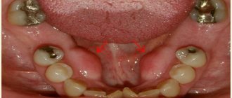
The oral cavity is the initial section of the digestive tract, in which food is chewed and saliva is produced to digest food. It is involved in the process of breathing, swallowing, articulation and speech.
The composition of the oral cavity includes:
- vestibule (lips, front side of teeth, inner surface of cheeks);
- gums;
- the bottom on which the tongue lies;
- two thirds of the tongue;
- teeth;
- retromolar triangle - the space on the lower jaw behind the third molar;
- hard and soft palate.
Benign odontogenic tumors
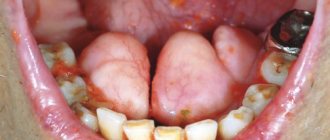
Ameloblastoma. Its characteristic feature is a pronounced change in the shape of the face associated with a violation of symmetry proportions as a result of the development of a tumor located in the lower jaw. The violation of symmetry can be mild or pronounced. The degree of distortion of the face shape is influenced by the size and position of the tumor. For example, the localization of a tumor along the body and ramus of the lower jaw is characterized by a change in the shape of the lower lateral part of the face. The color of the skin does not change, and it can be easily moved in the area of the tumor.
Inflammatory processes accompanying the tumor can give similar symptoms to phlegmon or mandibular osteomyelitis. During palpation, the body of the tumor is felt, which makes it possible to assess the degree of distortion of the shape of the face. The lymph nodes located directly next to the tumor do not change in size, and the deformed area is clearly defined. The formation has a thick filling and a wavy surface. Examination of the oral cavity reveals thickening of the alveolar ridge, soft tissue may be swollen, and teeth tend to shift or move.
Odontoma . Often this type of tumor is diagnosed in adolescence. The tumor has similar symptoms to other tumors localized in the jaw bones. The course of the disease is quite slow and ambiguous. During development, there is a gradual swelling of the jaw bones, which leads to delayed or absent teeth eruption. Large tumor sizes can change the shape of the jaw or contribute to the formation of a fistula. Despite the fact that the course of the disease passes practically without symptoms, the upper layer of the jaw may be damaged, and the tumor itself may contain teeth or their rudiments. When diagnosing, it is necessary to differentiate the tumor from adamantinoma. Odontoma can be simple, complex, soft or mixed.
Odontogenic fibroma . The nature of the development of this neoplasm is very slow; the tumor is mainly diagnosed in young children. A clear symptom of tumor development is impaired teething; pain is not observed during the period of tumor growth. Odontogenic fibroma can be located equally on both jaws and is rarely accompanied by an inflammatory process. It differs from similar neoplasms in its composition, which includes remnants of the epithelium that forms the teeth.
Cementoma . The hallmark of a tumor is the presence of cement-like tissue. The tumor grows quite slowly and is manifested by a change in the shape of the jaw. The tumor is clear and round, has pronounced boundaries, most often affects the upper jaw and is almost always connected to the root of the tooth.
Classification
Oral cancer is divided into three types:
- papillary. The nodule in the mucous membrane increases in size and hangs into the oral cavity. The neoplasm progresses slowly;
- infiltrative. The seal on the pinkish mucosa is distinguished by a whitish color, clear contours and shape, and thinning of the membrane around it. On palpation from the side of the cheek, a dense infiltrate is felt. The tumor tends to grow rapidly. The patient complains of unbearable pain;
- ulcerative The most common form of the disease. Ulcers on the mucous membrane do not heal, they grow, and the border around them turns red. The outline is torn and its edges are bleeding.
Tumor metastases appear quickly. Malignant cells grow into the mental, submandibular, and deep jugular lymph nodes. This process is influenced by the thickness and depth of the tumor. Thus, when the tumor deepens by 4-5 mm, metastases occur in 98% of cases. At the T1 stage of oncology, metastasis is detected in half of the cases, and when the T4 stage is reached, distant spread of cancer cells is observed in 85% of cases.
Malignant tumors of the jaws
Malignant jaw tumors are not observed in patients as often as benign ones. Oncological damage is accompanied by pain that has the ability to self-propagate. Teeth become loose and prone to rapid loss. Some tumors, due to their morphological manifestations, can cause fractures of the jaw bones. With the progression of a malignant tumor, erosion of bone tissue is observed, while the growth of the parotid and submandibular glands is noticeable, and the masticatory muscles increase. The source of the disease penetrates the cervical mandibular lymph nodes.
Some tumors affecting the upper jaw extend into the eye socket or nasal cavity. As a result, complications of the disease may occur in the form of bleeding from the nose, a suppurating one-sided runny nose, difficulty with nasal breathing, pain in the head, increased production of tears, bulging eyes and double vision.
Malignant tumors affecting the lower jaw quite quickly penetrate the soft tissues of the mouth and cheeks, begin to bleed, resulting in disruption and difficulty in closing the jaws.
Malignant tumors originating from bone tissue are characterized by rapid progression and penetration into soft tissues, which leads to disruption of facial symmetry, increased pain and the rapid appearance of foci of disease in the lungs and other organs.
Causes
The prevalence of oral cancer is growing and is currently diagnosed in 2% of patients among the total number of cases. Since 2009, the incidence has increased by 25%, with mostly squamous cell carcinoma being detected and only in isolated cases adenocarcinoma.
Most foci of oncology are observed in the tongue. Slightly less malignant formations on the floor of the mouth. Cancer of the soft and hard palate, gums and cheeks is detected in 20% of cases. Much less frequently diagnosed is damage to the alveoli of the lower jaw - 4%, the arches of the palate, retromolar region and vestibule - 3%.
Based on practice, men are more susceptible to oral cancer than women. This is due to bad habits, for example, the abuse of cigarettes or chewing tonic mixtures increases the production of saliva, which washes away beneficial elements from the mucous membrane. The risk group includes patients with HPV, elderly people, workers in hazardous industries, patients with lichen planus, people whose oral mucosa is systematically injured by fillings, prostheses, and metal objects.
Forecast criteria
The survival rate of patients with maxillary sinus cancer averages about 40% over 5 years.
At an early stage, when treated in European oncology clinics, tumors have a cure rate of up to 80%.
Patients with unresectable tumors treated with radiation had a survival rate of less than 20%. Survival rates have now improved slightly due to advances in skull base surgery.
In patients with squamous cell carcinoma of the nasal cavity or maxillary sinus, better survival outcomes are achieved when patients receive adjuvant radiation therapy, adjuvant chemoradiotherapy, or neoadjuvant therapy in addition to surgery.
In patients with sinonasal adenocarcinoma, better disease-free survival was associated with surgery followed by radiation therapy compared with surgery alone, for all tumor stages (T1-T4).
Symptoms
A malignant ulcer from ordinary stomatitis in the mouth can be identified by swelling and swelling of the cheeks, pain and constant discomfort even at rest. You should be wary of prolonged non-healing of the wound and its bleeding.
As the disease progresses, the symptoms intensify:
- swelling increases and spreads to the neck;
- the red or white spot on the oral mucosa intensifies;
- discomfort when chewing and swallowing;
- difficulty speaking due to friction of the mucous membrane on the teeth when moving the jaw;
- the appearance of bad breath;
- feeling of a foreign object in the throat;
- anemia of the mouth.
In the late stages of cancer, teeth fall out and body weight rapidly decreases.
Detection of pathology
Detection of tumors of the lower jaw is based on a thorough study of the course of the disease, as well as morphological and radiological data. The diagnosis is made by x-ray, since x-rays can detect destructive changes in the bone early.
In the initial stages of sarcoma or jaw cancer, x-rays show bone loss. Its changed area does not have clearly defined boundaries; they are blurred. If jaw cancer is localized in the area of the alveoli, then the cortical plates of its walls are destroyed, and an extensive zone of destruction of the spongy substance is visible around the circumference. If the process has spread, then the radiograph shows complete destruction of a certain part of the bone.
Histological analysis of the tissues of the surface of the extracted tooth during its mobility is informative. It is always necessary to strive to establish the cause of loose teeth. If the mucous membranes of the mouth have ulcerations and the area is clearly visible, a cytological analysis of the mucosa is performed to confirm the diagnosis.
Diagnostics
At the initial consultation, the doctor examines the oral cavity, examines ulcers, erosions, damage to the mucous membrane, and then takes a smear for examination. To confirm the inflammatory process, the patient is sent for a general and biochemical blood test.
The diagnosis is confirmed by the results of the examination:
- MRI and ultrasound of soft tissues of the neck. The images reveal the localization of the pathology, the depth of germination and the structure of the tumor, compaction from blood and lymph, decomposition of the cortical layer of the bone;
- if metastases are suspected, a fine needle aspiration biopsy of the lymph nodes under the chin, under the jaw and in the upper third of the neck is performed;
- positron emission tomography. Shows the depth of the tumor, as well as early metastases;
- osteoscintigraphy. Skeletal bones are examined to look for displaced cancer cells;
- CT scan of facial bones with contrast. The images show the tumor growing into the neck vessels, jaw or base of the skull.
Metastasis
The process of metastasis of pathology occurs through the lymphogenous route. Most often, metastases are observed in the submandibular zone and fuse with the lower jaw quite early, after which they infiltrate the skin.
If jaw cancer is in an advanced form, then metastasis to the spine, liver and other distant organs is noted. But often metastasis to the cervical lymph nodes and distant organs is not present. In the case of sarcoma of the lower jaw, metastasis to distant organs rarely occurs, and they, as a rule, do not form in regional lymph nodes.
Secondary (metastatic) tumor formations are detected much less frequently compared to primary ones, and they are more often observed in women. Pathological metastases occur in lung, breast, thyroid, and stomach cancer.
In case of metastases, they are resected together with excision of the cervical tissue: if there is one metastasis in the submandibular region, then an upper fascial-sheath excision of the neck tissue is performed on one side; if metastases are present at the site of the branching of the common carotid artery, then a Krail operation is performed, if necessary, fascial-sheath excision cervical tissue.
Treatment
The choice of treatment tactics depends on the stage and extent of the tumor. When the tumor grows rapidly, treatment methods are combined.
Operation
The doctor determines the principle of surgical intervention after determining the stage of the tumor and its spread. If cancer cells have penetrated the periosteum and surrounding tissues, a wedge-shaped, planar or sagittal resection of the jaw is performed. If the examination reveals the growth of cancer cells directly into the bone or the defect is noticed during surgery, segmental resection of the lower jaw is performed. The doctor assesses the lesion on site and determines the thickness of the excised layer.
The next stage of the operation is partial or complete excision of the cervical lymph nodes to prevent metastases if the thickness of the tumor is more than 4 mm or the location of the tumor in the floor of the mouth or on the tongue. If the tumor is located in the midline, then the cervical lymph nodes are excised on both sides. The operation ends with the immediate replacement of damaged tissue.
After removal, the tumor is sent for histological examination. Its size, thickness, depth, edges are assessed. Further treatment is affected by cell growth beyond the boundaries of the capsule of the removed lymph node, and the spread of cancer cells to neighboring organs.
Radiation therapy
Radiation after surgery is prescribed when diagnosing T3, T4, N2, T3 stages of the disease no later than six weeks after tumor removal. The need for radiation therapy increases with perineural invasion of the lymphatic vessels. The total focal dose for all sessions is 60 g, and the single focal dose for one session is 2 g. When metastases are detected on the neck, the SOD increases to 66 g, and if there is no risk of metastasis, the SOD decreases to 50 g.
As the main treatment, radiation therapy is used in a total focal dose of 60-70 g. The procedure is performed five days a week and is combined with chemotherapy. Every three weeks, 100 mg of cisplatin is administered.
Chemotherapy
Anticancer drugs are prescribed before surgery or along with radiation therapy to reduce the size of the tumor. Sometimes therapy is prescribed simultaneously with surgery.
Treatment involves the use of a 5-fluoroacyl regimen together with cisplatin or other agents - carboplatin, methotrexate, bleomycin. They cause a number of side effects, for example, vomiting or nausea, hair loss, decreased appetite, and increased bleeding. Symptoms disappear after treatment, but permanent hearing loss is sometimes observed after taking cisplatin.
The prognosis of oral cancer depends on the stage at which the disease is detected. If treatment is started at stage zero, the disease will stop. It is worth noting that smoking provokes relapse or degeneration of the tumor, so repeated surgery or radiation may be required. Surgery at the first stage increases survival rate to 80-85%, and the combination of radiation therapy with surgery at the second stage by 60-80%. Already at subsequent stages of cancer development, the survival rate is no more than 50%, and all three treatment methods are used simultaneously.
Benign non-odontogenic tumors
Osteoma . This tumor is not often diagnosed, and men are more susceptible to developing osteoma than women. It occurs mainly during adolescence. The development of the tumor occurs without pain, is quite slow and is localized in the nasal cavity, orbit or sinuses of the upper jaw. Tumor growth can occur both inside the jaw bones and on the surface. The mandibular location of the tumor is characterized by pain and a violation of the symmetry of the face, as well as the motor abilities of the jaw in this area. The maxillary localization of the tumor leads to failure of nasal breathing, doubling of the image perceived by the eyes, and bulging of the eyes.
Osteoid osteoma . The main symptom of the development of this tumor is the presence of pain, which intensifies with the progression of the tumor. It is noted that people with osteoid osteoma especially feel increased pain at night. Establishing a correct diagnosis is made difficult by the nature of the pain syndrome, which tends to spread, resulting in the activation of other diseases. In diagnosing a tumor, the action of medications (analgesics) that suppress the occurrence of pain helps. The affected areas appear swollen, and the motor function of the joints is impaired. The difficulty of making a diagnosis is due to the small size of the tumor and the absence of special symptoms.
Osteoblastoclastoma . The tumor is a single separate formation. It is extremely rare to see a double appearance of a tumor on adjacent bones. Mostly young people under the age of 20 are susceptible to developing the disease. The most pronounced symptoms are increased pain in the jaw, impaired facial symmetry and tooth mobility. The manifestation of the main symptoms depends on the location of the tumor. The peri-tumor tissues become pronounced, and fistulas begin to appear. Quite often, patients notice an increase in average body temperature, the cortical layer becomes thin, which can cause a fracture of the lower jaw.
Hemangioma . As an independent disease, it is relatively rare; a combination of hemangioma of soft facial tissues or the oral cavity with a jaw hemangioma is often diagnosed. The disease is characterized by a color change in the mucous membrane to bright red or blue-purple shades. This symptom is the main one at the time of diagnosis. However, diagnosis can be difficult in situations where the soft tissues of the oral cavity are not involved in the inflammatory and tumor process. Increased bleeding of the gums and root canals is considered to be a symptom of isolated hemangioma.
Dispensary observation
Since the tumor can recur and metastasize, after completing the course of treatment the patient is registered with the oncology clinic. The first year you should visit a doctor every month, the second year a preventive examination is carried out every 4-6 months, and then once a year or in case of any ailments. The examination involves an examination - ultrasound and contrast MRI of the soft tissues of the neck, PET, osteoscintigraphy. Consultation with an otolaryngologist, dentist and oncologist is required. The doctor may shorten the period of medical examination if there is a high risk of relapse.
List of references on the topic:
- Gantsev Sh.H. Oncology – M, 2012 – P.204-205.
- Golovin D.I. Errors and difficulties in diagnosing tumors, D.: Medicine. Leningr. department, 2015 305 pp.
- Selected lectures on clinical oncology/Ed. IN AND. Chissova, S.L. Daryalova. – M., 2010
- Matyakin E.G., Alferov V.S. Chemotherapy of head and neck tumors // Mat. 2nd Ros. oncol. conf. “Current trends in the development of drug therapy for tumors” December 8–10, 2021 – M., 256 p.
- Tumors of the head and neck: hands/ A.I. Paches. - 5th ed., add. And revised - M.: Practical Medicine, 2013. -478 p.
- Shine A.A. Oncology. M – 2014 365 pp.
- Encyclopedia of Clinical Oncology/Ed. M.I. Davydova. – M., 2014 –P.140-179.
- Bityutsky P.G., Kitsmanyuk Z.D., Trofimov E.I. Diagnosis and treatment of cancer of the oral mucosa // Medical consultations. - 2014. - No. 1. - P. 23-27.
- Byakhov M. Yu. Options for combined and complex treatment of locally advanced cancer of the oral mucosa and oropharynx: Dis. Dr. med. Sci. - M., 2013.
Tomotherapy for oral cancer
The use of external beam radiation therapy is accompanied by two problems: damage to healthy tissue and insufficient regression of the tumor. Side effects sometimes completely neutralize the achieved treatment result and increase the risk of developing postoperative complications. Intensity modulated radiation therapy (IMRT), and in particular TomoTherapy HD technology, can solve these problems and increase the effectiveness of radiological treatment.
TomoTherapy HD is a complex that combines a computed tomograph and a modern particle accelerator. The system allows you to accurately deliver a dose of radiation to a tumor, no matter how complex it is (localize the tumor, plan treatment and carry it out), protecting healthy tissue from radiation exposure. This approach can significantly reduce the risk of unwanted manifestations.
The ability to deliver a dose of radiation with minimal impact on healthy tissue allows for a more pronounced effect on the tumor and reduces the likelihood of severe side effects in the future
Forecast
An individual prognosis for the course of the disease is based on the size of the neoplasm and the depth of its penetration deep into the jaw, involvement of vessels and nerves in the conglomerate.
The degree of damage to the lymph nodes affects life expectancy; metastases almost halve the likelihood of cure. Much determines the aggressiveness of cancer cells; low differentiation is not in favor of the patient.
Specific percentages by years of survival are unknown because the incidence is low and statistically gum cancer is not distinguished from the group of oral carcinomas.