There are many health problems that modern people constantly face throughout their lives. Purulent inflammation in this series is by no means a rare occurrence. And if suppuration on the skin may not lead to many negative consequences, then, for example, an abscess in the mouth that is not treated in time threatens to develop into more complex diseases that carry serious complications.
An oral abscess is a wound surface of soft tissue that has become infected. Over time, it begins to develop, the level of leukocytes in the body increases, aimed at fighting harmful bacteria, as a result of which the body’s immune system, as it were, drives the infection into a limited space, where it begins to diligently destroy it. The result of such a struggle is the formation of pus, consisting of dead tissue, bacteria and blood serum.
Thanks to this process, the oral abscess takes on the final form of a pronounced inflammation of a round shape with clearly defined boundaries. Most often, an abscess in the mouth forms when there are problems with teeth, but the source of infection can be boils in the maxillofacial area, mechanical damage to the oral mucosa, and streptococci with staphylococci, which provoke a sore throat. Let's look at the most common cases of inflammation.
What is an oral abscess?
An oral abscess is an acute inflammatory disease characterized by the formation and accumulation of pus in the tissues of the gums, tongue or cheeks. The abscess is accompanied by local swelling and hardening of soft tissues, severe pain on palpation, fever and general weakness. The disease is diagnosed by a dentist after a visual examination of the tissue, after which immediate surgical intervention is required: opening the abscess, followed by cleaning and taking anti-inflammatory drugs.
Oral abscess is one of the most common complications in the practice of dental surgery. It can be observed in patients of different ages. Untimely treatment can lead to the transition of inflammation to the chronic stage. Against this background, sepsis and phlegmon can develop. That is why, if the slightest symptoms of an abscess occur, you should immediately visit a dentist.
Cheek abscess
The main danger of a cheek abscess is that it can spread to neighboring organs and parts of the face. Such an abscess can appear on both the outer and inner sides of the cheek. In addition to redness and hyperemia of the inflamed area, the swelling can be expressed by moderate pain, which intensifies when opening the mouth.
When formed on the inner mucous membrane of the cheek, such inflammation, in case of spontaneous opening, can infect the oral cavity in the presence of microcracks. In any case, you should not delay visiting a doctor, since even an open abscess can lead to many negative consequences.
Self-medication in such cases is dangerous, but before visiting a specialist, rinsing with antiseptic solutions, herbal decoctions at room temperature, as well as applying a cold bandage to the inflamed area will help relieve pain and stop the development of inflammation. It is important not to overheat the source of infection to avoid its further spread.
Types of abscess by location
An abscess is classified based on the location of the inflammation. The following types of pathology are distinguished:
Gum abscess The most common type, inflammation forms near a specific tooth. If left untreated, an abscess can provoke: leakage of pus from the formed fistula, putrid odor from the mouth and intoxication of the body. Abscess of the floor of the mouth Forms under the tongue, which causes severe pain and discomfort during communication or eating. When the abscess spontaneously opens, the infected fluid pours into the oral cavity and can provoke a new outbreak in the pharynx and neck. Abscess of the palate Occurs against the background of uncured or previous periodontitis of the teeth of the upper jaw. In the future, inflammation can spread to the peritonsillar region and other tissues of the palate, leading to osteomyelitis of the palatal plate. Cheek abscess The depth of damage to this area of the mucosa determines the localization of inflammation, which can sit inside the cheek or extend to the outer surface. Such abscesses are extremely dangerous, because the infection can affect nearby facial organs and tissues. Tongue Abscess Obvious signs include a swollen tongue, pain during meals, and difficulty communicating and breathing. This type of abscess, according to doctors, is the most dangerous and requires immediate treatment.
Periodontal abscess
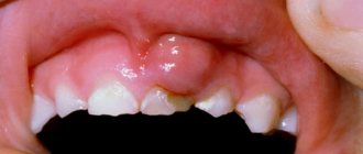
The causes of gum abscess (periodontal) are most often the gaps between the gum and teeth, the so-called periodontal pockets, into which the infection enters. Externally, the source of infection resembles a small red ball, painful when touched. Pockets can form for various reasons, in particular due to diseases of the oral cavity, constant injury from incorrectly installed dentures or fillings, mechanical damage (trauma, bruises, burns).
A gingival abscess that is not drained in a timely manner can damage one or even several teeth, which subsequently leads to their loss. Among other things, periodontal inflammation grows quite quickly and can increase to the size of a walnut, which not only deforms the jaw and facial contours, but also leads to severe intoxication of the body.
Treatment for this type of inflammation is standard: the abscess is opened with a scalpel, the pus is drained out, the cavity is drained and cleaned. Sutures are rarely used during such operations, since the size of the incision is usually quite small. However, if the tumor has reached an impressive size before the operation, the process of drainage and further recovery may be delayed. In some cases, doctors prescribe a number of physiotherapeutic procedures to speed up the patient's recovery.
Causes of development of abscess in the oral cavity
An abscess in the oral cavity occurs as a complication of advanced periodontal disease and periodontitis. These diseases are characterized by damage to the teeth and gums with the subsequent formation of pockets in the periodontium. They accumulate pathogenic microorganisms that provoke inflammation. Among other things, an abscess can occur due to infection in a damaged area of soft tissue (trauma, syringe needle, instrument). The cause of the disease is often staphylococcal and streptococcal sore throats, as well as boils on the face.
Inflammation in the oral cavity also appears as a complication after suffering from influenza or ARVI, which weaken the immune system, as a result of which the body is not able to fight infection.
We know how to cure ORAL ABSCESS
In the near future, a medical coordinator will contact you and advise you on the conditions and cost of treatment, select a doctor and make an appointment for you.
| Make an appointment | Or call us +375 29 699-03-03 +375 33 319-03-03 |
The cause of the disease is a mixed infection: streptococci and staphylococci, various types of gram-positive and gram-negative bacilli and often putrefactive bacteria.
Acute purulent periostitis is a complication of acute or chronic periodontitis in the acute stage. The inflammatory process can develop with difficulty in teething, suppuration of radicular cysts, and periodontal diseases. Sometimes the disease occurs after conservative dental treatment, during traumatic tooth extraction.
In acute and aggravated chronic periodontitis, the outflow of pus through the root canal or gum pocket is sometimes insufficient or impossible. Exudate spreads from the periodontium towards the periosteum. Often, an ulcer forms in the wall of the dental alveolus, through which purulent exudate penetrates into the periosteum. Sometimes microorganisms spread into the periosteum through the lymphatic vessels.
Sensitization and general factors play a certain role in the development of the disease: cold, overwork, stressful situations that reduce the body’s defense reactions.
Clinical picture. Characteristic complaints are pain, swelling of the soft tissues of the face, poor general health, and increased body temperature. At first, the pain is insignificant, then it intensifies and spreads to the entire jaw, radiating along the branches of the trigeminal nerve: to the ear, temple, eye. A swelling of the soft tissues appears, which progressively increases, while the pain in the tooth subsides. Complaints of headache, malaise, and poor sleep are possible. Body temperature is elevated - 37.5-38 °C, sometimes up to 38.5-39 °C; In some patients, general weakness, fatigue, loss of appetite, and insomnia due to pain are possible.
In case of acute purulent periostitis of the upper jaw, on the side of the vestibule of the mouth, upon external examination, diffuse soft swelling is visible due to collateral edema. Its localization and distribution depend on which tooth caused the disease. Periostitis of the upper jaw, associated with damage to the incisors, is characterized by significant swelling of the upper lip, spreading to the wings and bottom of the nose. The spread of the purulent process from the canine and premolars is characterized by the localization of collateral edema in the middle and lower thirds of the face, significant swelling of the eyelids, as a result of which the palpebral fissure is sharply narrowed. If the cause of the disease is premolars, then swelling of the buccal, zygomatic, and parotid areas occurs; from the third molar of the upper jaw, in addition to the listed areas, swelling spreads to the temporal region.
If the inflammatory process is associated with the incisors of the lower jaw, then swelling of the lower lip, chin and often submental areas appears, if with the canine and premolars, then there is collateral swelling of the lower cheek, the area of the corner of the mouth, descending into the submandibular region.
With inflammation from the molars of the lower jaw, collateral swelling of the lower part of the buccal, submandibular and parotid-masticatory areas is observed. The spread of the process to the periosteum of the lower jaw branch involves the masticatory and medial pterygoid muscles in the inflammatory process, which leads to their inflammatory contracture of the I-II degree. With the development of a purulent process from the third molar of the lower jaw, the swelling can spread down to the lateral parts of the neck. The patient experiences pain when swallowing and speaking.
Regional lymph nodes are enlarged, painful, and often fused. The inflammatory reaction of the lymph nodes is especially pronounced in children.
In acute purulent periostitis of the jaw, hyperemia of the mucous membrane over the affected area is noted on the vestibular side of the oral cavity. The vault of the vestibule of the mouth is smoothed due to inflammatory infiltration; palpation of this area is sharply painful. After a few days, the periosteum breaks through and pus penetrates under the mucous membrane of the alveolar process. Along the arch of the vestibule of the mouth, a roller-like bulge appears, covered with a thin mucous membrane; upon palpation, fluctuation is clearly visible. Percussion of the affected tooth may be painless, the tooth is sometimes mobile. Then the abscess can spontaneously open into the oral cavity. In such cases, the pain in the tooth subsides and the inflammation begins to subside.
With the development of the process in the lower jaw on the lingual side, collateral edema with a package of enlarged lymph nodes in the submandibular triangle is possible. Due to infiltration of the medial pterygoid muscle, mouth opening is painful and limited. In the oral cavity, from the alveolar part of the body of the lower jaw, swelling and infiltration of the periosteum are detected; palpation of this area is painful. Swelling and hyperemia of the mucous membrane can spread to the sublingual region, sometimes the anterior velopharyngeal arch and the pterygomandibular fold. Movement of the tongue becomes difficult.
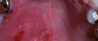
A palatal abscess develops with chronic or acute periodontitis of the lateral incisor, the root of the first premolar and the palatal roots of the maxillary molars. An external examination reveals enlarged, painful lymph nodes on the affected side, and a limited hemispherical or oval infiltrate on the hard palate. The enlargement of the abscess leads to smoothing of the transverse palatal folds. With the development of a palatal abscess associated with molars, inflammatory swelling from the hard palate spreads to the mucous membrane of the soft palate, palatoglossus arch, and pterygomandibular fold, causing pain when swallowing.
The accumulation of purulent exudate under the periosteum of the hard palate causes exfoliation of soft tissue from the bone. This is accompanied by pain, often pulsating in nature, aggravated by talking and eating. A week or more after the onset of the disease, the abscess breaks out and pus pours into the oral cavity.
Treatment. Complex treatment is carried out: surgical opening of the abscess, conservative drug therapy, etc. In the initial stage of acute periostitis of the jaw (acute serous periostitis), treatment can begin with opening the tooth cavity, removing decay from the canals and creating conditions for outflow, in other cases - with removing the damaged tooth that is the source of infection. All manipulations are performed under infiltration or conduction anesthesia. These therapeutic measures, together with lidocaine or trimecaine blockade with antibiotics, proteolytic enzymes along the transitional fold to the bone, and drug therapy can help to subside the inflammatory phenomena.
In case of acute purulent periostitis of the jaw, emergency surgical intervention is performed - opening the purulent subperiosteal focus and creating an outflow of exudate (primary surgical treatment of a purulent wound). This operation is usually performed on an outpatient basis, in some cases in a hospital.
Surgical treatment for acute purulent periostitis is performed under local anesthesia - conduction or infiltration anesthesia. For infiltration anesthesia, a thin needle is used, through which an anesthetic solution is slowly injected under the mucous membrane and the tissue is infiltrated along the intended incision line. The needle should not be inserted into the abscess cavity. Medicinal preparation of patients has a good effect. Sometimes the operation is performed under anesthesia.
If the subperiosteal abscess is located in the area of the vestibule of the mouth, then it is better to make the incision with a beak-shaped scalpel parallel to the transitional fold through the entire infiltration area; dissect the mucous membrane, submucosal tissue and periosteum to the bone, respectively, of 3-5 teeth. To prevent the edges of the wound from sticking together and to ensure the drainage of pus, a narrow strip of thin (glove) rubber is loosely inserted into the wound.
When the abscess is localized under the periosteum in the area of the tubercle of the upper jaw, the incision is made along the transitional fold in the area of the molars of the upper jaw, but to open the inflammatory focus, a rasp or a grooved probe should be passed from the incision along the bone in the direction of the tubercle of the upper jaw (back and inward). In the same way, a purulent focus is opened in case of periostitis of the upper jaw, which has spread to the canine fossa.
It is recommended to open the inflammatory focus during periostitis from the lingual surface of the lower jaw by making an incision in the mucous membrane of the alveolar part to the bone at the site of the greatest protrusion of the infiltrate. A grooved probe is passed down the surface of the bone and, pushing back the periosteum, allows the pus to drain out.
For palatal abscess, the incision is made in the area of greatest tissue bulging, slightly away from the base of the alveolar process, or at the midline of the palate, parallel to it. Then a wide strip of thin (glove) rubber is inserted into the surgical wound, which avoids sticking of the edges of the wound and creates conditions for good outflow of pus. The best results are obtained by excision of the wall of the abscess - a small section of the mucous membrane of a triangular shape, which ensures a freer outflow of pus.
To open the inflammatory focus in the area of the periosteum of the jaw branch, on its outer and inner surfaces, special techniques are used. In case of periostitis on the inner surface of the jaw branch, an incision is made with a sickle-shaped scalpel with a limiter or a regular scalpel to the bone, tissue is dissected in the retromolar region (at the base of the palatoglossal arch), and a rasp is passed to the inner surface of the jaw branch, creating an outflow of exudate from the source of inflammation.
The subperiosteal abscess along the outer surface of the lower jaw branch should be opened with an incision made from the vestibular side at the level of the second and third large molars along an oblique line to the bone, then with a rasp they pass subperiosteally in the direction of the angle of the lower jaw, retracting the masticatory muscle outward. After opening the lesion, a rubber strip must be inserted deeply into the wound for drainage. The lack of effect from such an intervention the next day is the basis for hospitalization and surgical intervention through external access. In children, indications for hospitalization should be expanded; children under 10 years of age are treated in a hospital.
The opening of periosteal lesions in children should be gentle. The periosteum must be peeled off well, but not to injure the bone with the instrument to avoid injury to the tooth buds.
After opening the purulent focus, it is advisable to let the patient rinse his mouth with a weak solution of potassium permanganate or 1-2% sodium bicarbonate solution, and it is also necessary to rinse the wound with a solution of chlorhexidine. A good effect is achieved by irrigating the abscess cavity with a solution of dimexide with oxacillin and applying a 40% solution of dimexide liniment to the wound for 15 minutes.
If the tooth that was the source of infection is destroyed and does not represent functional or aesthetic value, then it should be removed simultaneously with the opening of the subperiosteal abscess. This will improve the emptying of the purulent focus and will contribute to a faster elimination of inflammatory phenomena. Tooth extraction is sometimes postponed due to perceived technical difficulties or the unsatisfactory condition of the patient. In other cases, the tooth is preserved: its cavity is opened, the root canal is freed from decay products, and then conservative treatment of chronic periodontitis is carried out. It is especially important to remove the sources of infection in children - baby teeth, and if conservative treatment is impossible, permanent teeth.
Drug treatment of acute purulent periostitis consists of prescribing sulfonamide (sulfadimethoxine, sulfadimezin, etc.) and nitrofuran (furazolidone, furadonin) drugs, pyrazolone derivatives (analgin, amidopyrine, phenacetin, etc., as well as their combinations), antihistamines (diphenhydramine, suprastin , diazolin, etc.), calcium supplements, vitamins (multivitamins, vitamins C 2-3 g per day).
The patient is invited to an appointment on the 2nd day after surgery. During examination and questioning, the degree of subsidence of inflammatory phenomena is determined and, depending on this, additional treatment is prescribed. During dressing, local treatment of the wound is carried out, following the recommendations given earlier.
In case of acute purulent periostitis of the jaw, for a more rapid cessation of inflammatory phenomena on the 2nd day after opening the abscess, physical treatment methods should be prescribed: light and heat therapy (sollux lamp), warm baths of antiseptic or deodorizing solutions, ointment dressings (levomikol, levamisole, petroleum jelly, 20 % camphor oil, sea buckthorn oil, rose hips), UHF, microwave therapy, fluctuarization, laser therapy with helium-neon and infrared rays. Exercise therapy is recommended.
In most cases, inflammatory phenomena quickly (after 2-3 days) subside. If the subsidence of inflammation is delayed, then a blockade is carried out 2-3 times: intra-or extraoral infiltration with 0.25-0.5% trimecaine solution, 40-50 ml of lidocaine with enzymes, antibiotics (lincomycin). Weakened patients, patients with concomitant diseases, children and patients at risk, as well as persons with increasing inflammatory phenomena are prescribed antibiotics. A prerequisite for the effectiveness of antibiotic therapy is opening the abscess (primary surgical treatment). In a clinic setting, it is advisable to prescribe broad-spectrum antibiotics (semi-synthetic penicillins, tetracycline, oletethrin, oxacillin, macrolides). It is mandatory to prescribe metronidazole derivatives 200,000 units 3 times a day for 5-6 days, in the hospital - injections of these drugs 3-4 times for 6-7 days.
"Surgical Dentistry" edited by Robustova T.G.
Fourth edition. Moscow "Medicine" 2010
Symptoms
The abscess is characterized by rapid development. At first, the patient may be bothered only by minor attacks of pain, similar to the sensations that arise during caries or periodontitis. Subsequently, the pain is localized in one place and gradually increases. In a specific place there is swelling, sometimes a new formation on the gum, sometimes reaching the size of a walnut. If the inflammation is localized closer to the outer surface, then swelling and redness can be observed with the naked eye. If you notice the first symptoms, we recommend that you consult a dentist.
An abscess of the tongue is characterized by an increase in the organ's volume, difficulty swallowing, chewing, and even suffocation. Any abscess is accompanied by fever, deterioration and general weakness, insomnia, and loss of appetite. The progression of the disease leads to a breakthrough of the abscess, which is manifested in a decrease in temperature, a decrease in swelling and an improvement in overall well-being. However, there is no reason to stop treatment, since inflammation can continue and develop into a chronic form. This can lead to tooth loss, sepsis and phlegmon.
Causes
In 99% of cases, this disease develops due to dental problems. As practice shows, it is always a consequence of untreated caries. However, purulent melting does not develop immediately. This must be preceded by other pathologies, such as inflammation of the pulp, periodontium, development of a radicular cyst, or osteomyelitis. An obligatory element leading to infection must always be a source with microbial contamination.
Not always purulent-inflammatory processes can lead to the spread of infection. The human body’s immunity plays a huge role. When it decreases, pathogenic microbes in the purulent focus can lead to necrosis of nearby tissues. The head and neck area is rich in cellular spaces that smoothly merge into each other. Therefore, pus can freely move from the lower jaw area down the neck.
The following may be subject to melting:
- Subcutaneous fat tissue;
- Fiber of the intermuscular space;
- Interfascial tissue;
- The lymph nodes;
But there is another factor - an infected wound, which could be the result of injuries. The development of a purulent process is very rarely observed in this case, but with reduced immunity it occurs. Therefore, every wound must be subjected to antiseptic treatment.
Treatment of oral abscess
Treatment of an abscess requires surgery. In order to eliminate the source of infection and stop the spread of inflammation, the dental surgeon step by step:
- opens an abscess;
- drains the cavity;
- cleans the pocket;
- Rarely applies stitches if the size of the incision is large.
After removal of the pus, the patient’s well-being improves, and facial geometry is restored. Taking antibiotics, antihistamines, immunostimulants and a vitamin complex significantly speeds up the healing process. Sometimes the doctor may prescribe physiotherapeutic procedures (UHF therapy and fluctuarization).
For some time, the patient needs to exclude solid foods from the diet and adhere to healthy eating rules.
Diagnosis and treatment
The diagnosis is made on the basis of clinical studies:
- Collecting the patient's medical history. The doctor listens to complaints and asks questions about symptoms and other manifestations.
- Inspection. It is carried out to identify the causative tooth. The doctor evaluates the color of nearby tissues, the symmetry of the face, and checks the opening and closing of the jaw.
- Palpation of lymph nodes. Their increase indicates the presence of inflammation.
- Measurement of temperature and blood pressure. Deviation of indicators from the norm indicates intoxication of the body.
- X-ray (spot shot and orthopantomogram). It is carried out to establish the localization of the purulent focus.
- Electroodontodiagnostics. Necessary to assess the condition of the dental nerve in the causative tooth.
- Lab tests.
If necessary, general clinical studies are prescribed, such as an electrocardiogram and fluorography.
Material is taken from the opened lesion to assess the sensitivity of the microflora to the effects of antibiotics.
Prognosis and prevention of oral abscess
The success of treating oral abscesses depends on at what stage of the disease the patient seeks help and how strong the body’s defense mechanism is. With timely treatment, the prognosis for eliminating inflammation is quite favorable. In the absence of complications, strong immunity and a high-quality opening of the lesion, an oral abscess can be cured in a couple of weeks.
Prevention of the disease consists of following the following recommendations:
- regular teeth cleaning and professional oral hygiene at least once every six months;
- minimizing the risk of injury to the mucous membrane;
- timely treatment of caries and periodontal diseases;
- visiting a dentist for a preventive examination once every 6 months.
Odontogenic phlegmon
The cause of the development of this kind of disease is always a dental problem. Any pathology of the masticatory organs can lead to this outcome.
Problems may arise at the following stages:
- Before tooth eruption;
- At the moment when he is already in the row;
- After removing it.
The most common cause of this disease is untreated caries. The pathological process in hard tissues, having gone through certain stages of disease development (pulpitis -> periodontitis), can lead to the development of a radicular cyst. When immunity decreases, it can fester, and then the contents surrounded by the capsule have the opportunity to go beyond its limits.
Wisdom teeth can also cause problems as they are the most advanced teeth and may not have enough space in the row. Having appeared at least half, they become vulnerable to the carious process. Under the gingival hood, which is located above the tubercles, food begins to accumulate. Then cariogenic microorganisms join this substrate and begin to ferment leftover food into organic acids. As a result, a cavity forms in the hard tissues. The wisdom tooth is anatomically designed in such a way that it is very difficult to treat, and if preservation is not practical, then it is better to remove it.
A very rare phenomenon occurs when the disease develops after tooth extraction. The following reasons may contribute to this:
- Part of the organ was left in the socket;
- Food getting into an unhealed hole;
- Permanent injury to the socket with a blood clot, followed by infection.
If any element of the tooth remains inside, then in most cases it will lead to inflammation, since it will be perceived by its own tissues as a foreign body. In addition, local immunity will be activated, which will actively produce lymphocytes. When pathogenic microflora attaches, this phenomenon can spread to nearby tissues. Therefore, it is necessary to remove everything that remains in the hole after tooth extraction.
At first, a person complains of swelling in the area of the extracted tooth and painful opening of the mouth. Throughout the course of the disease, the puffiness of the face becomes more and more. Then, every day the person’s general condition begins to deteriorate. The temperature rises, and blood tests change, which begin to indicate an inflammatory process occurring in the body.