Flux is a common, non-medical name for a whole group of diseases that require immediate, and in some cases, emergency care. One of the main manifestations of flux is a violation of the shape of the face associated with a complication in an inflamed tooth.
The main groups of diseases that are commonly called flux: periodontitis, periostitis, osteomyelitis, abscess and phlegmon, lymphadenitis, odontogenic sinusitis.
Periodontitis
- inflammation of the tissues of the apical part of the tooth.
Periostitis
– inflammation of the periosteum in the area of the teeth.
Osteomyelitis
– inflammation of the jaw bone, which necessarily ends with necrosis (death) of the affected area of the bone.
Abscess
- This is a limited purulent inflammation of fatty tissue.
Phlegmon
- This is a diffuse purulent inflammation of fatty tissue.
Under the skin, under the mucous membrane, under and between the muscles there is fatty tissue. With an exacerbation of odontogenic infection, pus spreads under the periosteum, axillary and intermuscular spaces, causing inflammatory and edematous-infiltrative changes in the soft tissues of the face and neck.
Inflammatory diseases of the maxillofacial area (flux, abscess, phlegmon) and neck are divided into two main groups according to localization. The first is abscesses and phlegmons located near the upper jaw. The second is abscesses and phlegmons located near the lower jaw.
Lymphadenitis
– inflammation of the lymph node (group of lymph nodes) adjacent to the odontogenic (dental) focus of infection. Odontogenic sinusitis is often an inflammation of the maxillary (maxillary) sinus (sinusitis), caused by the anatomical features of the position of the roots of the maxillary teeth involved in the pathological process, in which the roots of the teeth either stand in the lumen of the maxillary sinus, or are located close to the bottom of the maxillary sinus. In this case, infection of the mucous membrane of the maxillary sinus occurs with the subsequent development of sinusitis.
Causes
Inflammatory (infectious-inflammatory) diseases of the maxillofacial area are caused by microbial microorganisms, which are usually part of the normal microflora of the oral cavity. Microbial pathogens are staphylococci, streptococci, enterococci, diplococci, gram-positive and gram-negative bacilli (intestinal, protea, etc.). In addition, during flux, fungi, mycoplasmas, treponema, and protozoa from the Trichomonas family are found in foci of odontogenic infection. According to various authors who analyzed the microbial flora in foci of odontogenic infection (flux), the microflora is represented by a monoculture of staphylococcus (aurus and epidermal) or streptococcus of groups D, F and G. Associations of the above microorganisms are often identified.
The question arises of how non-pathogenic or opportunistic microorganisms trigger the initiation of an infectious-inflammatory process (flux). For the occurrence of a disease, the presence of only non-pathogenic or pathogenic microorganisms is not enough. The answer to this question is given by the so-called Arthus-Sakharov phenomenon. According to the infectious-allergic theory, its essence boils down to the following: under the influence of a foreign serum protein, which has antigenic properties, antibodies are produced, which leads to sensitization of the body. Sensitization of the body is the acquisition by the body of increased sensitivity to foreign substances (proteins) - allergens. Against this background, repeated introduction of the protein into the vascular bed causes the formation of antigen + antibody complexes, which are fixed on the membranes of blood vessels and turn into target cells. Then the cell membrane is damaged, enzymes are released, mediators (from the Latin mediator - mediator) of inflammation are released. This is accompanied by activation of the third platelet factor and can cause intravascular coagulation, which leads to impaired microcirculation and necrosis (death) of tissue. This immunopathological reaction is also involved in odontogenic infection, leading to the occurrence of gumboil. The waste products of microbes participate in the role of antigen. This explains why in many patients non-pathogenic microorganisms are found at the site of inflammation.
The dynamic balance between the focus of chronic odontogenic infection and the body is ensured by the connective tissue capsule surrounding the focus. It limits the penetration of microbes into the tissues and vascular bed adjacent to the lesion, and limits the effect of immune factors on the infectious focus. The balance is also maintained by the fact that part of the waste products of microorganisms and tissue decay through the root canal of the tooth, fistula or periodontal fissure is released from the infectious focus into the oral cavity.
An imbalance can be caused by a disruption in the outflow of exudate from the lesion through the root canal due to food masses entering the carious cavity or when filling a carious cavity by a dentist. In the infectious focus, the concentration of microbes, their toxins, and waste products, which penetrate through the connective tissue capsule into the surrounding tissues, increases. Here their direct damaging effect on tissue can occur, and upon penetration into the vascular bed, the mechanism of the Arthus-Sakharov phenomenon is triggered. Clinically, this is manifested by an exacerbation of a chronic focus of odontogenic infection and the occurrence of gumboil.
Another mechanism of imbalance between the focus of chronic infection and the human body is associated with damage to the connective tissue capsule itself. This can happen when a tooth is removed, when there is increased load on the tooth while chewing hard food, etc. Damage to the capsule leads to the spread of microorganisms (their toxins, waste products) beyond the infectious focus, which in a sensitized organism leads to the occurrence of the Arthus-Sakharov phenomenon. These are the reasons and the main mechanism of exacerbation of odontogenic infection, leading to the development of gumboil.
How long does the flux last after extraction?
At first after removal, an inflamed and painful state is considered a normal reaction of the body. However, in situations where the infection penetrates deeply into the tissue structure, provoking pathological processes, even extraction of the problem unit does not guarantee relief of the negative syndrome.
Flux is the result of the development of the disease, and not its root cause, which makes it difficult to eliminate the pathology. Even with an independent breakthrough of suppuration, accompanied by the release of exudate, the resulting cavity can cause the development of an abscess or phlegmon, the treatment of which requires much more effort. In the most severe forms of development, flux leads to infection of the circulatory system, and, as a result, death.
Symptoms
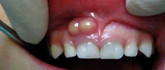
The main manifestations of gumboil disease are pain in the area of the causative tooth and the appearance of swelling in the soft tissues of the face. If at the beginning of the disease the swelling is mild, then as the process progresses the swelling increases and can spread to the eye area, scalp or neck. As the symptoms of flux increase, pus from under the periosteum can spread through the fatty spaces closer to the skin - this is manifested by the fact that the skin turns red, becomes dense and hot to the touch. Further, the skin in the center of the flux softens, becomes thinner, and a fistula with purulent discharge may appear. In this case, pain may decrease somewhat, which is associated with a decrease in pressure in the flux cavity during the formation of a fistula tract.
In addition to pain, symptoms such as limited mouth opening or inability to open the mouth, and pain when swallowing may occur. If the flux is located deeply, facial swelling may be mild or not expressed at all, and the main manifestations will be limitations in mouth opening, pain in the flux area and pain when swallowing.
Briefly about the disease
Periostitis is an inflammatory disease with purulent contents that causes infection. Pathogenic microorganisms often enter the pulp from carious cavities. An abscess forms at the root apex. Pus penetrates into the gum tissue, they become inflamed, and swelling develops. The sore spot looks like a lump on the outside. Cause of infection:
- Cyst;
- Untreated caries;
- Inflammation in the gum pocket;
- Jaw injuries;
- Periodontitis or pulpitis;
- Poor installation of fillings or crowns;
The clinical picture of the disease is clearly expressed. The acute phase of periostitis has the following symptoms:
- The patient experiences severe pain in the head, neck, and jaws. Discomfort intensifies during chewing.
- The temperature rises.
- The gum tissue turns red.
- A tumor forms on the cheek. It can spread to the nose, lips, eyelids.
- Malaise and general weakness appear.
- The lymph nodes located under the lower jaw become enlarged.
An alarming symptom is the rapid growth of a lump, increasing throbbing pain. The capsule with pus may burst, then the patient feels relief. The chronic form lasts several months. The disease will not go away on its own. The disease is eliminated by the dentist.
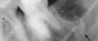
Diagnostics
To diagnose flux, methods such as questioning, examination, and additional research methods are used: radiography, ultrasound, computed tomography, magnetic resonance imaging, blood test data, culture of exudate, etc. An experienced maxillofacial surgeon who treats patients with flux, can, based on the patient’s complaints and examination, carry out an accurate topical diagnosis of the disease (localization of pus) and select the optimal surgical approach to ensure sanitation of the purulent focus and adequate drainage.
Does using traditional recipes help?
You cannot use only homemade preparations to treat an abscess. Otherwise, the flux will last for several weeks, which will lead to a large amount of pus. The result can be severe deterioration in health and blood poisoning. Traditional medicines can only be used in combination with medications after surgery. In this case, the disease will last about 14 days.
It won't hurt to rinse with baking soda, which helps relieve inflammation. 1 teaspoon of the product should be dissolved in 0.5 cups of warm water. The drug is used to rinse at least 5 times a day. Read more in the article: “Treating flux at home with soda.”
Salt will help at home
An alcohol solution of propolis helps well. It is diluted with water in a ratio of 1:10 and rinsed with mouth three times a day. The bactericidal drug Miramistin is also used. There is no need to dilute it. You should rinse your mouth with about 15 ml of the product and then spit it out. The procedure is carried out 3 times a day.
The use of warm compresses is strictly prohibited, otherwise the inflammatory process may worsen.
Treatment
Treatment for flux can be boiled down to a catchphrase in Latin: “Ubi pus, ibi incision,” which translates as “Where there is pus, there is an incision.” Treatment of flux begins with eliminating the cause that caused the flux. That is, from the removal of the causative tooth. Next, they begin to drain (empty) the purulent focus by opening (incision) in the area of the flux. The abscess is emptied, the abscess cavity is washed with antiseptic solutions, and drainage is installed through which pus will be released. Regardless of the type of flux, the principle of treatment remains the same: opening the lesion, drainage.
In the postoperative period, daily antiseptic treatment (washing) with antiseptic solutions is carried out. There is a peculiarity in the treatment of these diseases (flux, periostitis, phlegmon, etc.), which is that immediately after surgery and up to 1-2 days, facial swelling (flux) may increase. This should not be taken as a worsening of the disease. The fact is that any surgical intervention is perceived by the body as an injury, and the body responds to this by increasing tissue swelling in the area of injury. This may be accompanied by increased swelling of the eyelids, cheeks, etc. Painful sensations decrease and subsequently decline as acute inflammatory phenomena subside. As a rule, antibacterial drugs, antihistamines and painkillers are prescribed after surgery.
If there is an advanced case of flux, which is accompanied by a violation of the function of external respiration (the patient may suffocate), then hormonal drugs are prescribed, and even an operation is performed - a tracheotomy. The operation consists of making an incision in the area of the tracheal rings and installing a tube. As the swelling of the upper respiratory tract (pharynx, larynx) decreases, when the threat of asphyxia (suffocation) has passed, the tube is removed from the trachea, and the patient breathes through the natural airways. The duration of prescription of drugs is determined by the attending physician. The drainage in the wound is periodically changed, and the wound is removed when cleansed. The duration of drainage in the wound is also determined by the attending physician, based on the clinical picture of the disease. The drainage can remain in the wound for an average of 3 to 10 days.
If the incision for flux was made through the oral mucosa, then additional suturing of the wound is not required in the future. The wound in the oral cavity will heal on its own (by secondary intention). If the incisions were on the skin side, then after the acute inflammatory processes have subsided, the wound can be sutured. After suturing the wound (and this can be 2-4 weeks after surgery), the patient is also prescribed antibacterial drugs and painkillers, and the wound is drained.
With inadequate treatment, as well as in advanced cases of flux, complications such as suffocation, penetration of infection into the cranial cavity, onto the membranes of the brain, into the neck and into the mediastinum (part of the chest cavity where vital organs are located), penetration into the orbital cavity with damage to the optic nerve and the occurrence of a systemic inflammatory reaction (sepsis), which leads to multiple organ failure and even death.
Soft tissue incisions, which are used in the surgical treatment of flux, must ensure complete sanitation of the purulent focus and adequate drainage. Surgical treatment should not be replaced by the prescription of strong antibacterial drugs. Waiting tactics, delays in performing surgery, and small incisions that do not provide adequate drainage of the purulent focus are unacceptable. We must not forget that surgery for a purulent process in the maxillofacial area and neck is often an operation to save the patient’s life. The occurrence of complications is more likely when the patient has concomitant chronic diseases: diabetes mellitus, coronary heart disease, anemia, diabetes mellitus and others. Elderly and senile age are also factors that can aggravate the course of this disease.
What do experts think?
When patients ask their doctors how many days it should take for the flux to disappear, experts talk about 1-3 weeks. However, to do this, several important conditions must be met.
First of all, you should visit your doctor regularly and follow all his recommendations. In addition, you need to adhere to the correct treatment regimen and purchase only certified medications from pharmacies.
But even compliance with the above requirements does not guarantee that flux manifestations will disappear after 2 weeks. It is impossible to say exactly how many days periostitis lasts. We can only talk about approximate dates, since they are influenced by the individual characteristics of the body, immunity, type of disease and treatment. Let's look at this in more detail.