Types of lymph nodes in the neck
There are many groups of nodes in the neck that differ in size, location and protection provided:
- The anterior cervical, which is localized in the jugular part of the neck, is responsible for the condition of the tissues of the throat (posterior wall and tonsils) and the thyroid gland. They can be superficial or deep. The deep cervical lymph nodes cannot be palpated, even if they are inflamed.
- Posterior cervical are located on the back of the head and behind the neck. Inflammation of the occipital nodes can indicate bronchial diseases, as well as meningitis.
- Almond - palpated under the lower jaw and controls, like the anterior cervical, tonsils and the back wall of the throat.
- The submandibular lymph nodes of the neck are located along the lower jaw and become inflamed in the case of infectious dental diseases (periodontal disease, stomatitis, caries, etc.), diseases of the tongue, salivary glands, as well as diseases of the ENT organs (sinusitis, pharyngitis, rhinitis, tonsillitis, otitis).
- The lymph node in the neck behind the ear is sometimes classified as a separate group, and sometimes classified as cervical. It is small in size, about the size of a pea, and practically cannot be felt. Enlarged lymph nodes behind the ears may indicate the presence of pathology in the back of the head, in the parietal region.
- The submentals are located under the chin and control the cheeks, lower lip, and teeth.
- The supraclavicular muscles lie in the recess of the collarbones at the base of the neck and are divided into right and left. Controls the lungs, esophagus and heart. Inflammation of this group of lymph nodes in the neck is not a good sign, and often indicates the presence of a serious disease.
Self-diagnosis
The scheme for examining the lymph nodes in the neck depends on their location. You should start defining it like this:
- To palpate the tonsillar capsules, you need to place two fingers so that the tip touches the earlobe, and the middle of the finger touches the corner of the neck. The lymph nodes will be located in the lower part, on the side. The limited area is felt with the fingertips. The nodes are superficial, so they are not difficult to feel.
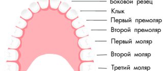
Diagram of the location of lymph nodes in the neck
- Finding the posterior cervical nodes is more difficult. May be hidden by subcutaneous fat in the presence of obesity. Palpation is carried out according to the following scheme: the head is tilted forward, the fingers are located behind the vertebrae, palpating the left and right sides of the neck. When pressed, tubercles are revealed. If it was not possible to identify the posterior nodes, this means that the lymph nodes are deep and small in size, which is considered a normal condition.
- To identify the anterior cervical lymph nodes, you need to tilt your head back and palpate the front on the left and right sides of the larynx. Small dense balls up to 2 cm in size are felt.
- To detect submandibular nodes, fingers are positioned 3 cm from the angle of the jaw, and the area towards the chin is palpated. It is difficult to detect if you are overweight.
Appearance of healthy tonsils
The healthy state of the tonsils is determined by several parameters:
- They should be medium in size.
- The color of the tonsils should be uniformly pink, without inclusions or furrows.
- The surface has small tubercles and depressions.
- The posterior pharynx is uniform in color and the follicles should not be noticeable.
- When examined with a spoon, no pus is released from the tonsils.
Often the gag reflex does not allow a thorough examination of the patient's throat. Most problems arise when examining tonsils in children due to their refusal to open their mouth wide.
- Choosing the right lighting will help you carefully examine your oral health. A beam of yellow direct light aimed directly at the throat will help give a more accurate picture of the patient's condition.
- During the examination, you can use a teaspoon or a device specially designed for this purpose, purchased at a pharmacy.
- When applying pressure to the tongue, it is important to do this carefully, without causing a gag reflex.
- The patient must breathe through his mouth. This will help make a better inspection.
The symptoms of a sore throat depend on the causes that caused it.
Nevertheless, the main and characteristic features are considered to be:
Other diseases
The main disease of the tonsils is tonsillitis. However, the tonsils can become swollen and inflamed due to other pathologies, including:
- sinusitis;
- otitis;
- stomatitis;
- caries;
- staphylococcal infection.
In the case of sinusitis, the tonsils become inflamed due to the entry of infected sinus contents into the throat. Otitis media in general is a consequence of adenoids, but can lead to inflammation of both the tonsils and sinuses.
Stomatitis is an inflammation of the oral mucosa. If the disease remains untreated for a long time, the infection can spread to the tonsils.
Advanced caries is a common cause of inflammation of the oral cavity and tonsils. This is due to a general decrease in immunity against the background of a chronic focus of infection, and the subsequent spread of bacteria throughout the nasopharynx.
Reasons for the increase
Soreness indicates the development of an infectious process in humans. The following diseases can provoke lymphadenitis:
- Inflammatory diseases of the neck, head: boil, erysipelas, herpes and fungal inflammation, osteomyelitis and otitis media, hematoma suppuration.
- Dental infections: caries, periodontitis, stomatitis, glossitis.
- Respiratory diseases: tonsillitis, rhinitis, laryngitis, scarlet fever and tuberculosis, tracheitis and influenza.
- Systemic infection: syphilis, AIDS, mononucleosis.
- Non-infectious disease: lupus erythematosus, sarcoidosis, lymphoma, allergy.
- Blood circulation system: lymphocytic leukemia, lymphogranulomatosis.
- Metastases of oncological tumors of the neck, head and other organs.
A common cause of inflammation in a child is colds. The same effect is exerted by weak immunity against the background of insufficient vitamins and unfavorable ecology.
You should not delay visiting a doctor if the following signs are observed:
- the lymph node has increased sharply;
- soreness to touch;
- in a calm state there is pain;
- the area around the lump turns red;
- purulent foci form on the skin;
- the temperature rises;
- the organ swells.
Timely treatment will help avoid complications. Enlarged nodes can be detected symmetrically.
Lymph nodes in the neck: which ones and when they increase
Since each group of lymph nodes is responsible for collecting lymph from a certain area of the body, by their enlargement it is possible to determine where the disease process began.
Very simply, we can say that:
- the mental lymph nodes collect lymph from the tongue, teeth and the floor of the mouth;
- submandibular lymph nodes are “responsible” for the bones and soft tissues of the face, as well as for the oral cavity;
- retropharyngeal lymph nodes collect lymph from the pharynx;
- supraclavicular lymph nodes collect lymph from the head, neck, and apexes of the lungs;
- subclavian lymph nodes specialize in diseases of the lungs and upper shoulder girdle;
- the anterior cervical lymph nodes are “responsible” for the tonsils, pharynx and tissues of the neck itself;
- posterior cervical lymph nodes collect lymph from the head and neck;
- The parotid lymph nodes mainly collect lymph from the organ of hearing and nearby tissues.
We suggest you familiarize yourself with Teeth whitening with tea tree, lemon, coconut oil, etc.
Palpation of the larynx and neck
Palpation of the larynx and neck is one of the diagnostic methods that are used in otolaryngology in the treatment of diseases of the ENT organs.
Palpation of the larynx and neck is a visual diagnosis of diseases of the pharynx and larynx, which allows you to assess the condition of the skin, the severity of the local vascular network, the shape and position of the organ.
Much becomes clear already during a conversation with the patient, for example, by the sound of his voice (the presence of nasality, rattling, shortness of breath and other characteristic symptoms), the complaints presented (their dynamics, concomitant diseases in the anamnesis). After examining and questioning the patient, the doctor proceeds to palpation of the larynx and neck. Depending on the indications, it can be superficial or deep.
Front
The anterior cervical lymph nodes, which allow the head to tilt and rotate, are located above and below the sternocleidomastoid muscle in front of the internal jugular vein. These are superficial jugular nodes. They are small, but there are many of them. The anterior cervical cleanses the lymph entering the pharynx, throat, tonsils and thyroid gland.
In turn, if you look at the figure, it is clear that among the anterior glands there are groups of preglottic, thyroid, paratracheal and pretracheal. These are deep nodes.
Palpation of the cervical node is difficult, it is impossible to find them, since they are small. In an adult they are smaller than in children.
The lymph node on the left or right side of the neck is inflamed. We need to find out what happened:
- The tonsils are inflamed.
- There was an infection in the oral cavity.
- Bacteria entered the respiratory tract.
The reason could be:
- decreased immunity;
- lack of vitamins;
- freezing of the body;
- long-term stressful situations;
- insect bites;
- inflammation in the ears.
A lump appeared on the front of my neck. Often it is not painful. The anatomy of the appearance of a lump is as follows: depending on the infectious lymph node that first caught the infection, the lump can be in the front, side or under the chin.
The submandibular glands are the first to fight infection of the pharynx, mouth and throat. Changes usually take place at the micro level. The neck remains unchanged. When an infection or viruses enter the gland, the node swells.
The neck can be seen with a lump under the chin. If the lump is on the neck when the glands have returned to normal, then the doctor can answer whether the lymphadenitis has developed into a chronic stage.
Categories, location
The body includes cervical lymph nodes, classified into the following categories:
- submandibular;
- chin;
- jugular;
- posterior cervical;
- anterior cervical;
- tonsillar.
First group
The submandibular lymph capsules are located under the lower jaw bone. They have a small volume. They can become inflamed due to diseases of the oral cavity, caries. Includes the following levels of lymph nodes: preglandular, postglandular, retrovascular, postvascular.
Second group
The mental lymph nodes are located under the tongue, these are the suprahyoid, thyroglossal-facial, and mental nodes. The location is deep and difficult to palpate.
Third group
Jugular nodules are upper, middle and lower. The jugular depression or fossa is located between the collarbones at the bottom of the neck. The top view is placed in front of the nerve, closer to the jaw and extends behind the nerve, closer to the neck. The group includes the retropharyngeal, jugular-digastric lymph nodes, and is classified as superficial.
The middle nodes are defined similarly to the top nodes. These include the superior thyroid and superior deep lateral lymphatic capsules.
The location of the lower ones goes from the bottom of the base of the larynx, going down to the collarbone. Includes the lower deep lateral, jugular-scapular-hyoid and supraclavicular Virchow lymph nodes.
Fourth category
The posterior cervical nodes run along the back of the neck, located on both sides of the spine. The posterior lymph nodes contain the lateral, intercalary, transverse cervical, lower deep lateral and above the clavicle (excluding Virchow) nodes.
Fifth group
The anterior cervical lymph nodes are located near the larynx, under the chin. Contains pretracheal, paratracheal, precricoid, perithyroid nodes. They are large in size and easily palpable.
Sixth group
Tonsillar lymph capsules are classified as facial and cervical groups. They are located on the neck, at the corners of the lower jaw. Susceptible to diseases of the tonsils (tonsils), throat.
Cervical lymph nodes not included in a specific list include the occipital, postauricular, parotid and retropharyngeal nodular capsules.
Causes
There are many prerequisites for the occurrence of inflammation of lymphoid tissue. Let's focus on the main ones.
- Acute respiratory infection. In the vast majority of cases, the disease is viral in nature, less often bacterial or fungal. It is obvious that the human pharynx is constantly exposed to attacks by foreign microflora. However, inflammation of the tonsils is provoked by a weakened immune system or the absence of antibodies to a particular pathogen in the body.
- Hypothermia. As you know, the height of infectious diseases occurs in the autumn and spring months. It is during this period that pathogenic microorganisms multiply rapidly and acquire resistance. Hypothermia of the body becomes a provoking factor for weakening the immune response from lymphoid organs, such as the lingual and palatine tonsils. That is why drinking cold drinks, being in a draft, and getting wet and cold feet often cause illness.
- The presence of foci of chronic infections. The constant penetration of pathogenic flora from the sinuses or teeth affected by caries into the pharyngeal cavity causes inflammation of the tonsils at the first signs of decreased immunity.
- Congenital or acquired anatomical abnormalities. Such changes in the structure of the nasopharynx interfere with the natural flow of air masses and make breathing difficult. Blood flow in the mucous membranes is disrupted, which causes them to dry out. As a consequence, the impossibility of performing the barrier function and the occurrence of an inflammatory process. Often, anatomical defects require surgical treatment.
Causes of hypertrophy
The tonsils or tonsils perform a protective function in the human body. The organ is located at the intersection of the nasal canals and pharynx, at the base of the tongue (one on each side). The tonsils are palpated from the outer part of the neck (under the jaw), especially if they are very enlarged. Their main function is to prevent infections, harmful bacteria and microorganisms from entering food, water, and air.
With an excessive number of pathogens, the lymphoid tissue cannot cope with its function, becomes inflamed and becomes an independent causative agent of the pathological process, which leads to enlargement of the tonsils. When a child’s tonsils are enlarged, the disease is called chronic tonsillitis or its acute form, in other words, tonsillitis.
Enlarged tonsils in a child are a sign of weakened immunity, dysfunction of the filtering organ, which accumulates pathogenic microbes and gradually becomes inflamed.
How to feel the tonsils in the neck
Tonsils
(palatine tonsils) are paired oval-shaped formations, consisting of lymphoid tissue and located in the recess between the soft palate and the tongue. The palatine tonsils, together with the pharyngeal tonsils (at the vault of the pharynx) and lingual tonsils (at the base of the tongue), form a lymphadenoid ring. It is believed that the tonsils, as an organ of the lymphatic system, are involved in the development of immunity, that is, they have a protective function. At the same time, they often turn out to be entry points for bacterial and viral infections, especially in children. Inflammation of the tonsils is called tonsillitis; it can be acute (angina) or chronic. Enlarged tonsils create serious breathing difficulties. Sometimes, in cases of chronic tonsillitis, the tonsils are removed.
Tonsils: remove or cleanse?
Tonsils, or, as we more often call them, tonsils, protect the body from various infections.
Naturally, sometimes they become inflamed. After all, they have to be a barrier to toxins that are released in diseases of the ENT organs, teeth, nervous, endocrine and cardiovascular systems, spine, digestive tract and many others. Previously, constantly inflamed tonsils were most often removed surgically. Today they do this only when other methods fail to solve the problem. However, even after tonsil removal, health does not always improve. Therefore, the best thing to do is not to bring them to a critical state. Many people today do not know that tonsils require constant care. They are similar in structure to a sponge. Imagine how this sponge absorbs germs and bacteria. When they begin to multiply intensively, the body defends itself by producing white blood cells, which begin to fight the infection in the tonsil tubes (lacunae). In this case, pus is constantly formed - these are the remains of bacteria and dead leukocytes. But when they die, these enemies of our body produce special enzymes that seem to “melt” the tissues of the tonsils themselves and form purulent sacs (cavities). If you stick your tongue out as far as possible, you can see a liquid coating of a whitish-yellow or even greenish color on it. This is how the tonsils are cleared of secretions. By the way, Eastern medicine considers such an exercise with the tongue sticking out as much as possible an excellent way to cleanse and even treat a sore throat. (Although it’s better to actually clean off the plaque, for example, with a silver teaspoon.) How to do the exercise correctly? Inhale and hold your breath. Open your mouth wide, stick your tongue as far forward and down to your chin as possible. At the same time, press your chin to your sternum. Concentrate on the muscles of the neck and larynx. Stay in this position for as long as you can breathe. Repeat the exercise at least three to five times. Then gargle with salted water (half a teaspoon of salt per glass of water). If the tonsils are not cleared naturally (for example, a person turns their head a little) or they are not cleared using the described exercises, the purulent cavities can become very large, go beyond the boundaries and begin to spread into the cervical tissues. When the tonsils are removed, the cervical cavities filled with pus become closed sacs and can turn into cysts that release toxins into the surrounding tissue. Hence - pharyngitis, loss of voice, cervical osteochondrosis and other troubles. Many people who already had their tonsils removed in childhood are familiar with them. Tonsils
can be well cleaned in another way - a special massage. Using the pads of the thumb and forefinger near the jugular cavity, we seem to grasp the trachea. Then we make squeezing movements up the muscle. To know exactly the “road”, turn your head to the side; in this position, the desired muscle will be clearly palpable. The fingers should not follow the muscle itself, but along its anterior edge. Direction - to the corners of the lower jaw or to the ears. Under the wisdom teeth, the fingers change direction and move along the soft tissues under the lower jaw towards the chin. It is important that the path passes through the tonsils themselves, and it is imperative to capture their back side. (The tonsils can be felt as two lumps at the angles of the lower jaw. Remember how a doctor checks inflamed tonsils.) During the massage, soreness may appear - this is the pus squeezed out of the tonsils that irritates the walls of the pharynx. Clear your throat and continue the procedure. After the massage, it is very important to rinse your throat with acidified (citric acid on the tip of a knife, diluted with water, or lemon juice in half with water) or salted (1 - 1.5 teaspoons of salt per glass) water. You can add a small amount of apple cider vinegar to the water until it tastes sour. Salty and sour solutions can be swallowed; it is harmless. One massage session is done from 30 seconds to a minute every morning and evening. Do not forget to lubricate your neck with Vaseline or cream before the procedure. Massage movements should be soft. Massage can and even should be done when your throat hurts. There are other ways to cleanse the tonsils, although they are less effective. Exercise “ring”: the mouth is closed, the tip of the tongue rests on the palate, turns back as far as possible and is fixed in this position - the muscles of the larynx and neck are tense. Concentrating on the muscles of the neck, larynx and tongue. Keep your muscles tense for as long as holding your breath allows. Do several approaches. Rinse your throat with salted water as described above. Smooth turns and tilts of the head forward, backward and to the sides, with the obligatory fixation of the head at the end point of the turn, also help in cleaning the tonsils. It is not for nothing that such exercises are included in many health-improving gymnastics. Moreover, regular tonsil cleaning exercises help get rid of headaches and reduce a double chin. When you feel better, the tonsils can be cleaned for prevention. Instead of gymnastics, you can use home physiotherapeutic devices. Or you can just sing more often. Sound vibration is an excellent way to keep your tonsils in good condition. At the same time, folk songs are considered the most “physiological” songs.
Professional treatment
The doctor will conduct an examination and take a swab from the throat to determine the nature of the disease. Then the patient is prescribed antiseptics and symptomatic treatment - immunostimulants, antipyretics, etc. In some cases, antibiotics are prescribed.
In the chronic form of the disease, as well as the formation of plugs in the lacunae, sanitation of the tonsils is prescribed - washing the recesses in which plaque accumulates. The procedure can be carried out with a special device or a curved syringe.
We suggest you familiarize yourself with Antibiotics for toothache - which ones are best to take (names of tablets)
In the chronic form of the disease, physiotherapy is also indicated - UV irradiation, warm compresses and mud applications, UHF therapy. Washing and physiotherapy are prescribed in a course lasting at least 5 procedures. As a rule, the standard course of treatment consists of 10 procedures.
In severe cases, when conservative treatment is ineffective and tonsillitis regularly worsens, the patient may be advised to remove the tonsils.
Indications for removal of tonsils:
- frequent episodes of sore throat - more than 8 times a year;
- the presence of stones in the lacunae;
- tonsil abscess;
- breathing problems due to pathological enlargement of the tonsils.
The operation is also recommended for patients who often experience sore throat and are allergic to antibiotics, which makes treatment ineffective. At the same time, in most cases, regular washing of the lacunae and strengthening the immune system is enough to get rid of the disease over time.
Medical diagnosis
To find out the cause of inflammation, it is necessary to undergo examination by specialists: a therapist or pediatrician, an otolaryngologist. To determine the pathogen, studies are prescribed:
- physical examination;
- biochemical and general blood analysis;
- throat swab (nose, mouth);
- Ultrasound;
- MRI;
- radiography;
- puncture.
A biopsy is performed to study the specific type of inflammation. Additionally, tuberculosis tests and fluorography may be prescribed.
Treatment is carried out with antiviral, symptomatic agents recommended by the attending physician.
Prevention of inflammation of the lymph nodes
In order to exclude inflammatory processes that occur in lymph tissues, it is necessary:
- promptly treat acute infectious diseases;
- maintain a healthy immune system;
- avoid body hypothermia;
- to harden;
- observe personal hygiene standards;
- use an individual gauze bandage during an epidemic of influenza and other viral infections;
- avoid injury and infection of the lymph nodes.
If all preventive measures are followed, a person reduces the possibility of inflammation of the lymph nodes several times. If you do not know where the lymph nodes are located in the neck, photos of which can be found in a medical reference book, then you can use specialized websites and medical textbooks.
If you have swollen lymph nodes in your neck, be sure to visit your local doctor. In this case, you cannot hesitate, as unpleasant consequences are possible, the treatment and elimination of which can cost a lot of time and money. But, as you know, it is better to prevent a disease than to treat it. Follow preventive measures and lead a healthy lifestyle!
Difference between tonsils and tonsils
The tonsils and tonsils are the same organ. The only difference between them is the area of use of the word. The tonsils are the popular name for the tonsils. And they are an accumulation of lymphoid tissues in the nasopharynx area.
There is a narrow circle of specialists who believe that the tonsils capture only the mucous membrane, and the tonsils also capture the lymphoid membrane. Such differences are not supported by all doctors, so it is not necessary to pay attention to this.
The photo shows plaque on the tonsils.
The question also often arises about what adenoids are and how they relate to the tonsils. If a person is healthy, then he does not have adenoids. This formation is a consequence of an increase in the mucous membrane.
When the adenoids become inflamed, they swell and partially block the nasal passage, which makes it difficult to breathe normally. Sometimes they interfere with the normal perception of sounds. This education brings great inconvenience to a person.
It follows from this that the tonsils are one and the same as the tonsils. And adenoids have their own differences. These are formations that are located on the nasopharyngeal tonsil and interfere with the normal functioning of a person. This is the difference between these tissues of the human body.
Types of palpation of the larynx and neck
Superficial palpation
This type of study is focused on obtaining objective data on the consistency and turgor of the skin in the neck area, which can be changed as a result of the development of a pathological process, for example, a malignant tumor. The patient is in an upright position.
The area is palpated in two positions - with the head in the normal position and slightly tilted back. The results make it possible to assess the nature of the relief of the skin covering the larynx and neighboring areas.
- hyoid bone;
- thyroid cartilage;
- cricoid cartilage;
- breastbone;
- prominence of the hyoid bone;
- the depression between the hyoid bone and the thyroid cartilage;
- Adam's apple;
- the cavity between the cricoid and thyroid cartilages;
- arch of cricoid cartilage;
- laryngeal prominence;
- depression above the breastbone.
Symptoms of tonsil disease
Tonsils are a protective tissue of a person, which contains a huge number of lymphocytes. They are the ones who are able to protect the body from the effects of pathogens that reach people with the air.
They are located on the roof of the throat on the sides of the tongue. The tonsils form the so-called pharyngeal ring. In their shape they are very similar to acorns, only pink. But this is in their normal state.
If the tonsils become inflamed, they change.
The signs that characterize the condition of inflamed tonsils are similar in both adults and children. When examining the patient, the following picture is observed:
- the color of the tonsils changes: from pink it turns into bright red;
- their size increases: when inflamed, the tonsils look not like an acorn, but like a nut;
- tonsils lose their solid structure and become loose;
- cicatricial adhesions form between the palate and tonsils;
- sometimes the tonsils become covered with a yellowish coating;
- ulcers appear on the tonsils, which have an unpleasant odor;
- cervical lymph nodes increase in size and cause discomfort;
Inflammation of the tonsils is accompanied by general symptoms:
- aches throughout the body;
- general weakness;
- headache;
- severe sore throat;
- increase in body temperature.
Inflammation of the tonsils has its own definition in medicine. It is called tonsillitis. This disease can have both acute and chronic forms. The difference between them lies in the severity of the disease and the frequency of inflammation of the tonsils.
We suggest you read: The gums at the end of the lower jaw hurt: what to do? ::
Deep palpation
The doctor examines in detail the area of the hyoid bone and palpates the angles of the lower jaw. The movements of the doctor's fingers are directed from top to bottom, going down along the edges of the sternocleidomastoid muscle. After palpation of the supraclavicular fossa, it moves directly to the larynx itself. The otolaryngologist touches all its elements with the fingers of both hands, excluding/establishing pain, determines the shape and consistency of the structures.
Moving the larynx from side to side helps assess the degree of its mobility. In case of injuries, for example, in the case of a cartilage fracture, when the organ is displaced to the right or left, a crunching sound becomes audible.
By palpating the larynx, you can also identify the isthmus of the thyroid gland, which is located close to the cricoid cartilage. Feeling this area, the doctor asks the patient to swallow saliva. If during such actions a kind of push is felt, this may indicate the presence of an ectopic area in the tissues of the thyroid gland.
To make an appointment with an otolaryngologist, call one of the numbers listed or leave an online request on the website. All specialists are accepted by appointment. In emergencies and emergency conditions, you can use the service of calling a doctor at home.
Degrees of tonsil enlargement
There are four stages of inflammation:
- The first stage is characterized by an increase in the tonsils by 1/3 of the space located at the edges of the anterior arch of the palate and the vomer (middle of the pharynx). The symptoms of this stage are not very developed. During the day, the child breathes normally, but at night some disturbances can be noticed: snoring and breathing through an open mouth.
- The second degree of inflammation is characterized by the enlarged tonsils covering ½ of the vomer. Breathing dysfunction becomes more noticeable.
- At the third stage, the vomer is almost completely covered by the tonsil. The child complains of discomfort during swallowing. He's not breathing well.
- The last, fourth degree, is characterized by complete closure of the pharynx, when the tonsils become greatly enlarged.
Every stage is dangerous. Firstly, the tonsils quickly enlarge with a constant source of infection. Secondly, inflammation develops at a rapid pace and can spread to nearby organs and enter the bloodstream, which spreads the infection throughout the body in a short time. Therefore, it is better to start treatment at an early stage. Otherwise, irreparable changes will occur in the child’s still undeveloped facial skeleton and body systems:
- incorrect jaw bite;
- underdevelopment of the chest;
- anemia;
- mental retardation.
Tonsillitis
When the disease occurs, the tonsils become covered with a white coating.
Sore throat, or tonsillitis, is the most common disease of the tonsils. It occurs in two forms - acute and chronic. In the vast majority of cases, the disease is associated with infection of the tonsils by pathogenic or opportunistic bacteria due to decreased immunity. Children suffer from this disease more often than adults.
Typical symptoms (for acute form):
- sore throat when swallowing;
- temperature increase;
- general malaise;
- redness, enlargement and swelling of the tonsils;
- light plaque on the tonsils.
Having figured out what healthy tonsils look like and knowing that children are susceptible to tonsillitis, every parent will be able to promptly detect the disease in their child.
The chronic form of tonsillitis has more mild symptoms. Plaque and plugs in the lacunae can be observed constantly, but acute pain appears when immunity decreases.
The problem with tonsillitis is that it quickly becomes chronic, which is difficult to treat. Sometimes you have to take radical measures - surgical removal of the tonsils.
Tubal tonsils
Another paired organ is the tubal tonsils. They are located on the lateral walls of the nasopharynx. These are small formations of lymphoid tissue, “hidden” in the pharyngeal recesses on the right and left.
Tubal tonsils can become enlarged and inflamed in response to infection. Swelling of this organ leads to breathing problems and can cause otitis and sinusitis.
This organ is located in the center of the upper nasopharynx. It can become inflamed and swollen, leading to breathing problems and wheezing. Pathological enlargement of this organ causes otitis media and sinusitis. Most often the problem occurs in young children. Enlargement of this tonsil is called adenoids. Starting from adolescence, the organ decreases in size.
Functionality and structure of nodes
The cervical nodes are classified as a system for purifying lymphatic fluid, which passes through the tissue layers and cells of the human body. Lymph nodes make up large groups of vessels of the lymphatic system. They look like beans, gray-pink in color. Located symmetrically.
The cervical node includes three zones:
- Cortical - located on the surface, includes small lymphatic formations (size no more than 1 mm), producing cells of the protective system that stop the spread of infections.
- Paracortical - located in the middle of the capsule, produces lymph cells to eliminate inflammation of viral origin. Creates a barrier that prevents further spread of bacteria.
- Brain - located in the central part, there are vessels inside. Promotes the formation of immune cells that protect against viruses and stimulate blood flow in the bone marrow.
A group of lymph nodes includes no more than 10 pieces. Children and adults have a similar anatomical structure. The main function is to protect the body from pathogenic infectious bacteria, preventing spread in the tissue layers.
Lymph nodes control the passage of lymph and are responsible for the production of lymphocytes (the main immune cell), macrophages, and monocytes.
A non-inflamed capsule is up to 2 cm in size and difficult to palpate. If independent palpation is possible, this indicates the onset of the inflammatory process.
Lymph nodes located on the neck close to the nose and oral cavity are more often susceptible to infections, which lead to disruption of protective activity. Lymph brings an increased amount of toxins and viral organisms into capsule formations. Pathogenic microorganisms accumulate, leading to the onset of inflammation of the lymph nodes.