Symptoms of complications after dental treatment
The first indication that complications have arisen after dental treatment is pain.
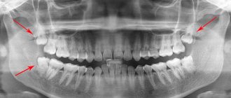
It can be colored differently: pressing, aching, shooting, pulsating, radiating to the eye, temple or cheek. In addition to pain, redness and swelling may occur if pus accumulates inside. Burning sensation due to allergies, fever and weakness. And also the feeling that the tooth rises above the rest. The deeper the complication develops in the jaw, the more pronounced its symptoms appear. At Stom-Firms.ru we look at what complications arise after treatment of the most common dental diseases, and what the patient can do to avoid them.
What complications arise after caries treatment?
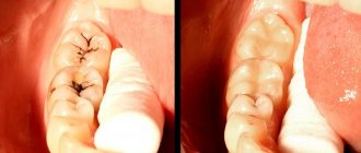
Caries is the disease that most often begins with most other diseases of the teeth and gums. At the appointment, the doctor drills out the damaged tissue and places a filling. Due to his mistakes or inadequate reaction of the body, the patient may experience:
- Prolonged pain after treatment . They occur when the filling technology is broken. If they do not subside, you will have to refill;
- Allergic reaction to filling material . The filling needs to be replaced urgently;
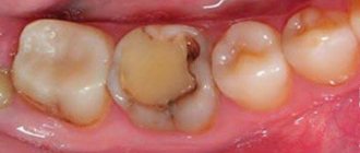
- Breaking and falling out of the filling most often occurs during chewing . The reason is errors when mixing cement, forming a cavity for a filling or modeling its surface. Refilling required;
- Pulpitis . Microbes managed to penetrate inside the pulp chamber; when drilling, the doctor accidentally pierced its bottom or overheated the dentin. Endodontic treatment required;
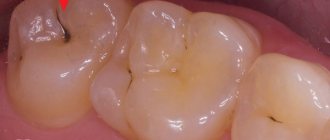
- Secondary caries under a filling . A time-delayed complication, when the destruction of poorly drilled necrotic tissue continues further. It is discovered accidentally in an image, when a filling falls out, or when pulpitis begins. Standard therapy is carried out.
Typically, the dentist gives a one-year guarantee for his work, but most complications after treatment appear in the first days or weeks.
How to treat caries at home
There is a clear answer to this question: “No way!” Dental caries cannot be treated at home; you can only carry out prevention to prevent it from appearing:
- Regular hygiene procedures using calcium and fluoride paste, rinses and dental floss are required
- once every six months - visit the dentist
- Protein foods should predominate in the menu, reduce the amount of carbohydrates
- Additionally, strengthening mineralized gels can be used
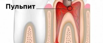
Complications after treatment of pulpitis
Pulpitis is inflammation of the pulp, a neurovascular bundle located inside the tooth. Therapy for pulpitis involves removing the pulp and subsequent filling of the canals. Sometimes, if the very beginning of the disease has been captured, the pulp is removed only from the coronal part. Poor endodontic treatment leads to the following complications:
- Periodontal irritation with potent antiseptic pastes . It may go away on its own, after electrophoresis or sedatives that the doctor will use to treat the canals;
- Acute periodontitis is inflammation of the tissues around the roots of the tooth, into which infection penetrates from unfilled canals. The doctor opens the canals, and the patient rinses his mouth for several days to remove the pus. After this, an antiseptic and medicine are alternately placed in the canals, and at the last visit they are finally sealed;
- A dental cyst is a variant of the chronic course of periodontitis. It grows at the end of the root or at the site of its perforation. The doctor injects medicinal paste into the canals and through them into the cyst itself for several visits in a row. If this does not help, cut it out through the gum along with the tip of the root.
- Periostitis or “flux” is an inflammation of the periosteum into which pus from the periodontal lesion has penetrated. If it is not practical to save the tooth, the dentist removes it, otherwise, drills it out to allow the pus to drain out. For the same purpose, it cuts the gum. For any type of treatment, antibiotics must be prescribed.
Darkening of the crown is an aesthetic defect that develops over the course of a year if there is blood in them during canal filling. You can lighten a pulpless tooth using intracanal bleaching: the doctor thoroughly cleans the canals and puts a bleaching agent into them several times. After achieving the desired result, the canals are filled and the crown is restored.
Rules for selecting and making a filling
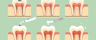
The key to success when applying a high-quality and durable filling is to follow the technology of its preparation. Even when using high-quality materials, violation of the manufacturing and application protocol sharply reduces its performance characteristics.
The choice of filling depends on the location of the tooth in the dental system, the strength of the chewing load, and the characteristics of the bite. So, if it is necessary to treat teeth along the smile line, the filling should not only be durable, but also aesthetically pleasing and inconspicuous. Dentists choose composite materials, silicates.
Filling a carious cavity as the final stage is performed only after complete cleansing of the prepared area, removal of dentinal fragments, and drying. The absence of moisture and organic sawdust increases the contact of the filling with the natural tooth tissue. After the filling is fixed and completely hardened, the doctor grinds and adjusts it to the anatomical shape (removes excess material, shapes the edges). Once the filling is comfortable for the patient when closing the jaws, it is polished.
Special attention is paid to polishing. The better the filling is polished, the lower the risk of developing recurrent caries. The uniformity and smoothness of the coating prevents corrosion, accumulation of plaque and tartar, and further destruction.
Complications after periodontitis treatment
Periodontitis is inflammation of the tissues near the roots of the tooth. Usually appears due to microflora that has developed in poorly treated root canals. Since there is no direct drainage for pus, it leaks into the surrounding tissue. As a result, the disease develops according to the following scenarios:
- Flux or cyst on the gum . Purulent fluid often breaks through the fistula opening, temporarily relieving the condition;
- Osteomyelitis . Purulent softening of bone tissue, which can lead to a bone fracture in this place and further spread of infection;
- Sepsis . The entry of infection and its waste products into the bloodstream is a life-threatening condition.
Local complications are treated surgically: an incision is made on the gum, and the tooth is most often removed. After manipulation, antibiotics are prescribed to suppress pathogenic microflora. In case of sepsis, hospitalize immediately.
Causes
Caries is the result of the activity of pathogenic microflora in accumulated plaque or tartar. A person’s immune status plays a major role in the development of carious defects. At particular risk are persons with autoimmune pathologies, AIDS, HIV, metabolic disorders, diabetes mellitus, and those taking immunosuppressive therapy for organ or tissue transplantation for a long time. The following factors can contribute to the development of caries:
- smoking, alcoholism;
- inadequate oral hygiene;
- natural aging (decrease in the body’s defenses, changes in the biochemical properties of saliva);
- metabolic disorders;
- endocrine pathologies;
- lack of food discipline (including prolonged fasting, poor nutrition, anorexia, bulimia);
- gum recession of various nature;
- excessive consumption of sugar and carbonated drinks;
- gastroesophageal reflux;
- bite pathologies;
- violation of hygiene rules when wearing braces and other orthodontic structures for the treatment of pathological occlusion.
Despite the variety of causes, eating disorders, bad habits and organ pathologies are the main etiological triad leading to the development of caries. Caries is not an independent disease. The pathogenetic link in the demineralization of enamel and destruction of the tooth body is a violation of the body's defenses.
How to avoid complications after dental treatment
In most cases, complications that arise after treatment of dental diseases occur due to medical errors. Some are due to an allergic reaction. But patients themselves can aggravate the situation by inappropriate actions, for example, starting to warm the sore cheek or suppressing the pain for a long time with analgesics, instead of immediately seeking help.
We have collected here the basic rules that will help reduce complications after treatment to a minimum:
- Visit the dentist regularly and do not delay the visit if the disease develops between visits;
- Contact a doctor immediately if there are signs of an allergy to the filling material;
- Come for an emergency appointment if pain, temperature and swelling of the gums or face increases;
- Take tablets only as prescribed by your doctor;
- Rinse your mouth thoroughly with recommended solutions to effectively draw out pus from the periodontal tissue or cyst. At the same time, plug the open crown with a cotton ball while eating;
- Do not self-medicate: do not heat, do not take “stronger” antibiotics, do not apply alcohol or garlic compresses, risking burning the mucous membrane.
In case of complications after treatment, it is better to return to your attending physician, since he is aware of the condition of the oral cavity, and the clinic stores images and information about the therapy performed. If you want to change clinic, request an x-ray and an extract from your card.
Classification and stages
The development of caries goes through several successive stages. The disease is classified not only by stage, but also by location and cause of occurrence. The stages of caries are of particular importance:
- Stage I. Formation of stains and clouding of the enamel layer. There are no visible destructions, the tissue structure is not changed. Sometimes the spot disappears on its own, which is associated with increased immunity.
- Stage II. Superficial caries. The enamel undergoes pigmentation, and when examined with instruments, a softening of the structure is felt. Often, superficial damage to a tooth covers all layers of enamel, but is limited. Dentin is still not involved in the pathological process. Visually, a carious defect looks like a dark gray spot with a rough base.
- Stage III. Average caries. The defect becomes pronounced, the lesion covers all layers of enamel and part of the dentin. Patients are increasingly noticing bad breath and discomfort when drinking or eating.
- Stage IV. Deep caries reaches the pulp membrane and is accompanied by tooth sensitivity, pain during chewing or at rest. The pain radiates to the temporal regions. The pain can be acute, throbbing, moderate, persistent. It intensifies and subsides involuntarily.
Typically, patients seek help from a doctor when symptoms worsen the quality of life and the dental-root system atrophies. When examining the oral cavity, caries is classified into:
- localized or generalized;
- acute or chronic;
- complicated or uncomplicated.
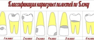
Carious lesions can be primary or secondary. In the first case, the disease occurs for the first time, in the second, caries develops again, under a filling, crown, bridge, or veneers. Clinically, there is another classification of caries according to Black:
- Class I or fissure caries - characterized by deepening of the natural grooves of the enamel on the chewing surface;
- Class II or carious defect of molars - the formation of defects on the contact surfaces of premolars, molars;
- Class III - damage to the canines and incisors without the defect extending to the cutting edge;
- Class IV - the edges of the canines and incisors are involved in the pathological process;
- Class V - the formation of cervical caries in any group of teeth.
Other types of caries are described in the international classification of diseases. There are unspecified or other caries, odontoclasia (atrophy of the root part of milk teeth), stopped caries after hygienic cleaning, preventive sanitation of the oral cavity.
Sections we recommend
On these pages you will learn how to solve the problem of complications after dental treatment:
- Dentist consultation
- Treatment under a microscope
- Treatment of caries with laser
Literature used for the article:
- Periodontitis: clinical picture, diagnosis, treatment. Textbook allowance / V.A. Kozhokeeva, K.B. Kuttubaeva, S.M. Ergeshov. ― Bishkek: KRSU Publishing House, 2011.
- Therapeutic dentistry. Dental diseases: textbook: in 3 hours / ed. E.A. Volkova, O.O. Yanushevich. - M.: GEOTAR-Media, 2013.
Author of the article: Natalya Aleksandrovna Kozlova
Copywriter of the information portal Stom-Firms.ru. Specializes in medical and dental texts.
Preventive actions
Prevention of caries consists of high-quality removal of tartar and plaque using special means: toothpastes, rinses, flosses, irrigators. Prevention of caries in children comes down to early instillation of oral hygiene skills. Popular recommendations:
- annual dental examination;
- professional teeth cleaning to remove tartar and plaque at least once a year;
- taking vitamins and calcium-containing medications;
- normalization of diet;
- improving food quality, balanced diet;
- timely treatment of chronic diseases of internal organs and systems.
Persons with chronic diseases of the ENT organs, prone to colds and respiratory infections, should take immunomodulators in courses. In case of a burdened dental history or hereditary predisposition, special pastes with a high concentration of fluoride are recommended. For recurrent caries, periodic application of mineralizing, antiseptic and protective drugs and varnishes to the enamel is indicated.
If you have braces, orthodontic aligners or plates, you should use an irrigator and special brushes. However, even careful care does not exclude professional cleaning of the product and teeth from plaque 2-3 times a year.
Timely treatment of dental caries preserves the integrity of the unit, prevents its early loss and complications. Regular sanitation of the oral cavity improves the quality of life, the aesthetics of a smile, and self-confidence.