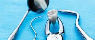
For a long time, the only procedure the surgeon performed was removal. Not so long ago, when you went to the dentist with a problem of deep caries, he would refer you to a surgeon to remove the unit. But now, more and more often, dentists stand guard over the health of teeth and the preservation of the patient’s family units. For this purpose, technologies and the experience of specialists who perform tooth-preserving operations are used. Tooth root amputation is a method of treating chronic inflammation of the tissues surrounding the roots (periodontitis).
Indications
The operation is performed in case of a complicated course of advanced carious process, which has already affected the root of the tooth. The procedure is prescribed in the following clinical cases:
- deep periodontal pockets have formed at the root level;
- pathological changes are observed in the structure of the root of the damaged tooth;
- perforation of the tooth roots occurs during poorly performed endodontic treatment.
The amputation method is most often used to remove the roots of teeth located in the upper jaw.
Hemisection of tooth root
Dentistry Apex-D / Surgical dentistry / Hemisection
Root hemisection is performed on the teeth of the molar group of the lower jaw, if one of their roots has significant destructive lesions.
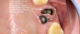
This surgical procedure refers to tooth-preserving operations, the essence of which is to remove one of the roots of a multi-rooted tooth along with the adjacent coronal part and inflamed periapical tissues.
The remaining part, not affected by the pathological process and capable of withstanding the functional load, is preserved.
Hemisection of the tooth root allows you to save a living unit in advanced (chronic) forms of the disease, which arose, for example, due to poor-quality endodontic treatment, as a result of which the remaining pathogenic microorganisms continue to multiply, localizing the source of inflammation in the area of the tooth root, while the inflammatory process spreads further, destroying the crown and infecting tissue in the periapical region.
In such cases, traditional methods of treatment do not allow access to the source of infection for proper treatment, so a surgical approach to eliminating the outbreak is considered effective.
In this case, technically the operation can be performed in the following ways:
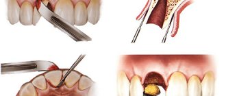
The first method involves accessing the root through the crown. To do this, the doctor cuts the coronal part to the bifurcation zone (i.e., to the place where the roots begin to diverge). After this, the damaged root is extracted.
The second method is to make an incision from the gum to the bone and peel off the mucoperiosteal flap. Next, a bone fragment is drilled into the root projection to form an operative approach. Through the formed hole, the root is cut off and removed. This technique is also used in case of detection of a cystic formation in the periapical area.
The perihilar cyst is excised through a perforated window, the cavity is thoroughly cleaned of remnants of pathogenic tissue, treated with antiseptic drugs and, depending on the size of the cyst, filled with osteo-replacing material. The flap is then returned to its place and the wound is sutured.
This patchwork technique is also used for tooth root amputation. This procedure also applies to tooth-preserving techniques and differs from hemisection in that only the part of the root affected by the inflammatory process is removed, and the crown is preserved.
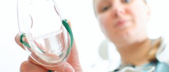
The operation is performed under local anesthesia. Preliminary treatment and filling of healthy root canals is carried out.
Indications for hemisection and root amputation include the following reasons:
- Obstruction of one of the root canals of a molar due to its atypical anatomy, which makes it impossible to treat it with conservative methods
- Diagnosis of cystic formation in the apical region
- Single root fracture
- If the infectious disease is accompanied by a purulent-inflammatory process
- Caries of one of the roots
- Late stage chronic periodontitis
- Deep periodontal pocket in the projection of one of the roots
- Perforation of the root canal wall
Contraindications include:
- Old age of the patient
- Merged roots
- Diabetes
- Diseases of the cardiovascular, circulatory and nervous systems
- Malignant neoplasms
Hemisection of the infected root allows you to preserve the functionality of the tooth for some time and prevent the destructive effects of pathogenic microorganisms on the tissue of the mandibular bone. This helps prevent bone tissue atrophy and maintain favorable conditions for further implantation.
The long-term success of the operation depends on how well the remaining molar roots can withstand mechanical stress as a support for the denture. Such factors include: periodontal condition, root anatomy, possible endodontic problems, and the likelihood of fracture of the preserved root.
The successful prognosis of the operation is facilitated by the maximum possible preservation of the structure of the unit, as well as the replenishment of the required amount of bone tissue at the site of root removal to preserve the original relief of the gingival margin.
Due to the violation of the integrity of the root system, the operated tooth will no longer have its former stability and in order for it to last longer, excessive chewing loads should be avoided.
The main advantage of hemisection is that the preserved healthy roots of the tooth can be used as a natural support for the crown and preserve the functionality of the resected molar without compromising the integrity of the dentition.
You can make an appointment at Apex-D Dentistry by calling the administrator at +7 and +7, or filling out an electronic form (the administrator will contact you at the specified phone number and agree on the date and time of the appointment).
How does the procedure work?
Tooth root amputation is a rather complex dental operation, which can only be performed in a dental clinic.
At the first stage, the dentist interviews the patient and collects anamnesis. Since the procedure is carried out under an anesthetic drug, a preliminary test is done to determine the tolerance of the selected anesthetic.
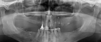
Additionally, to assess the condition of the root, an x-ray of the tooth to be treated is prescribed. If the patient has inflammatory processes in the oral cavity, they are first treated with antiseptic treatment and removal of inflamed or infected tissue.
The root removal procedure consists of several stages:
- Introduction of an anesthetic drug.
- An incision into the gum tissue.
- Detachment of gum tissue to provide access to the root system of the tooth.
- Cutting off the root at the junction with the crown and removing it from the soft tissue.
- Treating the hole with an antiseptic and filling it with a special osteoplastic material (imitation bone tissue).
If the hole is not filled with osteoplastic materials after surgery, then the cavity may become overgrown with gum tissue, which entails the development of infectious and bacterial complications. Sutures are placed on the cut gum.
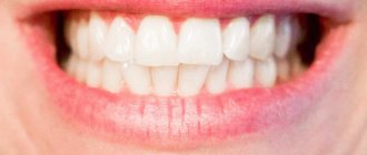
After the operation, the tooth retains its functionality for many years; in most cases, it is possible to preserve it without the need to install an artificial crown.
Preparation
Formation of a complete clinical picture requires a comprehensive clinical examination. The list of diagnostic procedures prescribed to the patient includes not only a visual examination and oral questioning, but also radiography, orthopantomography, and intraoral imaging. A prerequisite for the preliminary stage is sanitation of the oral cavity, eliminating the possibility of infections getting into the surgical wound.
Upon completion of the sanitation, the selection of instruments used for the operation, antiseptic treatment, and removal of stone deposits from the surface of the operated tooth are carried out. Surgical intervention involves the use of local anesthesia, after which the root part begins to take effect.
Rehabilitation period
After the procedure, the patient may feel pain. Painkillers prescribed by your doctor will help reduce it. It is also necessary to strictly follow all the dentist’s recommendations to make the healing process faster, take prescribed medications, and carefully observe oral hygiene. For three days after surgery, it is strictly forbidden to rinse your mouth.
For the first time after surgery, you can eat only soft foods, trying not to injure the damaged area. It will be important to stop smoking tobacco and drinking alcoholic beverages.
It is recommended to exclude baths, saunas, and not take hot baths for several days. If you heat the procedure site, bleeding may begin. If this does happen, it is necessary to apply a cold, wet cloth or ice, which will constrict the blood vessels and quickly stop the bleeding.
Orthopedic indications for tooth germ removal
Why do our orthodontists insist on complex extractions of teeth that are still in their infancy? This decision of the specialist is determined by certain features of the patient’s orthodontic condition:
- crowding of teeth (removal of the tooth germ as a preventive measure for crowding);
- before installing braces (to eliminate the risk of tooth displacement during treatment);
- the need to prevent relapse after orthodontic treatment;
- in cases where the rudiments are supernumerary teeth;
- the presence of a rudimentary dental tubercle that threatens to distort the normal position of full teeth;
- situations where the appearance of a new tooth can cause a defect in an even dentition.
In our work, we often encounter the need to remove tooth germs in order to reduce the time of orthodontic treatment, which often requires additional space in the dental arch. In some cases, excessive expansion of the dentition is undesirable, which negatively affects the maxillofacial parameters. These considerations may also be a sufficient basis for the removal of individual teeth in their infancy.
Possible complications
Amputation of a tooth root can provoke a number of complications, which are expressed in different symptoms:
- Damage to adjacent teeth.
- Violation of the integrity of soft tissues.
- Inflammatory process.
- Swelling of soft tissues.
- Loss of sensitivity in the operated area of the jaw.
- Increased bleeding.
If there is suppuration in the wound at the operation site, you should immediately consult a doctor. In some situations, it is still necessary to remove the diseased tooth, since it is not always possible to stop the destruction of dental tissue from the inside.
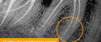
Retrograde root canal filling
Indications for retrograde filling:
1. Poorly sealed root canal with phosphate cement, resorcinol-formalin paste or other materials that make re-treatment impossible.
2. Poorly sealed root canal and the presence of a metal object in it: a) stump insert; b) anchor pin; c) fragments of an endodontic instrument.
3. Obliteration of the root canal.
4. Extremely long and curved root canal when conventional endodontic treatment is impossible or unsuccessful.
5. Canals with apical deltas or funnel-shaped expansion.
6. The presence of metal-ceramic or other crowns or fixed structures in combination with the above conditions.
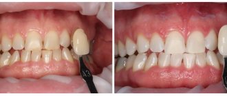
| A root apex resection operation was performed. A control photograph was taken after 3 months. The inflammatory process has been stopped. The tooth is saved! Leaky sealed canals of 12, 11, 21 and 22 teeth. Radicular cyst, core inlay of tooth 12, metal-ceramic crowns. | 2 years after retrograde filling with Trioxident cement. The cavity is filled with newly formed bone tissue. Teeth saved! In most cases, doctors would recommend such teeth to be removed and then made with a removable denture. |
Treatment with Damon braces
The patient visited the orthodontist every 1.5 – 2.5 months.
2.5 months after installing braces on the teeth, the gaps between the extracted teeth have noticeably decreased:
At the appointment, the orthodontist changed the arches according to the treatment plan, monitored the progress of treatment, and gave recommendations on dental care. Good hygiene is very important, since the health of the teeth, as well as the condition of the enamel after removing braces, depends on the thoroughness of removing plaque around braces and under the arches. If plaque is not removed well, the tooth enamel loses minerals and after braces are removed, white spots (foci of demineralization) become especially noticeable. Orthodontists advise anyone who is not confident that they can maintain good hygiene to get lingual braces, which do not leave marks on the outer surface of the teeth.
4 months after the start of treatment, the shape of the dentition became much smoother; there are still gaps from the extracted teeth:
8 months after starting treatment with Damon braces:
To create proper contacts between the teeth, the orthodontist prescribed wearing elastic bands. Orthoelastics are worn all day and overnight, only being removed for hygiene purposes. To completely close the gaps from extracted teeth, the bracket system is supplemented with special closing springs.
View of teeth after 1 year and 4 months from the start of treatment: