Dental granuloma is an inflammatory formation at the apex of the root. It is a proliferation of granulation tissue. Granuloma is formed as a result of the action of protective mechanisms in which the body localizes the source of infection and seeks to isolate it from other tissues. According to ICD-10, the disease was assigned code K04.5.
Typically, a granuloma is formed against the background of inflammation of the neurovascular bundle - the pulp. If pulpitis is not treated, its root part becomes inflamed, and the infection spreads beyond the boundaries of the tooth, into the peri-root tissue. As a result, a kind of pouch is formed, filled with decay products of dead cells.
A granuloma is considered to be a formation up to 0.5 cm in size, but it can grow, and as it grows, it transforms into a cystogranuloma, the size of which reaches 1 cm. With a diameter of more than 10 mm, we are talking about a tooth root cyst. There is no cavity in a granuloma; it is an area of tissue surrounded by a capsule. Due to the latter, the granuloma is firmly attached to the apex of the tooth root.
Reasons for the development of pathology
There are two reasons for the development of granulomas on the root of a tooth.
1. Untreated pulpitis. The development of caries leads to the appearance of a deep cavity in the tooth. Pathogenic microorganisms enter the pulp, it becomes inflamed, and severe pain appears. Lack of medical care leads to the gradual death of the pulp. Bacteria penetrate beyond the tooth through root canals. A focus of inflammation appears at the apex of the root. We are talking about periodontitis.
A deep carious cavity in this case is not always observed. Inflammation can develop internally when secondary caries appears under the filling.
2. Poor quality endodontic treatment. Granuloma can develop at the root of a tooth in which root canal filling was previously performed. Usually there is underfilling: the doctor has not completely filled the canals with material. In the remaining voids, pathogenic bacteria develop, and the tissues surrounding the root react with inflammation.
These causes cause most cases of granuloma formation. But there are others, less common:
- poor quality orthodontic treatment;
- previous dental trauma;
- other inflammatory diseases - tonsillitis, abscess, etc.
In the latter case, the infection enters the tissues through the blood or lymph flow.
Ask a Question
Why is granuloma dangerous?
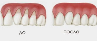
Granulation after tooth extraction, trauma, dental surgery is a natural phenomenon necessary for healing. This is a type of connective tissue that normally fills a wound and allows it to heal.
The process becomes pathological and leads to the formation of a granuloma if an infection gets into the wound. In this case, the granulation lesion begins to grow rapidly, replacing other tissues.
Even if the neoplasm does not manifest itself in any way and does not cause discomfort, granulation of the tooth and gums can lead to the development of serious complications:
- The growth of granuloma leads to the destruction of tissue at the root apex.
- The inflammatory process affects healthy tissue, which can result in the formation of an abscess, phlegmon, and osteomyelitis.
- There is a risk of developing fistulas, the channel of which can extend to the skin of the face.
- Since granuloma is asymptomatic for a long time, the infection can spread very widely, causing sinusitis, pyelonephritis, myocarditis, etc.
It is very important to diagnose and treat granuloma promptly. This will help avoid many serious health problems, and not only dental ones.
Granuloma symptoms and complications
Symptoms of dental granuloma are nonspecific. Often the patient is unaware of the disease, since there may be no signs at all. Usually the tooth does not bother you, but moderate pain occasionally occurs when biting, drinking hot drinks or eating food. Such symptoms are characteristic of all forms of periodontitis.
It is worth noting that from time to time the disease may worsen. For example, in case of hypothermia, an infectious disease, surgery - in all cases when the body's defenses are reduced. During an exacerbation, the following symptoms appear:
- sharp pain, intensifying when biting, tightly closing the jaws;
- swelling of the gums in the projection of the root apex;
- pain in the gums when touched.
An exacerbation can go away on its own, and the disease returns to its chronic form. But sometimes inflammation develops before the appearance of purulent contents in the tissues - periostitis or gumboil.
Inflammation can cause resorption or dissolution of a section of jaw bone tissue. The appearance of purulent complications is dangerous due to its consequences: from tooth loss and damage to surrounding units to tissue melting and sepsis. Therefore, it is important to get timely help from a doctor. Treatment of dental granulomas is carried out by a dental therapist, and if removal is required, you need to contact a dental surgeon.
Tooth granuloma: symptoms
The diagnosis of “apical granuloma of the tooth” can only be made by an x-ray, on which a slight rounded darkening will be visible at the apex of the tooth root (Fig. 1). Darkening at the apex of the tooth root on an x-ray always indicates the presence of a chronic focus of inflammation and the presence of bone tissue resorption. As for the patient's complaints and other symptoms, in most cases they are not pronounced or may even be absent.
Most of the time, the tooth may not bother you at all, or periodically there may be pain when biting on the tooth or aching pain from hot foods. Those. These symptoms are standard symptoms of chronic apical periodontitis, one of the forms of which is tooth root granuloma. However, periodically (during periods of decreased immunity) an exacerbation of chronic, low-grade inflammation may occur.
An exacerbation is usually accompanied by acute pain, especially pronounced when biting on a tooth. In addition, the gums in the projection of the root apex may swell at such moments and be painful when touched. An exacerbation of the process may spontaneously subside (return to a chronic course), but can also lead to the development of severe purulent inflammation, which is popularly called gumboil. Regardless of the outcome of acute inflammation, you will ultimately need the help of a dentist to save your tooth.
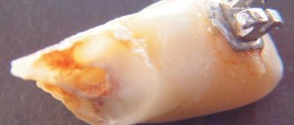
What does a granuloma look like on the root of an extracted tooth (video) –
Conservative treatment
For granulomas, conservative treatment is more often used. It consists of mechanical treatment of the root canals. After this, they are filled with temporary healing material - pastes based on calcium hydroxide. After 2-3 weeks, a control image can be taken, and if the inflammation is eliminated, the canals are filled with a permanent material - gutta-percha. A new permanent filling is placed on the crown of the tooth.
There are two treatment tactics depending on the initial condition of the tooth.
1. Treatment of granuloma of a tooth in which the root canals are not filled. In this case, treatment involves the following steps:
- removal of carious tissues, old filling on the crown, if any;
- mechanical treatment of canals - with the help of special tools they expand, the walls are smoothed;
- antiseptic treatment of canals.
Further actions depend on the size of the granuloma. If it is small, up to 3 mm, simultaneous filling is allowed. If the formation is more than 3 mm, then the root canals are filled with temporary paste. It helps the granuloma shrink or disappear completely.
You will have to wear temporary material for no more than 3 weeks. At the end of the period, the doctor will order a repeat x-ray, and if he sees positive dynamics, he will fill the root canals with permanent material. The restoration of the tooth crown is also carried out.
2. Treatment of a tooth in which the root canals have already been filled. In this case, the doctor will first remove the old material. If a tooth has a crown, it must be removed. The root canals must be resealed, and the treatment tactics correspond to those described above: sometimes the installation of a temporary therapeutic filling is required.
Symptoms
Granuloma can develop for a very long time without pronounced symptoms. At this time, granulation on the gum grows more and more, the size of the granuloma increases. The long asymptomatic period leads to the fact that the diagnosis is often made when the pathology is in an advanced stage. In order to avoid serious consequences that granulomatosis can lead to, it is important to pay attention to even the slightest manifestations of the pathological process:
- Swelling, puffiness, small protruding areas on the gums.
- A slight aching pain, usually not causing much concern.
- With significant development of the purulent process, the pain becomes acute, sharp, and severe.
- Change in color of the tooth crown in the affected area.
- Purulent discharge, usually appearing in the area between the gum and neck.
- Granulation after tooth extraction may be accompanied by persistent bleeding for a long period. The wound is healing slowly.
In the acute stage, general health deteriorates. The patient feels weak, unwell, many complain of a headache.
Surgery
Surgical treatment of dental granuloma may be required only in a few cases:
- obstruction of the root canals - complex, tortuous structure, too thin, narrow canals;
- impossibility of unsealing channels;
- the presence of a pin in the root canal - attempts to remove it may cause injury;
- patient's reluctance to remove the crown.
Many patients prefer granuloma removal because they do not want to resort to long-term treatment and remove a good crown. In this case, an apical resection operation is performed - part of the root is removed along with the granuloma through a small incision in the gums. Less commonly used is hemisection - removal of one root of a multi-rooted tooth along with part of the crown. In this case, further restoration of the crown of the tooth with a prosthesis will be required.
In rare cases, it is not advisable to preserve a tooth with granuloma. For example, if the crown is severely damaged and cannot be restored. In this case, when removing a tooth, the doctor must remove the granuloma from the socket in order to prevent the development of inflammation.
If purulent complications develop against the background of a granuloma, it is important to get help from a doctor immediately. The specialist will provide first aid: relieve acute pain by opening the tooth. Previously sealed canals are opened, and purulent contents are subsequently removed through them. In this case, relief comes instantly.
If severe swelling of the gums or cheeks appears, this may be due to the release of inflammatory contents under the periosteum or oral mucosa. In this case, a small incision is made to drain the pus. Further treatment is possible only after relief of acute symptoms. Drug therapy will also be required - the doctor will prescribe a course of antibiotics. You should not take them on your own. Moreover, it makes no sense to be treated only with antibiotics in the hope that the inflammation will go away - they are not able to eliminate the source of the disease or even reduce it, it is important to take local measures to eliminate the inflammatory process.
Is it possible to put a crown on a granuloma on the root of a tooth?
Dental prosthetics imposes special requirements on the doctor and the patient. If we are talking about installing a crown on a tooth of which no more than 1/3 remains, that is, only the root, then it must be carefully prepared: the canals are sealed, the infection is eliminated. A granuloma under a tooth is an indicator that an inflammatory process is occurring in the tooth, and before dental prosthetics it is necessary to get rid of it, or sooner or later the granuloma will still have to be treated, since it can become inflamed under the crown. So why not do this right away, before installing a denture?
Features of prevention
The main condition for the prevention of dental granulomas is timely assistance from a dentist when caries occurs. You should not allow severe tooth decay or the development of pulpitis. The peri-root tissues are healthy until the pulp becomes inflamed. Therefore, if symptoms of caries or pulpitis appear, it is important to immediately consult a doctor.
Endodontic treatment also increases the likelihood of developing periodontitis. Therefore, it is better to eliminate caries in the early stages and avoid the need for root canal filling. If this is unavoidable, it is important to carefully choose a dental clinic - the professionalism of a specialist will help eliminate possible mistakes and prevent complications.
The main stages of socket healing after wisdom tooth removal
Conventionally, the regeneration reactions after extraction of the “eight” can be divided into stages. Many of them remain invisible to humans, as they relate to changes occurring in the deep tissues of the gums.
- Stop bleeding. Normally, blood stops flowing within two (maximum four) hours after extraction. On the first day, the patient may still detect a small amount of bloody impurities in the saliva. There's nothing wrong with that.
- Formation of a clot. Occurs on the first day after surgery. Usually at the time of its appearance there is an increase in pain in the wound area and increased swelling. Body temperature may rise slightly. Headaches are also possible. These negative symptoms disappear in one to two days. They usually do not require additional treatment.
- Formation of a thin epithelial layer on top of a blood clot. This stage occurs on the second to fourth days after dental surgery. Then the person sees that the hole has turned white or gray. The state of health at this point usually becomes satisfactory. Toothache no longer bothers me.
- On the fifth to eighth day, the area of gray connective tissue increases rapidly . The clot is visible only in its center. The swelling goes away. If stitches were placed after removal, now is the time to remove them.
- The surface of the wound is covered with granulation tissue. This occurs two to three weeks after removal. As a result, the gums begin to look normal.
- In the period from the third or fourth week to six months, bone tissue grows in the socket.
If the removal was carried out against the background of progressive periodontal disease or periodontitis, then the patient’s recovery period increases by about ten days.
Thus, a month after extraction, a person can chew food on the affected side without restrictions . Nothing will remind him of the previous dental intervention. Since the “eight” is practically not involved in the process of grinding food (that is how far it is located in the dentition) and does not change the appearance of the smile, there is no need for prosthetics in the vacated area.
Granuloma and cyst: what do they have in common?
To summarize, all granulomas are divided into simple and complex. The first type is characterized by a slight compaction of granulation tissue without the release of purulent exudate. Developed forms have a pronounced focus of inflammation and large sizes (up to 10 millimeters in diameter). Some experts do not share the concepts of cyst and granuloma. This needs to be sorted out. A classic granuloma does not have clear boundaries and shape, while a cyst is a capsule with purulent fluid, much larger in size, usually with less pronounced symptoms. On the other hand, a cyst is a developed form of granuloma, i.e., in fact, its more complex type.
PREVENTION
Preventive measures must be carried out in a complex manner. They should be aimed at preventing the occurrence of the disease. Preventive actions include the following:
- Constantly maintaining cleanliness of the oral cavity. That is, this is daily, high-quality cleaning and rinsing.
- Treat bleeding gums.
- Scheduled visit (2 times a year) to the dentist.
- Change your toothbrushes regularly to avoid spreading infections in your mouth.
- The slightest pain in a tooth should prompt the patient to immediately go to the hospital. The process cannot be delayed.
- Pay special attention to diseases such as caries, pulpitis and periodontitis, which are common causes of granuloma.
- Use only medicated toothpastes as a preventive measure.
- Regularly rinse your mouth with herbal decoctions.
- Eat food with the maximum content of calcium, trace elements and vitamins.
WHAT IS THE FORECAST
The prognosis of treatment always depends on the severity of the disease. Everything influences: the stage of development, complexity and methods used in treatment. Treatment in the first stages of the disease always gives only a positive prognosis. Antibiotics and therapy always help. Granuloma in childhood also responds well to treatment.
If the granuloma is at the stage of pus appearing, success depends on where the source of inflammation appears. If it is the root of a tooth, then most likely the prognosis will be unfavorable and the tooth will have to be pulled out. If suppuration occurs only in the gum, then the contents are cleaned out using drainage. The patient is also prescribed a course of antibiotics.
Failure to treat granuloma may well lead to death. Like this? The resulting pus penetrates the muscles and enters the heart area. The result is sepsis, which leads to death.
Yes, granuloma is a completely unpleasant and dangerous disease. If this is the initial stage, then there is no need to worry. You should go to the hospital without hesitating for a minute. The patient must try to avoid extreme stages.
TREATMENT OF GRANULOMA
From the above it is clear that treatment should begin at the first stage. The patient’s first action is to come to the clinic. The second is to follow all the doctor’s recommendations and undergo an x-ray examination. Third, remaining calm during treatment.
Treatment can be of three types:
- Conservative or therapeutic.
- Surgical.
- Folk.
The most effective are the first two types. They are performed in a hospital setting. Traditional treatment is more supportive in nature.
CONSERVATIVE TREATMENT
Such therapy includes taking antibacterial, sulfonamide drugs and tooth filling. Such treatment stops the progression of granuloma and saves teeth from the process of destruction.
Antibiotics help to immediately stop the disease, remove painful microflora and rid the tooth of infection. While taking medications, the doctor also prescribes additional rinses for mouth antiseptics. Anesthetic medications help relieve pain.
In case of deep caries, the pulp is inflamed, the dentist cleans the canals and removes the source of infection. Next, the doctor applies medicine deep into the tooth and installs a temporary filling. After some time, a permanent filling is placed in this place.
In severe cases, therapeutic treatment is pointless. Then the tooth affected by granuloma is treated surgically.
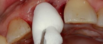
SURGERY
The essence of this intervention is to open the gums, from which pus is subsequently removed using drainage. As a result, the tissue becomes non-inflamed. In parallel with surgery, antibacterial drugs, painkillers and antiseptics are prescribed.
The prognosis after surgical treatment is always favorable.
Surgical treatment consists of one of the following procedures:
- Opening followed by drainage.
- Resection of the apex of the tooth root.
- Hemisection of the tooth.
- Removal of a tooth.
If there is a pocket on the gum or a gap on the tooth, then the cyst is dissected, from which the substance located there is removed.
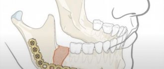
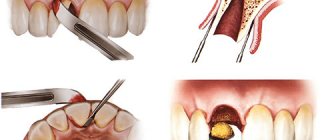
ROOT RESECTION
Root resection consists of the following steps:
- Opening the shell.
- Channel cleaning.
- Filling with medicinal solution.
- Removal of the granuloma itself and the affected apex of the tooth.
- Replacing the inflamed tissue that has been cleaned out with new artificial tissue.
- Filling a tooth.
HEMISECTION OF TOOTH
The operation is performed if the tooth has many roots and the disease has reached such a stage that it is impossible to save the root. Hemisection of a tooth consists of the following stages:
- Complete removal of the root and the coronal part that is adjacent to it.
- Filling an empty cavity with a special dental material.
- Installation of a crown.
- Control of the tooth after surgery using x-rays.
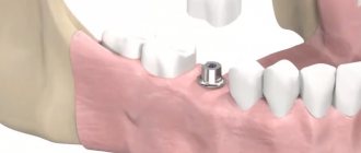
REMOVAL OF A TOOTH
When a tooth cannot be cured, it is removed. Removal is carried out in the following cases:
- If the patient has prolonged illness.
- If a gum pocket begins to form.
- If there is a vertical crack on the tooth.
- If the tooth is completely destroyed or the crown is destroyed.
- Perforation of the root is visible.
- The root canals are impassable.
FOLK TREATMENT
People use several recipes to alleviate the condition of granuloma:
- Preparation of tinctures with propolis and calamus. You will need 30 g. dry propolis and 30 gr. calamus roots The ingredients are poured with vodka and infused for 2 weeks. Used for rinsing.
- Herbal decoctions: eucalyptus, chamomile, sage. Used for rinsing.