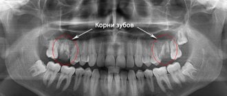
Root resorption is one of perhaps the most mysterious phenomena in dentistry, which occurs in people regardless of age, gender and state of oral health. No matter how well you fill the root canals or perform prosthetics, all this is meaningless if root resorption occurs. It occurs even when at first glance the patient is dentally healthy. The questions are why does it arise? and how to eliminate it? dentists have been fighting for over a century. This article discusses the types, diagnosis and treatment of root resorption.
Types of root resorption
The following types of root resorption are distinguished: physiological resorption of the roots of temporary teeth and pathological resorption.
Depending on the site of occurrence, resorption is divided into external and internal.
Due to the reason for their occurrence, the external one is conventionally divided into
- Inflammatory
- Superficial
- Substitute
Cervical resorption is added to the types of external root resorption.
Internal resorption may be complicated by perforation.
Concomitant factors of occurrence include trauma, whether acute or chronic - constant mechanical pressure, inflammation of the pulpal and periodontal complex, cysts, tumors and unknown etiology.
Physiological resorption of the roots of primary teeth
Physiological resorption of the roots of primary teeth begins during the period of replacement of temporary teeth with permanent ones. The underlying mechanism is the selective activation of osteoclasts through receptor activator of nuclear factor kappa B (RANKL ligands). However, the literature is very vague in explaining how pulp and periodontal resorption occurs.
It is known that resorption of primary teeth occurs evenly, unevenly and in the furcation area.
Uniform resorption is characterized by the simultaneous resorption of all tooth roots, slightly affecting the furcation zone.
Uneven, in turn, occurs on the root that is closest to the follicle of the permanent tooth.
It is easy to guess that the third type of resorption begins in the furcation area and only then moves to the roots of temporary teeth. In the field of bone resorption, the role of osteoclasts is taken over by pulp cells and odontoblasts.
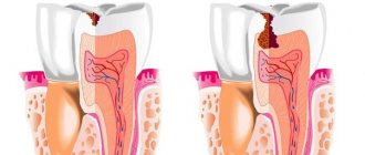
Internal root resorption (IRR)
Internal root resorption is an asymptomatic phenomenon and is almost always detected accidentally.
The cause is often inflammation in the pulp and trauma. Damage to predentin and dentinal tubules by inflammatory mediators (interleukins 1B, tumor necrosis factor) stimulate the RANKL system. This system selectively activates osteoclasts in periodontal and/or pulp tissues.
Since, due to internal resorption, normal pulp turns into granulomatous tissue, it is visible through the tooth tissue and a so-called “pink spot” appears. As a result of further necrosis of the pulp, the pink color changes to gray.
As resorption progresses, the patient complains of pain, and periodontal damage leads to increasing tooth mobility.
Internal root resorption and perforation
It proceeds similarly to VRC without perforation, but reaches the cement and periodontal ligament. Based on this, the treatment and prognosis of the disease is complicated and depends on the size of the defect.
Superficial root resorption
This type of resorption is considered a physiological process due to a reaction to damage during trauma or orthodontic treatment - ischemia and necrosis of cementoblasts occurs, so it is not of particular clinical interest. It operates within the cement and rarely goes beyond it. The defects are usually small and rarely detected, especially on the vestibular and oral sides. There are no functional impairments.
Once the trigger is removed, new structures are immediately built, so no treatment is required.
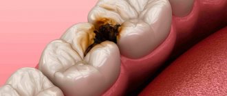
External inflammatory resorption
The fastest and most aggressive type of external root resorption. The occurrence is associated with bacterial infection from the root canals, trauma (especially complete tooth luxation), pulp necrosis and wide dentinal tubules due to incomplete root formation. The process is accompanied by extensive destruction of root tissue, loss of the central nervous system and dysfunction.
Replacement resorption or, as it is called, ankylosis
The latter type is external resorption, but in terms of the severity of tissue destruction it does not lag behind the inflammatory one. It also occurs during injuries, especially with an impacted or completely dislocated tooth. Usually the outcome of inflammatory resorption, even with its treatment. Replacement resorption is a chronic process that occurs at the site of damage to the periodontal ligament on the outer surface of the root, can never stop on its own and almost always leads to tooth loss.
Due to the strong pathological impact, damage to the periodontal ligament occurs, and as a result, inevitable bone formation in this place. The tooth becomes immobile.
If replacement resorption stops, they speak of a transient form. If it comes to tooth loss, such resorption is called progressive.
Cervical resorption
No matter what you call this type of resorption, external cervical, invasive or peripheral cervical resorption, it remains an idiopathic variant of external resorption. The development of trauma is initiated by orthodontics, bruxism, and scaling. Some studies have presented cases of the influence of intracanal bleaching on the development of external resorption. It is believed that 30% hydrogen peroxide can penetrate through the tubules onto the surface of the cement and destroy it and the periodontium.
It does not always occur in the cervical area, which depends on the depth of the pathological pocket and the level of marginal tissue. Resorption is maintained by infection in the gingival sulcus and gradually goes around the pulp chamber, which is not damaged. Cervical resorption is asymptomatic until periodontal and pulpal infections occur. With a deep defect, temperature sensitivity occurs, the walls of the cavity are hard, creak and bleed slightly upon probing.
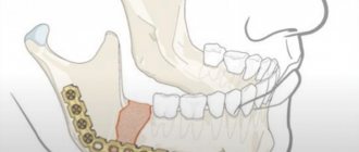
When is it recommended to remove part of a tooth root?
Most often, resection is performed when:
- the formation of a cyst on or near the root of the tooth;
- proliferation of granulomas or multiple granulomas on the root part or in the area nearby;
- anatomically incorrect position of the tooth canals.
It is important! To successfully perform the operation and rehabilitation after it, a prerequisite is the presence of a sufficient volume of bone tissue in the operation area (approximate tissue thickness - from 5 mm). If these indicators are lower, there is a high risk of bone injury during the removal of granulomas or cysts.
What is important, the manipulation is performed under local anesthesia; If the patient is very distressed, premedication may be administered. In any case, the procedure is not accompanied by discomfort and is easily tolerated.
Dentistry for those who love to smile
+7
Make an appointment
Diagnosis of root resorption
The problem with diagnosing internal root resorption is that nothing bothers the patient, and this can last for years. Therefore, X-ray diagnostics should be given its due.
| Internal resorption | External resorption |
| Smooth, clear focus of destruction | Asymmetrical, corroded lesion |
| The defect is always located within the root canal | The defect is located on the root surface and can change its position |
| Fuzzy, barely noticeable contours of the root canal | The contours of the root canal are clear, superimposed on the outlines of the defect |
X-ray examination also helps in diagnosing external resorption. Defects usually have uneven edges, can go deep into the dentin and are detected on any root surface. However, there are cases when the focus of clearing has clear contours, as with internal resorption; in this case, you should take an x-ray in several projections and make sure that the contour of the root canal overlaps the contours of the defect, or even better, do a CBCT scan.
But it is more difficult to distinguish replacement resorption from inflammatory resorption. Radiologically, with replacement resorption, there are no foci of clearing and periodontal cleft due to bone formation. Ankylosis occurs more easily due to the chronicity of the process.
The diagnosis of cervical invasive resorption is established on the basis of clinical and X-ray diagnostics. The condition is asymptomatic, as with VRC we see a “pink spot” on the enamel, periodontal inflammation, but the pulp is not damaged.
Upper first molar
Average age of teething: 6-7 years
Average age of root formation: 9-10 years
Average length: 20.8 mm
The largest in size, with a complex anatomy of the root and root canal system, the so-called “6-year molar” is the most frequently treated, while presenting the greatest difficulties in treatment among the posterior teeth. During its treatment, the largest number of endodontic errors and complications arise, and it is undoubtedly one of the functionally important teeth.
The three separate roots of the maxillary first molar form a trifurcation: the palatal root is the longest, and the distal buccal and mesiobuccal roots are approximately the same length.
The palatal root in the apical third often curves in a buccal direction. Of the three channels, it has the largest diameter and is the easiest to access. Its mouth is shifted to the palatal wall of the crown. The root deviates sharply from the median axis of the tooth. In cross section, the root is flattened and has a ribbon-like shape, which requires special attention when cleaning and instrumenting it. Fortunately, it rarely has more than one apical foramen.
The distal buccal root is conical and usually straight. It always has one channel.
The mesiobuccal root of the first molar has generated more research, clinical inquiry, and frustration than any other root in the oral cavity. Green showed that 14% of the mesiobuccal roots of the maxillary first molars studied had two apical foramina, and 36% of the roots had two orifices. Pineda reported that 42% of these roots had two canals and two apical foramina. Slowey confirmed Pineda's data within a few percent difference. The fact that nearly half of these roots have two canals, whether they end in a single opening or not, is reason enough to always assume two canals until careful examination proves otherwise.
The additional orifice lies centrally, between the orifices of the mesiobuccal and palatine canals. The search is facilitated by using fiber optics and by identifying the anastomosis between the orifices of the mesiobuccal and palatal canals. The second canal in the mesiobuccal root will always be narrower than the other canals, so it is more difficult to clean and shape. Access to the main mesiobuccal root canal is easier when a straight entry is properly created.
All carious tissue, leaking fillings and denticles must be removed before endodontic treatment begins.
After treatment, complete closure of the approach is necessary to prevent vertical coronal or crown-root fractures. If indicated, internal reinforcement with intraradicular pins is recommended.
Care after tooth resection
- Do not chew on the operated side.
- Until the wound heals, avoid irritating, solid foods (food should be warm and soft).
- Do not use a hard toothbrush, aggressive pastes, or mouthwash.
- Take antibiotics prescribed by your doctor to prevent infection.
- Rinse your mouth after eating with an antiseptic solution.
- For the first week after surgery, limit physical activity to moderate, and exclude extreme, contact sports.
- Do not visit the bathhouse, swimming pool, sauna until the wound is completely healed.
- Avoid eating too hard foods until the bone is completely restored (at least 3 months).
- Visit your surgeon regularly to evaluate the results of the operation.
Resection stages
- Formation of access
- the dentist cuts through the gum, exposing the jaw bone. Forms a small hole in the bone tissue (in the projection of the root apex), gaining access to the pathological focus. Often, the bone tissue has dissolved in the projection of the cyst; there is no need to cut out a hole. - Excision of tissues and correction of the apex
- the doctor removes the source of inflammation along with the dead apex of the root, cutting it off perpendicular to the upper dental axis (to the level filled with the filling mass). The empty space is filled with osteoplastic material (bone volume is restored faster). - Suturing
- suturing the wound area is sometimes performed with the installation of drainage for the outflow of ichor. Drainage is installed between the seams and is removed after 1-2 days.
Root resection takes 40-60 minutes, depending on the location of the tooth. It is easier to intervene on canines and incisors than on multi-rooted units.
Possible complications
Root resection is a micro-operation, the purpose of which is to preserve the full functionality of the affected tooth. Usually, the risk of complications is minimal, but they cannot be completely excluded. Sometimes the following complications are possible:
- Bleeding due to injury to nearby vessels;
- infection of a postoperative wound;
- recurrence of cysts, granulomas (if all affected tissues were not removed);
- perforation of the maxillary sinus, nasal passage;
- damage to the nerve passing along the alveolar ridge;
Sometimes complications are caused by structural features of the jaw, for example, the proximity of the roots to the maxillary sinus. Complications caused by non-compliance with medical recommendations include swelling and tissue inflammation. Ignoring advice on limiting physical activity is fraught with the development of bleeding, and failure to maintain hygiene can lead to wound infection.