Dr. Kizim
>
Articles
>
Occlusal plane of teeth in dentistry
To determine the position of teeth in the dentition, the concept of the occlusal plane is used. Its definition is extremely important, since it is this concept that best describes a person’s bite.
The occlusal plane of the teeth is an imaginary line that runs along the cutting edges of the frontal and occlusal surfaces of the chewing teeth. Starting from the plane of the upper dentition, you can determine its maximum closure with the lower one. Thus, dentists can judge the fissure-tubercle contact of the teeth and talk about their mutual fit.
Varieties
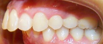
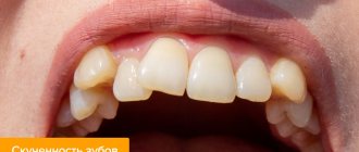
Incisal disocclusion is a general concept. It combines various phenomena in which the dentition in the frontal part does not close correctly.
Orthodontists distinguish the following types of incisal anomalies in the sagittal plane (divided into right and left sides):
- sagittal disocclusion – there is no contact between surfaces, this can occur due to abrasion or a change in the angle of inclination;
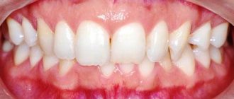
- direct occlusion - contact of the incisors of the lower and upper jaws with their cutting surfaces;
- reverse occlusion - the lower incisors are pushed forward, but the contact between the upper and lower dentition in the frontal zone is maintained;
- reverse disocclusion - the lower incisors are pushed forward so much that they do not contact the upper ones.
Incisal closure anomaly in the vertical plane:
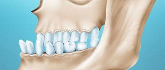
- vertical disocclusion - the upper incisors are pushed forward, there is no contact between the antagonist teeth;
- deep occlusion – vertical overlap in the frontal area is more than 1/3;
- deep disocclusion - the overlap is the same as in the previous case, but there is an open gap.
In addition, there is the so-called false incisal disocclusion. It occurs due to injury, loss of teeth during an accident, etc.
Types of malocclusion in adults and children
Dentists usually divide anomalous occlusions into planes.
- Sagittal - characterized by elongated/shortened rows of teeth.
- Transversal - narrowed/expanded dentition is visible.
- Vertical - the presence of shortened/elongated certain areas in the dentition.
In addition, in dental practice it is customary to use the following classification of occlusion.
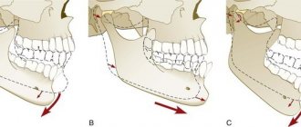
- Distal.
Sagittal occlusion with the upper jaw pushed forward. - Mesial.
Also a sagittal variety, but with the lower jaw moving forward. - Cross.
Transversal pathology with displacement of the jaws, which can only be partially formed, in one direction or another. - Open.
Vertical occlusion occurs with both partial and complete non-occlusion of teeth. - Deep.
The so-called traumatic, contributing to damage to the enamel. Characteristic is almost maximum overlap of the lower rows with the upper ones. - Vertical occlusal anomaly.
In addition to the above listed occlusion disorders, some experts identify 2 more types of anomalies:
- dystopic bite - displacement of one or more teeth;
- reducing - formed due to damaged and (or) lost teeth.
Symptoms
The orthodontist sees all of the above anomalies during a visual examination of the patient’s oral cavity. The absence of normal closure in the frontal or lateral region is determined; a vertical gap may be observed.
Anatomical features cause various functional abnormalities. Patients with incisal disocclusion experience a number of symptoms:
- Violation of the parameters of the lower third of the face. Disproportions can be noticeable both in profile and in frontal view, but in profile they are usually more pronounced. The lower third of the face can be significantly reduced, an effect that makes the supramental fold appear enlarged. The nasolabial folds are smoothed out, and the angle of the lower jaw becomes larger, sometimes approaching 90°.
- Various diction disorders. The most strongly distorted dental consonants are hissing and whistling sounds. If the occlusion is maintained, speech is less affected.
- The process of swallowing saliva and food is disrupted, so overstrain of the muscles involved in lowering and raising the corners of the mouth develops; the orbicularis oris muscle becomes enlarged due to the constantly increasing load.
The stronger the degree of pathology and the larger the size of the vertical gap, the more pronounced all of the listed signs are. Even if they are not developed at the initial stage, over time all symptoms gradually worsen.
Causes
The appearance of malocclusion in the incisal zone occurs due to an increase in the angle of incisor inclination, reduction of one jaw in the frontal zone, lengthening of the lateral sections of the dentition, as well as due to protrusion of the lower or upper teeth.
All causes of these disorders are divided into primary and secondary. Primary ones are those that appeared at the time of fetal development, and secondary ones develop after birth.
Primary reasons:
- Hereditary factor - inheritance from one of the relatives or arising due to an unsuccessful combination of characteristics (for example, having a small jaw, like mom, and a large slope of teeth, like dad).
- Disruption of the formation of dental buds due to chromosomal mutations or unfavorable pregnancy - maternal illness, the effects of heavy medications, unfavorable environmental conditions, certain diseases during gestation, etc.
- Macroglossia is an increase in the size of the tongue.
- Shortened lip frenulum or short tongue frenulum.
Secondary causes:
- Bad habits - sucking fingers, sucking various objects, biting the upper or lower lip, etc. In preschoolers, the presence of such bad habits can lead to the development of pathology in record time, starting from one month.
- Mouth breathing, which becomes constant due to adenoids, chronic nasal diseases, allergic reactions, aggravated by rhinitis.
- Incorrect placement of the tongue in the oral cavity, which displaces the floor of the mouth and leads to impaired load on the muscles.
- Deep or multiple caries of the incisors, which leads to tooth protrusion.
- Metabolic disruptions – disturbance of mineral metabolism, rickets, osteomyelitis, etc.
- Hypovitaminosis.
- Weakened immunity due to frequent or chronic diseases - dyspepsia, exudative diathesis, staphylococcal infection, etc.
Diagnostics
Despite the fact that incisive disocclusion is determined by the dentist even during a routine examination, it requires detailed diagnosis. This need is necessary in order to identify the causes of the pathology and prescribe an effective course of treatment.
During diagnosis, the patient may be prescribed:
- X-ray of the frontal zone to detect unerupted teeth or other anomalies hidden in the mucous layer or in bone tissue.
- Orthopantomogram images allowing to assess the condition of the dentition in all areas.
- Profile and frontal photographs to measure the patient’s anthropometric parameters.
- Casts of the dentofacial apparatus. On the model, which is manufactured in a technical laboratory, more accurate measurements of all the necessary parameters are performed.
Measurements of the size and length of the gap between the upper and lower teeth are of decisive importance, since these parameters indicate the severity of the disorder.
Treatment
The course of treatment is prescribed based on the clinical picture and age of the patient. The approach differs at the time of primary, replacement and permanent set of teeth.
Temporary kit
Often, to completely restore the physiological state of the dentition and eliminate incisal disocclusion, it is enough to eliminate bad habits. A return to normal may occur within four months or six months.
In almost all cases, treatment in children includes myogymnastics, which, when used temporarily, demonstrates the highest effectiveness. It is based on performing individual exercises that tone the facial and chewing muscles. This technique can significantly change the proportions of the dentofacial apparatus. Myogymnastics can be prescribed as an independent remedy or in combination with other methods.
If the cause of the pathology is shortening of the frenulum of the lips or tongue, its plastic surgery is performed. This surgical treatment is not considered difficult and is performed under local anesthesia. After the procedure, the patient is immediately sent home and his condition is monitored on an outpatient basis.
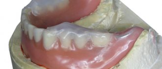
For incisive adentia, the use of therapeutic and prophylactic children's dentures is indicated. An automatic sliding prosthesis can be used, which increases as the child grows. Fixed structures must be changed at intervals determined by the doctor.
If the cause of the incisal closure anomaly is protrusion, the damaged teeth are restored. Sometimes silicone mouth guards are prescribed, which activate the development of the jaw and palate, while simultaneously leveling the angle of the teeth.
Replacement kit
During the replacement set period, disocclusion is eliminated relatively quickly. There are several most common treatment methods:
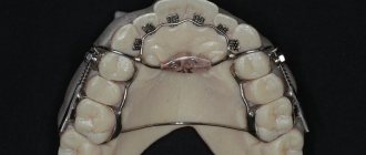
- Katz plate with bite pad in the incisal area. The base rests on the palate, and clasps are attached to it. The size, angle and thickness of the bite pad are determined by the orthodontist. Its function is to increase pressure and load on the alveolar process of the jaw.
- Muleman Propulsor . This is a two-jaw functional appliance that combines the functions of a vestibular plate for the upper jaw and an activator for the lower jaw. The support falls on the teeth and alveolar processes. The Muleman Propulsor is a removable device. Most often recommended for ages 7-9 years for use during nighttime sleep. In addition to normalizing the condition of the dental system, it helps to get rid of bad habits - finger sucking, lip biting and mouth breathing, and forms the correct position of the tongue.
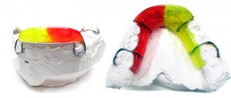
- Frenkel function regulator . Types I and III designs are used. They are complex devices based on a rigid metal frame and plastic shields. In most cases, it involves use at night plus a few hours during the day.
- Functional apparatus Myobrace . Constructed from a rigid inner frame and soft outer silicone. Using Myobrace eliminates the need for braces. The advantage is comfort and convenience, gums and soft tissues are not chafed or irritated. The devices are available in standard sizes, from which the orthodontist selects the appropriate ones.
Permanent kit
During this period, restoration of occlusion lasts longer than at a younger age. It can be performed using orthodontic, orthopedic or surgical methods.
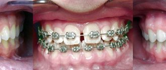
The most commonly used orthodontic treatment is wearing braces. This approach is aimed at gradually returning the physiological position to the incisors of the upper or lower jaw. The doctor determines the force with which the arch should press on the teeth. Braces are made according to individual parameters and can be worn from six months to three years. After removal, a retention period follows, during which the result is secured using silicone retainers.
In the case of the traumatic nature of the pathology, prosthetics are necessary - the installation of prostheses or implants is prescribed.
In severe cases, surgery is necessary to enlarge or reduce the palate or eliminate pathologies of the alveolar process. Such operations are performed in a maxillofacial surgery hospital under general anesthesia. Surgeries of this type are quite serious, so after them the patient must remain in the hospital for some time. Sometimes these operations precede the wearing of braces. Surgical methods can also be used to remove an impacted tooth.
How to correct a child's malocclusion
- Children under 7 years of age
are shown a set of gymnastic exercises and massage that will help solve the problem. - Children under 10 years of age
are already prescribed trainers, with the help of which they can set the desired direction for their teeth. They must be worn for a certain number of hours during the day. But, if the pathologies are more advanced, removable plates and mouthguards are used. Correction of the anomaly takes approximately 2 years. - For children aged 10-12
years, braces are used to correct their bite - special orthodontic structures consisting of a power arch and clasps that set an individual direction for each tooth. They cannot be placed at an earlier age; it is necessary that all milk teeth be replaced by permanent ones. How long to wear braces for malocclusion is determined by the treating orthodontist.
How to correct malocclusion in an adult
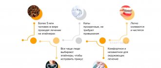
A very common treatment method for adults is wearing braces. Transparent aligners (aligners) are also very popular now. They are made of transparent plastic material. Aligners are effective in correcting impaired occlusion, are easy to use and look very aesthetically pleasing. All details about this technique can be found here.
In cases where the patient is not in the mood for long-term bite correction with aligners or braces, there is another solution - microprosthetics. In this case, special overlays are installed on the teeth - veneers, with the help of which you can correct uneven teeth and gaps between them. But if there are serious malocclusions, this technique is not used.
There are bite defects for which only surgical treatment is indicated. Examples include: severe malocclusion, facial asymmetry due to trauma or hereditary causes, and chin dysplasia.
Each method of orthodontic treatment has both indications and contraindications. Only an orthodontist can determine the method of treatment after a thorough examination and full diagnosis.
Refusal of treatment
Malocclusion in the incisal region leads to various complications. First of all, conversational speech is disrupted, which entails the appearance of complexes in a child or an adult. The compensatory mechanism causes the facial muscles to be in constant overstrain, which causes myalgia or inflammation of the facial nerves.
Due to poor quality of biting and chewing food, various digestive disorders are possible. Dysfunction of the temporomandibular joint may develop, which causes blocking of the movements of the lower jaw. This phenomenon causes frequent or even constant headaches.
The periodontium in the frontal group is constantly under overload, so it begins to quickly wear out. Improper load distribution leads to injuries to the soft tissues of the upper palate.
To prevent the development of all these complications, treatment should be carried out at the initial stages of the development of the anomaly.
Consequences of malocclusion
It is a mistake to think that a pathological bite concerns only appearance and only leads to an unattractive smile. This is where the problems arise that are more serious. For example, in 90% of people with abnormal occlusion, incorrect posture is also detected. There is a logical explanation for this: with a broken bite, the center of gravity of the head shifts. This in turn affects the compensation mechanisms of the musculo-ligamentous apparatus of the maxillofacial system. The result of all this is an increase in the pathology of teeth closure.
Aesthetically, occlusion abnormalities lead to facial asymmetry. A weak-willed chin becomes pronounced, and lips protrude unattractively.