Each person goes through the stages of the eruption of the first teeth, the development of milk teeth and their subsequent replacement by permanent ones. Despite their similar appearance and function, temporary and permanent teeth have differences, which we will talk about, at the same time we will consider the timing of the appearance of the main teeth, possible problems with them in the process of their development.
The photo shows a diagram of the structure of human teeth
The structure of human teeth
Teeth are not only intended for mechanical processing of food, but are also necessary for the formation of speech, breathing, and influence facial features. To navigate what dentists advise, how to take care of your teeth, and what the risks of disease are, it is useful to know how they work.
Anatomical structure
3 parts that make up a tooth:
- Crown. The visible part of the tooth used for chewing. The outside is covered with durable enamel, which protects it from bacteria, chemicals contained in food, water, and saliva. The surfaces have their own names: Facial (vestibular) - in contact with the lip or cheek.
- Lingual (lingual) – the opposite of the facial, involved in the formation of speech.
- Occlusion – the upper surface in contact with the tooth of the opposing jaw.
- Contact (approximal) – contacts with adjacent teeth.
Milk teeth, having a largely similar structure, also have differences in anatomy:
- They are noticeably smaller in height than permanent ones.
- The crown is much wider than the root.
- Enamel is thinner and more fragile.
- The roots are more round.
- The wear of baby teeth, as well as their spontaneous loss, is a normal physiological process.
Histological structure
The structure has several layers:
- Enamel is the most durable fabric. When a tooth just erupts, a cuticle is located on it, which is gradually, under the influence of saliva, replaced by a pellicle.
- Dentin is a highly mineralized tissue that resembles bone, but has better mechanical strength. Instead of enamel, the root part of the dentin is covered with cement.
- The pulp, the central part of the tooth, is a soft connective tissue containing a large number of blood vessels. Caries and inflammatory processes “owe” pain to the pulp with its large number of nerve endings.
Milk teeth are distinguished by dentin with a lesser degree of mineralization, which weakens their protection against caries. The volume of pulp occupies most of the tooth, and small protective layers (enamel and dentin) provide less protection against the penetration of bacteria and the development of inflammatory processes.
Types of teeth
There are 4 groups:
- Incisors. 4 chisel-shaped cutters. The largest are a pair of upper central incisors, and the situation is opposite from below - the lateral incisors are slightly larger than the central ones.
- Fangs. 2 on the upper and the same number on the lower jaw. Their length is longer than the others, the front wall is convex.
- Premolars. There are 8 in total, prismatic in shape, the upper surface with two tubercles (buccal and lingual). Premolars have 2 roots. The second premolar has a larger buccal surface. There are no primary premolars.
- Molars. The first molar (molar) is the largest tooth in the upper jaw. The chewing surface has four tubercles, 3 roots. The cubic-shaped second molar is smaller, and the buccal tubercles are larger than the lingual ones. The third (“wisdom tooth”) is in many ways similar to the second, but not everyone has it.
Dental formula
In order to improve the convenience of describing each tooth, numbering them, and filling out cards, it is customary to record the order of the teeth using a special formula. There are several varieties of it.
Zsigmondy-Palmer system (quadratic-digital)
Arabic numerals are used, numbering starts from the central incisors in each direction:
- 1 and 2 – incisors.
- 3 – fang.
- 4, 5 – premolars.
- 6-8 – molars.
Milk teeth are designated differently - using Roman numerals:
- I and II – incisors.
- III – fang.
- IV and V – molars.
Two-digit Viola system
Teeth numbering uses 2 digits. The jaws are divided into 4 quadrants. The first digit shows its number.
For adults this is:
- 1 – upper jaw on the right.
- 2 – upper jaw on the left.
- 3 – lower jaw on the left.
- 4 – lower jaw reference.
For a similar description of baby teeth, numbers 5 to 8 are used.
So, there are 8 teeth in each quadrant, its number is shown by the second digit. Thus, the first molar of the lower jaw on the left is designated 35, and the child’s canine from the lower right is designated 43. Therefore, the phrase that “treatment of the 48th tooth is required,” or, for example, the 55th, does not indicate the doctor’s lack of qualifications or what - or pathology in your child, who suddenly acquired so many teeth.
Dental development
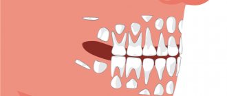
The differences between primary and molar teeth begin with their number - only 20 primary teeth, 8 incisors and molars, and 4 canines. This is explained by the fact that children simply have nowhere to fit more teeth. In this regard, there are no primary premolars. By the time the permanent teeth appear, the adolescent's jaws are already sufficiently developed for all teeth to appear.
The formation of tooth buds in humans begins at the 6th week of intrauterine development, and at the 14th week hard dental tissue appears. The crown develops first. The development of the rudiments of permanent teeth begins in the 5th month.
By the time a child is born, the formation of the rudiments of both milk and permanent teeth is almost complete. The process of development of permanent teeth, which have no analogues among milk teeth, begins a year after birth.
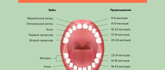
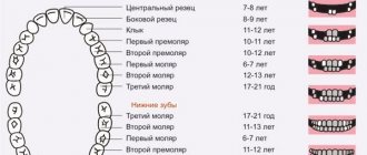
While the first teeth may appear at 4 months, and their eruption may be delayed for up to a year, permanent teeth erupt in everyone at approximately the same age. The sequence of their eruption is the same as in the case of milkweeds:
- 6-7 years. The central incisors appear from below.
- 7-8 years old. The central incisors on top and the lateral incisors on the bottom are replaced.
- 8-9 years old. The lateral incisors of the upper jaw appear.
- 9-12 years old. Canines and premolars are replaced.
- From the age of 12. From this age, molars begin to change, and from about 14 years of age, teeth appear, which were not among the milk teeth.
When should I take my child to the dentist?
The Ministry of Health of the Russian Federation recommends showing a child to the dentist once a year starting from 12 months.
The purpose of the first trip to the dentist is prevention. The doctor checks whether teeth are growing correctly and gives recommendations on oral hygiene. It is better to do this no later than your first birthday, so that the specialist does not miss caries and possible problems with your bite.
The first trip to the dentist should not frighten your child.
Here are some tips to make it painless:
- Do not pay too much attention to this event, do not prepare your child on purpose;
- Don’t worry yourself so that your baby doesn’t get nervous along with you;
- If possible, arrive early to your appointment to give your baby time to look around.
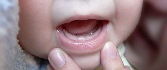
Signs of the imminent appearance of molars
You can determine when you should soon wait for the baby teeth to begin replacing with permanent teeth based on several signs:
- The gradual growth of the baby's jaws leads to increasing gaps between the teeth.
- The tooth begins to wobble. This is due to the fact that the already small root begins to gradually dissolve, causing the fixation of baby teeth to be significantly weakened.
- A fallen tooth indicates that the formed permanent one, which is about to appear, pushed it out.
- Swelling and redness may appear on the gums at the site of the eruption of a permanent tooth.
- Pain in the gums, where the permanent tooth erupts, increased temperature, and poor health of the child indicate problems have arisen, and it is necessary to see a doctor. The process of erupting molars should be painless.
How to help your baby when teething
- Give your baby something to chew on, such as a teether, which can be pre-chilled in the refrigerator.
- Massage your baby's gums with a clean finger. You can first wrap your finger in clean gauze. This will relieve the pain for a while.
- If your baby is already eating solids, give him cool foods (applesauce or yogurt).
- You can give your child something hard to chew on, such as a cracker. Just make sure your baby doesn't choke.
- If all of the above does not help, you can give your child a baby pain reliever or a special teething gel - strictly after consulting a doctor.
Vomiting, runny nose and diarrhea are not always symptoms of teething. If your baby appears unwell, call the doctor.
Possible problems
At the moment the molars appear, certain dental problems are possible. In order to take timely measures to eliminate them, parents must have an idea about them.
Molars do not erupt
A situation is possible in which baby teeth do not fall out in a timely manner, or they have fallen out, but molars have begun to appear in their place. The reason for this must be determined by the dentist, who must be visited without delay. A general x-ray is usually taken to show the degree of development of the molars.
Among the options for the lack of eruption of molars in due time can be indicated:
- Hereditary predisposition, which is the cause of a possible delay in the appearance of molars. If the x-ray shows that the process of forming the rudiments of teeth is underway, then you will just have to wait a little for their appearance.
- Adentia. Disturbances in the processes of formation of tooth germs during the intrauterine development of a child, inflammatory processes can lead to a similar pathology - the absence or death of tooth germs. The solution is prosthetics.
Pain
The first time after teething, the tooth is poorly protected from caries and the effects of various bacteria. This is explained by the low degree of enamel mineralization at the initial stage. Almost nothing interferes with the development of caries; tooth tissue is destroyed, pulpitis occurs, with the subsequent risk of its transition to periodontitis. Severe pain, changes in body temperature and deterioration in well-being may occur.
It is highly advisable not to let the situation get worse, not to cause severe pain, but to visit a dentist as soon as painful sensations appear. If a child is predisposed to caries, it is better to carry out preventive procedures, for example, fissure sealing. The folds on the chewing surface are covered with a composite material that protects such natural cavities from the accumulation of food debris in them, the development of bacteria, and inflammatory processes.
In the worst case, you can lose a tooth.
Teeth grow crooked
A common situation is when the molar has already begun to erupt, but the baby tooth does not want to fall out. The result is that the new tooth seeks alternative growth paths, which leads to its displacement and change in the direction of growth. Hence the malocclusion and the alignment of the dentition. Treatment by an orthodontist will be required.
If this situation occurs, you should not remove or loosen a baby tooth yourself; you should visit a doctor.
Loss of molars
An alarming symptom of the presence of diseases (caries, etc.) in the oral cavity, or there are problems with the entire body (connective tissue diseases, diabetes, etc.). A visit to the doctor is mandatory.
This is necessary to develop a strategy for restoring a lost tooth. This is necessary for the proper growth of the remaining teeth and the formation of the maxillofacial system. Considering that the jaw tissue is still in the process of growth, prosthetics are only possible temporary, which must be adjusted as the jaws develop. Permanent prosthetics will be available only after their formation is completed.
Injuries
The first few years after teething, teeth are at increased risk of injury from impact. Sports injuries, falls, and blows can lead to chipping of parts of the tooth and cracks. Be sure to contact a dentist who will restore the lost part with modern materials.
"Dairy" and "meat"
The first teeth are called “baby teeth”. They appear first, fall out and are replaced by permanent ones - “meat”. There are young parents who believe that all their child’s teeth are changing. And since milk deposits fall out anyway, there is no point in treating or even caring for them. Primary teeth do not have permanent roots. The roots holding the tooth in the gum tissue are resorbed. Teeth are easily pulled out by babies when the time comes. They are given to the Tooth Fairy to bring back a healthy new tooth. But not all teeth fall out, or rather, not all the first teeth that a child grows are rootless.
Important! Some teeth erupt with permanent roots and do not change throughout life. It is important to take care of your baby's teeth from the moment they appear and before. It is recommended to start hygienic oral care at two to three months.
Despite the rather individual timing of eruption, it must follow a certain pattern provided by nature. If the pattern is violated, it makes sense to talk about an anomaly that can lead to the development of serious pathologies.