26.11.2019
Wisdom teeth are a problem that can affect anyone between the ages of 18 and 25. In most half of humanity, they are preserved only in the form of rudiments and never erupt, but the remaining half are much less fortunate. 25% of people suffer from their appearance, some have 1 tooth erupting, some 2, and some all 4. What does this depend on and why not everyone grows such teeth, we will look into this article.
Number of teeth and age
The number of teeth a person has depends on age, health and physiological characteristics.
By the age of 12-14, people finish replacing their baby teeth, and on their jaws you can count 14 molars ( 28 teeth )!
By the age of 25-30, two more teeth should erupt from above and below - a total of 32 teeth! But here everything is individual: for some it is earlier, for others it is later! And it happens that they don’t grow at all!
These are molars ( wisdom teeth ) - 4 teeth that appear last (by the age of 30). But the absence of molars is also considered normal! It turns out that 28 teeth in the mouth is also normal?
Location of wisdom teeth
They always grow in places other than where baby teeth were previously located.
The wisdom tooth is located in each jaw quadrant, is the third molar and the 8th tooth in the jaw arch. Unfortunately, when the wisdom tooth erupts, it rarely fits into the dental arch and is involved in chewing; most often it deviates to the side and contributes to the occurrence of various complications. If it deviates towards the cheek, then it constantly irritates its mucous membrane, which causes the formation of trophic, poorly healing ulcers.
If a tooth deviates towards the tongue, it injures the mucous membrane of the tongue and causes erosion and inflammation.
Molars - normal or abnormal?
Speaking of molars! Why do some have them and others don't? And why is both their absence and their presence considered the norm?
The study of the evolution of molars shows that they are facing imminent extinction - complete disappearance from the human mouth. Once upon a time, the human jaw could accommodate as many as 44 teeth, today there are 32, but a tendency towards reduction is observed, which is due to soft food. A few hundred years and molars can completely disappear. Some modern people never have them!
It turns out, from the point of view of modern dentistry, the number of teeth in a person’s mouth from 28 to 32 is considered the norm. If you have a full set of teeth (32), we congratulate you on reaching the norm. But in practice, only a select few reach the standard of a complete set!
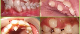
The eruption of “figure eights” is almost always associated with complications
Difficulties in eruption of third molars are caused not only by lack of space, but also by the fact that they do not have deciduous predecessors. Therefore, they often grow only partially or at an angle. But even the appearance of units growing correctly in most cases causes complications.
Most often found:
- destruction or displacement of adjacent teeth;
- bite problems;
- pericoronitis.
Very often, the eruption of the figure eight is accompanied by a purulent-inflammatory process. This happens because a hood appears over the tooth, and around it there is a pocket in which bacteria, plaque and food debris accumulate. They create favorable conditions for the development of inflammation (pericoronitis).
Often, when the eighth tooth appears, severe pain is felt, which occurs due to oppression of the roots of neighboring units.
Why is it difficult to calculate the exact number of teeth?
Dental X-ray allows you to find out the exact number of teeth.
How many teeth do you have? This question may also seem simple: now let’s calculate and find out! But a surprise may await you. After all, the number of roots and canals may differ due to the fact that the latter very often bifurcate near the pulp.
It also happens that several canals are located in parallel in one root and only an x-ray will help you calculate the exact number of teeth in your mouth!
To accurately count everything in a tooth, dentists at YuliSTOM clinics use x-rays. This allows you to avoid incidents during dental treatment!
Removing "eights" is always difficult
If the unit is located correctly and does not have serious pathologies, it will not be difficult to remove it. To carry out the removal without the risk of complications, before the procedure the patient is prescribed an X-ray of the tooth.
However, most often the third molar grows incorrectly, partially erupts or becomes intertwined with neighboring roots. In this case, the intervention will be carried out in a complex way. The final decision on the choice of method is made by the doctor after examination and obtaining the results of an x-ray of the tooth.
When using a complex method, manipulations are performed in the following sequence:
- The gum is cut and peeled away from the bone.
- To access the roots, a hole is drilled into the bone.
- The unit is extracted (in whole or in parts).
- The wound is treated and stitches are applied.
The procedure can take from 20 minutes to 2 hours. Everything is individual and only an experienced doctor can professionally perform the removal. Therefore, you should be careful when choosing a clinic and specialist.
After surgery, it is recommended to eat soft foods and refrain from physical activity for several days. Antibacterial therapy may be prescribed for prophylactic purposes. Pain and swelling persist for 3-4 days and then gradually disappear.
Visiting the dentist immediately after the figure eight begins to erupt will save you from many serious complications.
Complex dentistry "Sanident" invites all residents of Ivanteevka and Shchelkovo to use the services of experienced dentists who can cope with any dental disease.
What if there are more than 32 teeth?
Hyperdontia is an anomaly when there are more than 32 teeth.
If there are more than 32 teeth in a person’s mouth, this is already an anomaly, and in dentistry the term hyperdontia , and the teeth themselves are called supernumerary .
Supernumerary teeth (hyperdontia) are not so rare: 5% of people have more than 32 teeth. Moreover, in 70% of cases one supernumerary tooth grows, in 25% of hyperdontia - 2 teeth, and only 5% of people - 3 and more. Among carriers of supernumerary teeth, the number of women and men is the same.
Unfortunately, modern medicine does not know the exact answer to why and how a malfunction occurs in the human body and supernumerary teeth appear.
Moscow metro station Zvezdnaya, Danube Avenue, 23
Bottom line
Today we shed a little light on such concepts as how many wisdom teeth grow or can grow in a person throughout life, how the upper eights differ from the lower ones, and what problems the dentist may encounter when removing them, and the consequences after the operation for the patient? Unfortunately, the extraction of these teeth often leads to pain and is fraught with unpleasant consequences if you ignore the doctor’s recommendations during the rehabilitation period.
I hope this article was interesting and informative enough to find out the basic patterns of the functional purpose of wisdom teeth, their position in the oral cavity and the importance of maintaining hygiene in this part of the dentition.
You can share these materials with your relatives, friends and work colleagues, since the problem of treatment or removal of wisdom teeth will always be relevant. After reading the article, you now know exactly how many wisdom teeth a person has and where they are located?! In our blog you can find other interesting topics. Have a good day!
Oral hygiene during tooth loss
Losing teeth leaves an unprotected wound in the mouth. The mouth has rightfully earned the nickname of the dirtiest place in the human body. And the baby explores the world with his eyes, ears, hands and tongue. When a tooth falls out, a hole opens, representing a deep wound. The risk of developing infection during the period of filling the hole with a new odontopagus is high. To avoid suppuration and inflammation, it is necessary to help the baby maintain oral hygiene.
- Cleaning your mouth should occur after breakfast, afternoon snack, lunch and dinner. Leftover food can clog into the hole and rot there, becoming an ideal breeding ground for harmful microbes.
- Rinsing your mouth after eating will help completely clean the holes from food debris. As a rinse, you can use an infusion of chamomile or marigold flowers, which has an antibacterial effect.
- Twice a year you need to carry out a preventive examination by a dentist in order to identify dental growth disorders.
Modern dental medicine offers an effective remedy for caries of primary teeth - fissure sealing. Fissures are the space between baby teeth. The dentist fills them with a special solution to prevent the accumulation of food particles.
You should pay attention to the speed at which bleeding stops after tooth loss. With normal clotting, the blood should stop within 5 minutes. Otherwise, the child may have bleeding disorders and should consult a doctor.
Anatomical features
The G8s have no external features .
Their structure is absolutely identical to other molars: crown, root system, neck. There can be up to 5 roots, and they are curved in such a way that it is problematic to treat and remove the “eights” .
A molar with one root can only grow if its development has been disrupted.
However, it is worth mentioning the features of another plan.
Firstly, eights do not have milk predecessors (after all, there is no space at all on the dental arch).
And this not only makes growth and eruption difficult, but also leads to various pathologies . These include malocclusion, pericoronitis, pathologies of the masticatory muscles, and many others.
Secondly, the blood supply of the “eights” is much poorer than that of the other teeth. As a result, they grow more fragile, prone to caries, age early and wear out quickly.
For your information! The molar can grow not only vertically, which is the norm. There are 4 types of growth pathologies: medal inclination, distal, buccal and lingual.
Linguistic – the figure eight grows towards the tongue . Buccal – towards the cheek. Distal – leans back, which interferes with eating (in addition, the patient has chronic pain syndrome).
Medial inclination - the molar grows towards the neighboring tooth , while resting against it.
This leads to the fact that the enamel of the “seven” begins to deteriorate, the gums periodically become inflamed, and caries soon begins to develop .
A traumatic effect on the tongue or buccal mucosa leads to the formation of wounds.
If this happens constantly, non-healing ulcers form at the site of the wounds, which can become infected.
If the situation is left to chance, there is a high risk of developing cancer.
At what age do adults lose teeth?
Returning again to the issue of dentition development, the process occurs individually for each person. The presence of a specific amount is determined by heredity, irregular care, and unfavorable conditions.
Elderly people experience tooth loss, which is explained by their age and this is a natural process, but this can happen to a person who is far from old age.
In addition to the above reasons, loss occurs in the following cases:
- injury resulting from mechanical impact,
- periodontitis, causing loosening and loss.
The disease is characterized by bleeding gums; if left untreated, their condition worsens and they move away from the necks of the teeth. Complications soon arise in the form of the formation of periodontal pockets where food ends up.
You won't be able to clean it yourself; you'll have to see a dentist. If you ignore the problem, tartar and ulcers will appear under the gums. The disease at the last stage contributes to the loosening of the tooth and its falling out of the socket.
Possible problems
The third molars have exactly the same structure as the rest of the teeth. they still have some features
- extreme position in a row;
- not clamped by other teeth;
- there are no previous dairy products;
- third molars are larger;
- The roots of eights are usually crimped and irregular in shape.
These features contribute to the problems that the appearance of third molars often causes:
- Inflammation of surrounding tissues. When teething occurs, a shell of mucous tissue appears on the surface of the gum - the so-called hood. Bacteria often accumulate underneath it, causing inflammation (pericoronitis). The disease is accompanied by severe pain radiating to the temple, throat or ear, fever, headache, and swelling.
Eights often grow incorrectly, so they bring many problems and must be removed.
If any of these problems are present, the third molars must be removed. This process is more complex than the extraction of other teeth, again due to the remote position of the figure eights and convoluted roots. Recovery after surgery is usually quite long, but in most cases, if the doctor decides to remove it, there is no other option.
Where to look for them?
You don't need to be a dentist to determine the location of wise teeth. They are located at the end of a row of teeth on the lower and upper jaw. They are the only ones that do not grow in place of the old dairy ones. Of course, these teeth are no longer needed for chewing. However, they often make themselves known and begin to erupt.
Their main feature is the structure of the roots. They are woven and very curved. Because of this, root canal treatment becomes a significant problem. It is for this reason that experienced dentists recommend getting rid of these teeth as soon as they manifest themselves.
Practice shows that such “newcomers” can develop with “ready-made” carious cavities, which will negatively affect the health of other teeth.
Structure of permanent teeth
An adult tooth consists of the following parts:
- The root is a process with which the tooth is securely attached to the alveolar tissue. The roots vary depending on the load on the tooth - the greater it is, the more developed the root system; The molars have the largest branched roots.
- The neck is the space between the root and the crown, located just below the edge of the enamel.
- The crown is a jaw socket in the alveolus. The surface part of the crown performs a chewing function. The crown is covered with enamel on top, which prevents pathogens from penetrating into the inside of the tooth.
- The pulp is the inner part, it makes up the dental canals and the pulp chamber, it is surrounded by dentin, which in turn provides support for the enamel.
This is interesting: What is teeth whitening Zoom 4 and how does it work?
How does a human tooth work at the tissue level?
All human teeth have a similar internal structure. The diagram below shows all the dental layers - enamel, dentin, cement, pulp.
The figure shows the histological structure of the tooth
The composition of the enamel layer includes:
- minerals – 96%;
- organic substances – 1.2%;
- liquid, water – 2.3%.
The enamel is covered by an outer shell, the cuticle, which covers the chewing surface of the tooth. Most of the tooth is occupied by dentin, located immediately under the enamel. It has less strength, its hardness is 58.9 kg/mm.
Cement – covers the root part and in most respects is similar to bone tissue. Depending on the type of structure, there are two types of cement:
- acellular or primary - consists of collagen fibers;
- cellular or secondary - the composition includes cemenoblasts.
The internal dental cavity is occupied by the pulp - soft tissue, including:
- numerous nerves;
- blood vessels;
- connective tissue.
Sectional structure of a tooth
Despite the differences in the purpose and structure of the root system, all teeth have the same internal structure:
- Enamel. A very hard surface layer that protects teeth from environmental influences. It is designed as a structure of glued prisms. Thanks to the large amount of mineral salts in its composition, enamel is the strongest tissue in the human body. Its main task is to preserve the dentin of the crown. The presence of a pellicle ensured the tissue's resistance to the effects of acids.
- Dentine. It consists of coarse fibrous tissue, the basis of which is collagen fiber. The structure is similar to porous bone. There are two types of dentin: superficial and internal. The first has a higher density and prevents infection from entering the dental cavity.
- Cement. Consists of collagen fibers saturated with limestone salts. Layer thickness - from 50 to 150 microns, depending on location. There are two types of cement: cellular and acellular.
- Cavity. It has a shape identical to the crown and is located under the dentin. The entire area of the cavity is filled with pulp, a type of tissue responsible for nourishing the tooth and performing a connecting function. The presence of blood and lymphatic vessels ensures high sensitivity of the pulp.
Photo: this is what a tooth looks like in cross-section
Features of the structure of dental canals
The root canal is an anatomical space, which in its structure has a pulp chamber. That, in turn, is connected by channels.
All of them are divided into the following types:
- Type I – one canal with an apical foramen.
- Type II, III - often observed in premolars. Their feature is branching at different levels of the root.
- Type IV - has in its structure a single mouth and two separate root canals ending in two apical foramina.
- Type V, VI, VII - often found in the lower incisors and are distinguished by the types of both fusion and branching of the canals.
- Type VIII - three-channel with three apical openings.
The structure of root canals varies not only in type, but also in its shape and quantity.
They can be divided into the following groups:
Frontal (front)
These include:
- Upper central and lateral incisors, upper canines. Consist of one root and canal. It is extremely rare to observe both a two-channel and a two-root type. The type of structure of the lateral incisors shows a distal bend. The apex of the canines has a buccal curve.
- Lower incisors and canines. 37% are two-channel, the channels of which are often merged with each other. The gap in the mouth of the root is clearly visible on x-rays, but after branching it is barely visible.
Lateral
These include:
- Upper first premolars. 20% are single-canal and single-rooted teeth, 79% are double-canal and double-rooted, and 1% have three roots with three canals: one palatal and two buccal.
- Upper second premolars. In their structure, 56% are single-rooted, 46% are double-rooted and 2% are three-rooted, having a rather complex morphology.
- Lower first premolars. According to a study conducted in 1955, the majority of mandibular first molars—81%—are single-canal. The rest are two-channel. The 1979 studies are different - 70% single-channel and 30% dual-channel. 0.5% accounted for three-channel. The separation of canals on multi-canal teeth usually occurs in the middle part of the root.
- Lower second premolars. Most second premolars are single canal. Cases of a two-channel or three-channel structure are extremely rare.
- Upper first molars. In two out of three cases they have two channels, in the rest - one. The buccal root is medial, wide and flat; it is this structure that causes the two-channel structure. The orifice of the medial buccal canal is located under the medial buccal tubercle.
- Upper second molars. They are characterized by a variety of different types of structure. There are both three roots and three canals, and four canals with the same number of roots. They have a C-shaped canal structure at the confluence of the palatine with the medial-buccal root or the distal-buccal root. Cases of a two-channel and two-root structure are quite rare; cases of a single-channel structure are even more rare (no more than one percent of all observed cases).
- Lower first molars. A two-channel structure is often observed in the medal root, and in the distal root in two thirds of cases. Moreover, in 48% they are four-channel. With a three-channel structure, the third is distal-lingual.
- Lower second molars. Often their root is conical in shape, but variants with a more complex canal structure (crescent structure) are not uncommon. The most commonly observed is a two-root, three-channel structure.
When should you see a doctor?
Let's consider the main indications for surgical removal:
- “eights” interfere with the normal growth of nearby teeth;
- provoke an inflammatory process;
- incorrectly positioned;
- caries.
There is no need to worry about removal. Remember that their presence in the mouth can cause a lot of problems and discomfort. In any case, you need to consult a specialist who will determine a plan for further action.
Note!
Wisdom tooth pain - causes of pain and tips on how to relieve pain at home- A wisdom tooth is inflamed - a step-by-step description of the process of inflammation, treatment and postoperative recovery
Wisdom tooth roots - removal features, growth symptoms, treatment options and rules for caring for wisdom teeth
Are they needed today?
Scientists have proven that thousands of years ago all adults had “wise” teeth. If we look at modern statistics, they show that 15% of people do not even have the rudiments of “eights”.
However, this cannot be said that they are not needed at all. Additional teeth perform several important functions:
- Act as a restraining element if there are loose problem teeth nearby;
- They are used as a support in the process of prosthetics (a “bridge” is fixed to them);
- They are considered spare when the main chewing teeth fall out; accordingly, they continue to be responsible for chewing functions.
Of course, healthy and normal “eights” are extremely rare. If they erupted without any problems or complications, then there is no need to get rid of them. Perhaps in the distant future they will be useful to you.
It is worth noting that there is another reason. Japanese scientists have found that wisdom teeth contain unusual tissues that make it possible to obtain stem cells. Their value for modern science is difficult to overestimate.
How many incisors, canines, and molars are there in each jaw in the mouth of a healthy person?
- As already mentioned, a person has 32 permanent teeth if his wisdom teeth (of which there are only 4) have erupted and are intact.
- They are located absolutely symmetrically, each having its own shape. When we smile, the first thing we show is our incisors (also called front or anterior teeth ). Four incisors are located in the upper jaw and the same number on the lower jaw, i.e. only 8.
- Next to them are fangs , of which there are 4. They are each located on the right and left sides of the outermost incisor on both jaws. The fang has the longest and most powerful root, holding the outlines of the facial oval; these same teeth support the facial expression of a smile.
- Next comes the location of the lateral teeth . These include premolars, which are located on both sides of the upper and lower canines, and there are 8 of them in total. Behind the premolars, at the very ends of the dentition, there are molars (12 of them: 3 each on the left and right on the upper and lower jaws).
Human teeth: location diagram
Location and views
- Dentists' teeth are numbered according to a single scheme, created so that any doctor can help a patient based on his medical record, which already indicates previously performed actions with a particular tooth.
- The number assigned to each tooth is determined by its location on the jaw line, and the names are determined depending on the function it performs.
Numbering begins from the middle of the dentition, so the incisors located in the center are numbered 1,2. Further in both directions there are canines (No3), premolars (NoNo4,5) and molars (NoNo6, 7, 8).
- In addition, to make it immediately clear whether we are talking about the upper or lower jaw, the tooth with a certain number is located on the left or right, and the jaw segments are indicated. The rules for calculating them are as follows: 10 is the segment located on the top right, 20 is on the top left. The same goes for the lower segments: 30 is the left, and 40 is the right.
- The numbering is arranged clockwise to make it easier to imagine, remember and count. Thus, when a dentist talks, for example, about the 11th tooth, he means the incisor located on the right side of the upper jaw.
- By the way, children have a different dental pattern from adults; it is created in order not to confuse baby teeth with permanent ones. With the same numbering of teeth, segments are counted from 50 to 80, also clockwise.
This is interesting: The best teeth whitening products that work
The described scheme is international and is called the Viola system . There are also other systems that dentists can use: an American development that uses both numbers and letters; in the Zsigmondy-Palmer system, a special plate-diagram is used in which data about the teeth is entered; according to the Haderup system, in the same table next to the tooth number, pros or cons are indicated, indicating whether it is an upper or lower jaw.
What does the dental formula look like in a medical record?
The dental formula of an adult, as well as a child, in the medical record of a dental patient looks in the form of a schematic table (Fig. 5), which will reflect only the serial numbers of permanent or baby teeth. Directly in this formula, the doctor will mark the missing teeth (in this case, the number is crossed out with a cross), which teeth are affected by caries, pulpitis or periodontitis, as well as which of them have crowns or bridges. Dental formula in the form of a table in the medical record –
Should I save?
You will have to decide for yourself whether to save or delete the “eights”.
Unfortunately, in addition to the fact that their growth is accompanied by a lot of unpleasant sensations, subsequent problems can also be very serious .
Firstly,
eights often begin to grow crookedly .
If there is no room for him, he begins to rest against his neighbor or his cheek. This slowly but surely loosens the adjacent tooth, causing gum inflammation.
Secondly, the growth of a molar can take a year, or even two, or even three . During this time, initial caries may “mature.”
If left untreated, the gums will begin to swell, which will require surgical intervention. And it’s difficult to clean such teeth properly, only in a doctor’s office.
That is why caries spreads to the entire jaw, healthy teeth, causing inflammation of the dental nerve - pulpitis, halitosis, gingivitis, pericoronitis.
origin of name
Naturally, it is also interesting where this name came from. But here everything is extremely simple. Teeth are called “wise” teeth because they appear in adulthood. “Eights” are formed quite quickly. But they can cause discomfort for several years.
It is impossible to say for sure how long wisdom teeth take to grow. However, this process will be accompanied by painful sensations, increased body temperature, weakness throughout the body, numbness and immobility of the jaw. In severe situations, the lymph nodes begin to become inflamed.
It is not recommended to self-medicate or let things take their course. If the tooth does not erupt for a long time, then go for a consultation with a qualified doctor. The specialist will examine the problem area, if necessary, cut the gum and remove the tooth.