According to antiplagiat.ru, the uniqueness of the text as of October 16, 2018 is 97.5%.
Key words, tags: caries, periodontitis, periodontitis, deep caries, tooth extraction
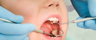
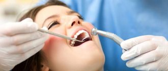
According to WHO statistics, every fifth inhabitant of the Earth has at least once encountered such a problem as pulpitis. Pulpitis is the case when the disease itself “pushes” the patient to the doctor, and rarely anyone succeeds in ignoring these signals, because almost always this problem signals its appearance with pain, often unbearable. Therefore, even those patients who are ready to drink “packs” of painkillers just to bypass the dental office, with such symptoms still strive to get an appointment with a dentist as soon as possible. Pulpitis requires immediate medical intervention, since further development of this disease promises serious complications, including complete tooth loss. This disease has been sufficiently studied and, thanks to modern technologies and techniques, can be effectively treated.
Historical reference
In ancient times, humanity did not yet know the term “pulpitis,” but they were familiar with toothache in different parts of the world. The main means of getting rid of it was tooth extraction. In some countries, conspiracies and rituals with sacrifices were used “for medicinal purposes.” In ancient Egypt, according to information found in ancient papyri, doctors looked for ways to help the patient using anti-inflammatory ointments containing the juice of various plants, and pastes made from myrrh, ash, pumice and eggshells.
In the 1st century AD The personal physician of the Roman Emperor Trajan, the surgeon Archigenes, drilled a tooth for medicinal purposes. Around 150-160's. the famous physician and philosopher of antiquity, Claudius Galen, described in his works the differences between pulpitis and periodontitis, but this knowledge was forgotten for a long time. In the 9th century in the Middle East, the physician and pharmacist Muhammad al Rashid advised using arsenic to destroy the dental nerve that was causing pain to the patient. But in European countries this method became known much later.
In the 11th century, in some European countries, caries and the pulpitis caused by it were “treated” with laxatives and enemas, and if this did not help, the pulp was burned with a hot iron with “anesthesia” in the form of using alcohol-containing compounds before manipulation or even hitting the head with a board. , the so-called Rausch anesthesia. In the 15th century, a professor at the University of Bologna repeated the experiment described by Archigen - he removed the affected dental tissue by drilling, after which he cauterized the pulp and filled the tooth cavity with gold.
Pierre Fauchard, a French doctor who lived in the 18th century, learned to identify 102 types of toothache, studied and practiced various methods of eliminating it, and became the founder of the “dental” patient position. Before him, the patient was placed on a table or seated on the floor, pressing his head between his knees, and P. Fauchard insisted that the patient in this position experiences unwanted nervousness and it is necessary that he sit in a chair, and the doctor stands next to him.
After 1871, when James Morrison patented the dental drill, therapeutic dentistry began to develop rapidly. Tools, equipment, drugs for pain relief, technologies began to appear, some of which are still actively used by dentists. Today, modern dentistry has effective methods, modernized instruments, and improved technologies with the help of which dental diseases, including pulpitis, can be effectively treated.
Pulp anatomy
In the depths of the tooth, under the dentin layer, there is a pulp, which consists of soft, loose, fibrous connective tissue, dotted with blood and lymphatic vessels, as well as nerve endings running from the jaw along the root canal through the apical foramen.
Pulp (lat. pulpis dentis) is the “heart” of the tooth, reliably protected by powerful dental walls of bone tissue from the effects of external factors, nourishing the tooth with minerals, ensuring its growth, restoration, and vitality. It is important to note that the pulp is not only a space of soft tissue (pulp chamber), but also the dental canal connected to it. The pulp chamber is a loose, amorphous colloidal system containing loose, fibrous connective tissue, as well as a large number of elastin and collagen fibers. The cellular composition of this system contains histocyotes, mast cells, macrophages, as well as fibroblasts that produce collagen and provide intercellular communication. The surface layers of the fibrous structure of the pulp contain odontoblasts - cells with long processes located in the dentinal canals. These processes make dentin sensitive to any irritants. Stellate cells are located a little deeper, and the central layer contains collagen and nerve fibers and blood vessels. If an inflammatory process begins in the pulp, then leukocytes appear in the structure, lymphocytes and plasma cells are activated.
In addition to providing the tooth with nutrition, the pulp performs several other important functions. Plastic, responsible for the delivery of “building” proteins, is ensured by the activity of odontoblasts involved in the formation of dentin: before the eruption of the primary tooth, after eruption - the secondary one. The protective function of the pulp is carried out thanks to macrophages, lymphocytes and fibroblasts. Macrophages “recycle” dead cells and, together with lymphocytes, are responsible for immune reactions, and fibroblasts produce and maintain the necessary balance of the intercellular substance of the pulp, which is responsible for metabolic processes in it. In general, the protective function of the pulp is to create a barrier for the penetration of pathogenic bacteria that have entered through the dentin further, along the root canal into the periodontium, and then to the soft tissues surrounding the tooth. In addition, the protective function includes the regeneration of the so-called replacement (tertiary) dentin: when caries occurs, this dentin prevents its spread deep into the tooth. The trophic function of the pulp, which affects the metabolism and nutrition of the tooth, supporting the vital activity of tooth enamel, is ensured by the activity of a developed vascular system, characterized by thin vessel walls, high blood flow speed and, accordingly, higher pressure than in other organs. The sensory function of the pulp is carried out due to the activity of a large number of nerve fibers, which, like a fan, diverge from the apical foramen to the periphery of the pulp.
Very often the pulp is called the “dental nerve” because its sensitivity to any irritants is so high that inflammation, as a response to a bacterial, viral, or infectious attack, occurs almost immediately. Such inflammation in medical terminology is called pulpitis.
Odontoblast is a multifunctional cell
The most obvious function of odontoblasts is the formation and maintenance of dentin. Like many other tissue-sparing cells, odontoblasts also contribute to protection against external factors. Localized at the periphery of the pulp and with processes located in the dentin, they are at the forefront of the fight against aggressive agents that penetrate dentin from the oral cavity (Fig. 2.4).
Rice. 2.4. Dentin, predentin, pulp tissue with peripheral odontoblasts. Staining of the preparation with hematoxylin-eosin.
When necessary, odontoblasts synthesize and release many molecules involved in the fight against invading microorganisms. In addition, it activates special receptors located on neighboring cells, vessels and nerves, as well as located directly on the odontoblasts themselves (see "Advanced Aspects"). Thus, odontoblasts, together with local protective resident cells and specific blood cells, have a wide range of response modes and play an important role in the activation of both innate and adaptive immune responses of the pulp (Fig. 2.5).
Rice. 2.5. The functions of the odontoblast change during the development, maturation and damage of the teeth, (a) Receptor: 1 – damaged from the outside by antigens, mechanical and thermal stimuli; 2 – exposed from the inside to circulating hormones, paracrine and autocrine substances. (b) Secretory cell: a – for the formation of replacement dentin, b – for the preservation of dentin, participates in immune reactions, (c) Neurotransmitter of pain: acts as a transducer between external stimuli and sensitive nerve fibers of the pulp.
Pulpitis: definition, causes, signs, consequences
Pulpitis is an inflammation of the pulp that occurs due to the entry of one or another infection into it through the crown of the tooth (intradental infection) or through the apical foramen located at the apex of the tooth (retrograde infection). Most often, pulpitis is a consequence of long-term caries.
But there are other factors that provoke the appearance and development of this disease. Modern dentistry divides them into 3 main groups:
Physiological ones include overheating of the pulp and/or accidental opening of the dental cavity during preparation, fracture of the coronal part with opening of the pulp chamber, the presence of decalcified formations in the pulp - calcifications (denticles and petrification), which, deposited in it, irritate the nerve endings, compress the blood vessels, disrupt blood flow, cause swelling, discomfort and pain.
Chemical factors include iatrogenic factors caused by doctor errors during the treatment process: the use of strong antiseptic solutions for treating a carious cavity, incomplete removal of the etching gel, etc.
Biological factors include factors that create conditions for infection to enter the pulp: secondary caries, spread of infection from the carious cavity along the dentinal tubules, retrograde pulpitis, when the infection enters the pulp through the apical foramen in sepsis, osteomyelitis, through the lateral branches of the root canal - in periodontitis (after curettage).
The most common symptom of pulpitis is an intractable throbbing pain as a reaction to one or another irritant: temperature, chemical (eating sweet food), mechanical (brushing teeth, etc.). Such pain does not go away on its own, and painkillers often do not help. However, such pain is characteristic not only of pulpitis, but also of other diseases. Therefore, if pain occurs, it is very important to immediately contact your dentist to determine the cause of the pain and begin treatment. If the disease is not treated, the inflammatory process becomes more intense and spreads to the periodontium, resulting in periodontitis. Remember: acute toothache requires immediate consultation with a doctor, without attempting self-medication.
Causes of development of chronic periodontitis
As already mentioned, one of the main causes of periodontal inflammation is untreated caries. However, the disease can develop for other reasons.
- Low quality filling materials. Some types of root fillings (especially those based on clove oil) can dissolve over time, forming empty cavities in the tooth tissue, which become a favorable breeding ground for anaerobic microbes. Other filling materials may shrink or crack, which can lead to the same result with bacteria and periodontitis. Partial filling of the root canal. Very often, when removing the pulp and cleaning the canals of the tooth, a small area remains at the apex of the root. If the canal is not completely sealed, then an infection develops in these tissues, which ultimately leads to periodontitis.
- Perforation of channels. When installing pins or cleaning channels, they sometimes break through. If a small hole in the root of a tooth is not disinfected and sealed hermetically, then inflammation will develop on the part of the periodontal tissue. This disease is characterized by the occurrence of periodontitis on the puncture side.
- Inflammation of the gums or periodontitis can also cause periodontal disease. There is a high probability of infection spreading if the patient has chronically inflamed gums with deep pockets of soft and then bone tissue. Particularly advanced cases can even lead to periodontitis of the apex of the roots.
- Tooth injuries. There is a category of people who do not consider it dangerous to crack a nut or open a beer bottle with their teeth. This bravado lasts until the tooth becomes inflamed or breaks off. Even such a “harmless” action as the habit of chewing a pen can turn into chronic periodontitis.
In addition to the obvious provocateurs of this disease, there are also secondary factors that indirectly cause or contribute to the development of incipient periodontitis in the dental tissues.
- Failure of metabolic processes in the body.
- Pathogenic microflora in the oral cavity.
- Malocclusion, which leads to injury to dental crowns.
- Chronic diseases of internal organs.
- Reduced immunity.
- Frequent infectious and viral diseases.
- Diabetes.
- Disturbances in the endocrine system.
A separate category of such factors should include poor nutrition, which leads to multiple imbalances in the body, and poor oral hygiene.
Types of pulpitis
Today, the main classification that defines diseases, including pulpitis, and its types, is the International Classification of Diseases and Related Health Problems, developed by the World Health Organization, tenth revision (ICD-10). Also considered an authoritative classification among Russian dentists is the MMSI, developed in 1989 at the Research Institute named after. N. A. Semashko.1
According to the clinically and legally accepted international classification ICD-10, pulpitis (K04.0) as a disease is divided into several types, but this classification has some discrepancies with the classification according to MMSI:
K04.00 – initial (pulp hyperemia) / according to MMSI – deep caries
K04.01 – acute / according to MMSI – acute focal pulpitis . Acute pulpitis is a common complication of deep caries and is characterized by severe pain, aggravated by impact on the tooth. According to the MMSI classification, the first stage of acute pulpitis is focal pulpitis, lasting no more than 2 days. Due to the proximity of the carious cavity to the pulp, a sharp “shooting” short-term (10-30 minutes) pain of a spontaneous and cyclical nature occurs: it arises arbitrarily, without affecting the tooth, and also disappears arbitrarily, only to reappear after some time. It “envelops” one tooth without spreading to adjacent teeth and tissues.
K04.02 – purulent (pulp abscess) / according to MMSI – acute diffuse pulpitis . This is the next stage of the disease, when inflammation spreads to the root part of the pulp. The pain becomes radiating - spreading along the branches of the trigeminal nerve, “radiating” to the area of other teeth, to different parts of the jaw, to the cheekbones, to the temples, to the back of the head, to the ears, its attacks become more frequent (especially at night), and the intervals between them become shorter (30-40 minutes) – diffuse pulpitis. If the patient notes that hot food and drink increase pain, and cold food and drinks relieve pain, this often indicates that the purulent stage of pulpitis or a pulpal abscess has occurred. This stage lasts maximum 14 days, after which pulpitis enters the chronic stage.
K04.03 - chronic / according to MMSI - chronic fibrous pulpitis : this is a long-term inflammatory process that lasts from 2-3 weeks to several years. Toothache at this stage becomes less pronounced, “dulls”, worsening during chewing, bleeding of the pulp and fragility of the hard tissues of the tooth may appear. This also correlates with the first stage of chronic pulpitis according to the MMSI classification - fibrous pulpitis, which often occurs secretly, without showing itself or is signaled by mild discomfort and pain. When examined at this stage, a large carious cavity is almost always discovered, which in many cases is connected to the pulp chamber. The pulp is painless, pain appears only when you touch it, and minor bleeding is possible.
K04.04 - chronic ulcerative / according to MMSI - chronic gangrenous pulpitis . This stage of the development of the disease is characterized by atrophy of the nerve fibers of the pulp, a change in its color to dirty gray, increased pain, and the appearance of bad breath. The examination also reveals an extensive and deep carious cavity.
K04.05 – pulp polyp / according to MMSI – chronic hyperplastic pulpitis. From the stage at which the connection of the carious cavity with the pulp is always detected, the growth of tissue, the formation of a painful and bleeding polyp when pressed, filling the free space of the pulp chamber.
K04.08 – other specified pulpitis (retrograde, traumatic, residual)
K04.09 – pulpitis, unspecified
K04.1 – pulp necrosis (pulp gangrene). It is considered to be the final stage of chronic pulpitis, which is characterized by signs of acute and chronic disease at the same time. Attacks of acute pain intensify and become more frequent, soft tissues are affected by necrotic changes, the bone tissue of the tooth is intensively destroyed, and periodontal infection is often detected during examination.
K04.2 – pulp degeneration (dentals, pulp petrification)
K04.3 – improper formation of hard tissues in the pulp (secondary or irregular dentin)2.
Pulpitis, like most diseases in principle, progresses and worsens, moving from one stage to another, but nowadays modern dentistry has methods that, in some cases, can treat this disease while maintaining the viability of the pulp. Chronic pulpitis in 90% of cases is irreversible and the only solution is removal of the pulp.
Periodontitis by location
There are two main divisions in the classification of periodontitis based on the location of the disease: apical and marginal. The first option (apical) is also apical, based near the apex of the tooth root. This type is the most common, since periodontal infection in most cases occurs through a descending channel: from caries to inflammation of the pulp, and then through the root canal the infection descends into the periodontal tissue. This process most often becomes chronic, since the protective mechanisms of the periodontium are much more powerful than those of the pulp. Therefore, the infection can enter the deep tissues of the tooth for years without causing signs of disease.
Marginal chronic periodontitis is located on the lateral walls of the tooth root and has the etiology of microtraumas.
Periodontitis, the treatment stages of which are a complex, complex process, requires immediate attention to a dental clinic. It is safe to say that such a diagnosis requires highly qualified specialists with extensive experience in solving such problems. Dentists of the LeaderStom network of clinics are rightfully considered the best in this area of dental therapy. The latest technologies, progressive techniques and extensive practice allow them to cope with the most complex, advanced cases.
Treatment methods for pulpitis
All methods of treating pulpitis can be divided into two main ones - biological, aimed at treating and restoring the pulp, and surgical, involving its partial or complete removal in order to preserve the tooth. Only a qualified dentist can determine which method to use in each specific case based on a thorough diagnostic examination.
The biological method is a method of conservative treatment, with the help of which the inflammatory process is eliminated and the pulp retains its viability. Thus, the affected pulp is subjected to alkalization, after which secondary dentin begins to be produced again. The biological method is effective only if the patient comes to the dentist immediately after the onset of a pain symptom. Treatment of pulpitis with a biological method is more effective in people at a young age (up to 30 years), when the pulp is capable of self-healing, in the absence of chronic diseases and sufficient caries resistance (resistance to caries). The treatment follows the following algorithm: the pulp is opened, treated with an antiseptic solution, a bandage with calcium hydroxide is applied on top, the cavity is closed with a temporary filling, which after some time is replaced with a permanent one.
The biological method is quite complex to perform and requires high professionalism of the attending physician. In general, this technique is characterized by low predictability of a positive treatment outcome. And even with extensive clinical experience, this method is not always effective. Because of these reasons, this treatment method is not very popular, and often doctors, bypassing it, immediately proceed to a more radical and predictable surgical method of treating pulpitis.
The surgical method consists of removing the affected pulp, cleaning the canals, sanitation from infection and subsequent filling of the tooth canals. The surgical method combines several techniques.
Amputation is prescribed in cases of acute pulpitis or accidental trauma to the pulp and involves removing the coronal part of the pulp while maintaining the viability of its root section. This technique is only suitable for treating pulpitis of multi-rooted teeth. Amputation can be vital (“life-saving”) - this is when part of the “dental nerve” is removed immediately under anesthesia. In this case, a necessary condition for the operation is a completely healthy periodontium. And devital (“life stopping”) - when the pulp is mummified using a special paste. After this, one section of the “dental nerve” is removed, and the second is mummified so that in the future this part does not become a source of recurrence of pulpitis. This technique is used extremely rarely in clinical practice, since this method is quite controversial and the possibility of relapse cannot be excluded. Therefore, for more effective treatment of pulpitis, a more radical method of treating pulpitis is often used - extirpation.
Extirpation
is the complete removal of the pulp when it is impossible to maintain its viability. Extirpation, just like amputation, is of two types - vital and non-vital. During vital extirpation, which is performed under anesthesia in one visit, the pulp is not mummified before being removed from the cavity. The dentist removes carious dental tissue, then penetrates the canals using special thin needles and removes the affected “dental nerve,” after which he treats the cavity with antiseptics. This technique is used for all forms and stages of pulpitis.
During devital extirpation, the pulp is first mummified using a paste containing arsenic, paraformaldehyde or another substance with a similar effect. On single-rooted teeth, the paste should remain for at least 24 hours, on multi-rooted teeth - at least 48. Some mild pastes can be left on for 7-14 days. The tooth cavity is closed with a temporary filling. After the paste expires, the doctor removes the pulp, cleans the canals and installs a permanent filling.
Using the devital extirpation method, pulpitis can be cured in 2–3 visits, depending on the number of roots in the diseased tooth. Devital extirpation is suitable for the treatment of all types and stages of pulpitis except purulent and necrotic, and is also not used in the treatment of baby teeth. With the development of technology, this method in modern dentistry is also becoming less popular and, perhaps, can be found in remote areas from regional centers.
The final stage of endodontic tooth treatment is filling (obturation) of the tooth canals, which requires a highly qualified doctor. Regardless of what method of vital or devital extirpation the pulp is removed, special attention is paid to filling the canals. After all, the main task is to prevent periodontal infection. Obturation of the root canal of a tooth can be carried out using the following methods - filling with one paste without pins and using gutta-percha pins in various variations, filling using heated gutta-percha with its vertical compaction, on a carrier (thermophile), using the “System B” device, a combined technique , or filling with gutta-percha from a syringe. The choice of methodology and materials always remains with the attending physician based on his preferences, clinical experience, level of training and capabilities of the clinic.
After all medical manipulations with the tooth have been completed, the final step is the application/installation of a permanent filling in accordance with aesthetic, individual and clinical characteristics.
Attention:
A temporary filling that closes the tooth cavity, where the active “dental nerve-killing” substance is located in the root canals, can be very durable and can last for several months. In this case, the pain that bothered him goes away and the patient does not experience any discomfort, and therefore postpones the next visit to the doctor indefinitely. Remember, in no case should such drugs be allowed to remain in the pulp chamber for a longer period than was determined by the doctor. You must come to the dental office on the appointed date and complete your treatment!
Also, every patient must remember and know that it is mandatory to attend a follow-up appointment after depulpation, strictly on the day prescribed by the attending physician. Since the patient himself is unable to independently distinguish the normal physiological state of the tooth after treatment of pulpitis from the pathological one.
Complications: pain of a pulpless tooth
After removing the pulp, followed by filling the canals and restoring the crown of the tooth, the patient may still experience pain, especially when biting. If pain goes away within a week, this is normal. If the pain continues to bother you after 5-7 days, this may indicate poor quality treatment and/or filling. For example, the filling material was carried beyond the root apex and got into the soft tissues, or during the removal of pathologically affected tissues, the tooth root was accidentally damaged, or the patient is allergic to filling materials. Also, if the treatment and/or filling of the dental canals is not thorough enough, pulpitis can turn into periodontitis.
Symptoms and manifestations
If pulpitis is diagnosed by acute pain upon contact with cold or hot food, then the characteristic symptoms of periodontitis are painful tapping or pressing on the tooth.
This diagnosis may be indicated by:
- swelling of the cheek or gum near the tooth;
- a feeling of fullness in the tissues of the organ;
- the occurrence of a fistula with the outflow of pus into the oral cavity;
- increased pain when eating or pressing on the causative tooth;
- the feeling of an “overgrown” tooth (due to compaction of the tissues under the root, the organ begins to bulge);
- pulsation in the root area;
- increased temperature in the area of the causative tooth.
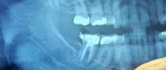
The chronic type of this disease can be asymptomatic and detected only with the help of an X-ray examination. Therefore, the insistent recommendations of the dentists of the LeaderStom clinic to undergo a dental examination by a doctor once every six months are so important for the early detection of such diseases.
Methods used in the clinic
We are adherents, first of all, of effective and then modern treatment methods that can quickly, efficiently and radically eliminate the cause of pulpitis and its consequences. But, in our work, we always try to “save” the pulp and preserve its viable properties using conservative methods and use them in all cases where possible.
At the same time, if removal of the dental nerve seems to be the only solution according to the indications, in many cases we use effective anesthesia of the “dental nerve”, after which we carry out its removal. We are convinced that advanced methods do not deny classical methods, but only complement, optimize, and improve them. That is why in our clinical practice we always try to follow the “classical” treatment algorithm. Its first stage is a complete comprehensive clinical diagnosis.
Treatment is carried out using a dental microscope and using perhaps the most modern instruments, which makes it possible to eliminate carious lesions without missing a single micron of the affected tissue, flexible and thin endodontic needles for the most effective cleaning of canals, and, of course, the safest filling materials. It is worth noting that filling includes work in the canals and in the crown of the tooth. If suddenly the patient experiences some deviations from the normal course of the adaptation process, patients may be prescribed conservative anti-inflammatory therapy, physiotherapy with ozone or laser treatment.
Age restrictions
Pulpitis can occur in anyone at any age. The conservative method of treating this disease has no age restrictions. When choosing a surgical method in patients over 45 years of age, it is necessary to take into account the condition of periodontal tissues.
Treatment of pulpitis in children with baby teeth has its own characteristics. Thus, the inflammatory process in baby teeth arises and spreads rapidly and does not always depend on the depth of carious lesions and visible tissues affected by caries. In this case, it is extremely important to stop the spread of infection to the periodontal tissue, since the rudiments of molar teeth are formed in this tissue. However, removal of teeth affected by pulpitis is used only in rare cases, since the absence of each dental unit has a negative impact on the formation of the bite. In the treatment of pulpitis of baby teeth, filling pastes are used that do not affect the rudiments of the molars, but are absorbed along with the “milk” roots when teeth change begins. Anesthesia must be carried out taking into account possible allergic reactions.
How long does it take for teeth to grow?
The formation of milk elements begins during the period of fetal development - the dental plate is formed in the second month, and by the middle of the period it is possible to identify the cells responsible for the structure of the structure of enamel and dentin. The pulp and dentinal layers are outlined towards the end of the second trimester, paralleling the process of formation of the collagen fibrous structure. The eruption of baby teeth begins 9-11 months after the birth of the child, and by the age of two or three years a complete set has already been formed. The radical units are located directly behind the dairy ones - initially they are separated by a small partition, which gradually collapses as the permanent elements grow.
The starting point in changing the bite is considered to be the age of 6-7 years: the process begins with the frontal incisors of the upper jaw, and lasts until adolescence, ending at the age of 13. It is worth noting that molars take longer to form and develop than primary ones - this is especially true for “eights”, or wisdom teeth, which in some cases bother their owners at an older age.
Price
The cost of treating pulpitis is influenced by many factors. First of all, these include the form and stage of pulpitis, diagnostic measures that make it possible to establish an accurate diagnosis and choose the most appropriate treatment method. In addition, the medications, materials, equipment and instruments used during treatment are important. Not the least important role is played by the qualifications of the doctor, additional consultations with specialists, as well as medical measures accompanying the main treatment, if necessary.
Many patients think that toothache is a temporary “little thing in life” that can be overcome with the use of modern painkillers. But this illusion quickly dissipates as soon as a person experiences unbearable pain... Remember that the sudden appearance of toothache is in all cases a serious signal warning of the presence of some kind of pathology in the maxillofacial system. In many cases, this pathology turns out to be pulpitis - a disease that, if not treated in a timely manner, can lead to many complications, including tooth loss. But only a qualified doctor can determine the exact cause after conducting a thorough diagnostic examination. Therefore, visit the dental office as soon as possible. Your efficiency, combined with modern treatment methods and the professionalism of the doctor, is a guarantee that the disease that caused the pain will be completely cured and will not deprive you of the beauty of a full smile.
According to antiplagiat.ru, the uniqueness of the text as of October 16, 2018 is 97.5%.
Key words, tags: caries, periodontitis, periodontitis, deep caries, tooth extraction
1 Therapeutic dentistry. Dental diseases: textbook: in 3 hours / ed. E.A. Volkova, O.O. Yanushevich. - 2013. - Part 1.). 2 https://mkb-10.com * Images: - Domenico Ricucci, Jose Siqueira, “Endodontics. Clinical and biological aspects”, Publishing house “Azbuka”, Moscow, 2015. A book for dentists and endodontists. Edition in Russian, translated from English, 415 pages, 1682 illustrations, hardcover. The original edition of the book “Endodontology: An Integrated Biological and Clinical View (Ricucci, Domenico and Siqueira Jr, Jose)” was published in 2013. — Database of clinical photo protocols of the Dental Clinic Dr. Edranov; Personal archive of S.S. Edranova.