27.08.2020
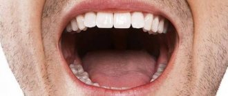
At Unit dental clinics, we educate our patients about oral hygiene. There is an opinion that if there are no bacteria and microbes in the oral cavity, then caries and other diseases cannot occur. Is it so?
Our body, including the oral cavity, is inhabited by millions of bacteria. They form a harmonious microflora. When the balance of healthy and pathogenic bacteria tilts toward the latter, then a person begins to get sick.
What is the normal and pathological state of the oral microflora?
Detailed description of the study
Microbiocenosis of the oral mucosa and nasopharynx, or the totality of populations of different types of microorganisms that constantly live in the human body, is a diverse and multi-level system.
Microorganisms inhabiting the human body under normal conditions create a protective barrier and inactivate pathogens entering from outside. They are found in the intestines, on the mucous membranes of the mouth and nasopharynx, as well as on the skin.
Normally, the mucous membranes of the human oral cavity and nasopharynx are populated by a variety of microorganisms that interact with each other and maintain a relatively constant composition of the flora.
Representatives of normal microflora:
- Actinomycetes;
- Veillonella;
- Lactobacilli;
- Bacteroides;
- Bifidobacteria;
- Campylobacter;
- Fusobacteria;
- Staphylococcus;
- Streptococci.
The microbiome of the mucous membranes of the oral cavity and nasopharynx can change due to local and general medicinal influences, including taking antibiotics, glucocorticosteroids, as well as various diseases of the mouth, teeth, nose and sinuses. The qualitative and quantitative composition of microorganisms is influenced by:
- Integrity of mucous membranes;
- Temperature and nature of food consumed, its acidity;
- Composition of saliva;
- Pathologies of teeth and gums;
- Pathologies of the mucous membranes of the mouth and nasopharynx.
Most often, the following diseases are diagnosed, caused by changes in the composition of microorganisms: sinusitis, sinusitis, laryngitis, tonsillitis, adenoiditis, etc. Restoring the microbiocenosis of the mucous membranes in the case of the listed pathologies is one of the important directions in the treatment of such conditions and the prevention of their relapses.
Microbiota analysis according to Osipov allows one to identify the composition of microorganisms living on human mucous membranes quickly and with high accuracy. The method is based on chromatography-mass spectrometry (CMS).
Infections are detected by detecting specific microbial markers (proteins) in the test sample. These markers are produced by microorganisms during their life processes; they differ from human cells in chemical composition.
A distinctive feature of the method is its high separation efficiency, sensitivity down to micro- and nanograms, as well as the speed of analysis.
Determination of skin microbiocenosis according to Osipov allows:
- Determine the qualitative and quantitative composition of microbial communities;
- Determine the microecological status of the organism and the possible degree of deviations;
- Identify or clarify the origin of the infectious-inflammatory process in any clinical manifestations of the disease.
Thanks to this analysis, a specialist can determine which flora predominates on the patient’s oral and nasopharyngeal mucosa, detect microbial imbalances and prescribe appropriate therapy to restore balance.
Scrapings from the mucous membranes of the mouth and nose are used as the test material.
The results of the study indicate the number and types of identified microorganisms. The patient must pass the results obtained to the doctor, who will make an appropriate diagnosis and, if necessary, prescribe treatment.
Oral microflora: what you need to know
When patients ask what causes tooth decay, do you usually tell them “sugar” or “germs in the mouth”? We know that oral microflora plays an important role in maintaining oral health. Understanding the relationship between the body and the bacteria that inhabit it can help you teach your patients how to prevent dental disease.
What is oral microflora?
The microflora of the oral cavity is the totality of microorganisms found in it; As noted in a study published in the Journal of Bacteriology, more than 700 species and strains of bacteria have now been found in the oral cavity. Bacterial microflora in the mouth forms a structure that covers the teeth, dentists call this plaque or dental biofilm.
Many oral bacteria are associated with dental diseases that result from dysbiosis. An imbalance of different types of bacteria creates conditions for the excessive proliferation of potentially pathogenic microorganisms, playing a leading role in the development of a number of diseases. The current hypothesis is that the entire biofilm may be involved in this process. This is supported by recent research and the theory of a key pathogenic factor.
The main causes of dental and gum disease are two types of bacteria: Streptococcus mutans (S. mutans) and Porphyromonas gingivalis (P. gingivalis); In addition, researchers note a connection between lactobacilli and the occurrence of tooth root caries. Publications in the journal PloS One note that S. mutans consumes easily fermentable sugars and, as a result of metabolism, produces acid, which leads to demineralization of dental hard tissues, which causes caries. S. mutans also produces extracellular polysaccharides early in the biofilm formation process, essentially providing the “glue” for its formation and attachment to dental and oral soft tissue surfaces. According to the authors of Frontiers in Microbiology, P. gingivalis is the main microorganism involved in the development of chronic periodontitis.
Causes of changes in oral bacterial flora
The microflora of the oral cavity of each person changes throughout life. As children grow, bacteria take up residence in their mouths. S. salivarius colonizes soft tissue shortly after birth. As teeth erupt and gingival sulci form, microorganisms such as S. sanguinis and S. mutans colonize tooth and soft tissue surfaces. As a person grows and ages, the composition of the intraoral environment changes and the number of strains present increases.
Imbalance of the oral microflora can occur for many reasons, for example due to a diet high in fermentable carbohydrates, which changes the pH, or due to the formation of a dense biofilm, which is particularly protective of microorganisms located in its thickness (that is, it prevents the penetration of antimicrobial agents). Finally, opportunistic microorganisms such as Helicobacter pylori can penetrate the oral cavity. This microorganism is present in patients with gastroesophageal reflux disease (among other stomach diseases) and is involved in the development of periodontitis, according to publications in the International Journal of Clinical and Experimental Medicine.
Helping patients control oral microflora
How can you help patients balance their oral flora? The main thing is not to allow the balance to shift in any direction favorable to the development of diseases. Provide patients with several practical steps to help avoid creating unfavorable conditions and developing oral dysbiosis.
- Reduce your sugar intake. This will reduce the risk of creating a cariogenic environment by bacteria that metabolize carbohydrates. Instead, encourage patients to drink unsweetened and non-acidic beverages, as well as milk, and consume dairy products as part of a healthy diet that includes adequate calcium.
- Brush your teeth twice daily with an optimal fluoride toothpaste to break down biofilm and protect tooth enamel.
- Floss daily or use other interdental cleaners, such as interdental brushes, to remove plaque buildup between teeth and in the sulcus. Simply brushing your teeth will not eliminate bacteria from all of these hard-to-reach areas. Finish with an antibacterial mouth rinse. This can help remove food debris from your mouth and also reduce bacteria.
- Probiotics have been found to be beneficial for digestive disorders in healthy patients, and preliminary evidence from several studies suggests that probiotics may also help protect teeth and gums.
- Encourage patients to visit your office regularly for examinations, with the frequency of visits based on your and their own assessment of the risk of developing tooth decay and other oral diseases. Despite proper oral care at home, some people are at very high risk for dental disease and will need more frequent visits.
Understanding the role of the intraoral microflora is the first step in teaching patients how to control its balance to maintain oral health. Educating patients about the role of bacteria in dental disease can help them achieve overall oral health.
References
- Pomazanov, V.V. Microecological status of a person and its definition. — Prospects for the implementation of innovative technologies in medicine and pharmacy, 2021. — pp. 167-184.
- Gostry, A.V., Simonova, A.V., Mikhailova, A.N. and others. Chronic pharyngitis: etiology, pathogenesis, treatment. New approaches to assessing etiopathogenesis. - Archives of Internal Medicine, 2021. - No. 1 (45). — P.32-43.
- Man, W., Clerc, M., Pieters, W. et al. Loss of microbial topography between oral and nasopharyngeal microbiota and development of respiratory infections early in life. – American journal of respiratory and critical care medicine, 2021. – Vol. 200(6). — P. 760-770.
1-mini lecture “Microflora of the oral cavity, esophagus and stomach.”
The oral cavity and pharynx carry out preliminary mechanical and chemical processing of food and assess the bacteriological danger of bacteria penetrating into the human body.
Saliva is the first digestive fluid that processes food substances and affects the penetrating microflora. The total content of bacteria in saliva is variable and averages 108 MK/ml.
The normal microflora of the oral cavity includes streptococci, staphylococci, lactobacilli, corynebacteria, and a large number of anaerobes. In total, the oral microflora includes more than 200 types of microorganisms.
On the surface of the mucosa, depending on the hygiene products used by the individual, about 103–105 MK/mm2 is detected. Colonization resistance of the mouth is carried out mainly by streptococci (S. salivarus, S. mitis, S. mutans, S. sangius, S. viridans), as well as representatives of the skin and intestinal biotopes. At the same time, S. salivarus, S. sangius, S. viridans adhere well to the mucous membrane and dental plaque. These alpha-hemolytic streptococci, which have a high degree of histadhesis, inhibit the colonization of the mouth by fungi of the genus Candida and staphylococci.
The microflora transiently passing through the esophagus is unstable, does not show histadhesiveness to its walls and is characterized by an abundance of temporarily present species that enter from the oral cavity and pharynx. Relatively unfavorable conditions for bacteria are created in the stomach due to increased acidity, the influence of proteolytic enzymes, the rapid motor-evacuation function of the stomach and other factors that limit their growth and reproduction. Here microorganisms are contained in quantities not exceeding 102–104 per 1 ml of content. Eubiotics in the stomach primarily colonize the cavity biotope; the wall microbiotope is less accessible to them.
The main microorganisms active in the gastric environment are acid-fast representatives of the genus Lactobacillus, with or without a histagesive relationship to mucin, some types of soil bacteria and bifidobacteria. Lactobacilli, despite their short residence time in the stomach, are capable, in addition to their antibiotic effect in the gastric cavity, of temporarily colonizing the parietal microbiotope. As a result of the combined action of the protective components, the bulk of microorganisms that enter the stomach die. However, if the functioning of the mucous and immunobiological components is disrupted, some bacteria find their biotope in the stomach. Thus, due to pathogenicity factors, the Helicobacter pylori population is established in the gastric cavity.
A little about stomach acidity: The maximum theoretically possible acidity in the stomach is 0.86 pH. The minimum theoretically possible acidity in the stomach is 8.3 pH. Normal acidity in the lumen of the body of the stomach on an empty stomach is 1.5–2.0 pH. The acidity on the surface of the epithelial layer facing the lumen of the stomach is 1.5–2.0 pH. The acidity in the depths of the epithelial layer of the stomach is about 7.0 pH.
Small intestine
- this is a tube about 6 m long. It occupies almost the entire lower part of the abdominal cavity and is the longest part of the digestive system, connecting the stomach with the large intestine. Most of the food is already digested in the small intestine with the help of special substances - enzymes.
The main functions of the small intestine include cavity and parietal hydrolysis of food, absorption, secretion, and barrier protection. In the latter, in addition to chemical, enzymatic and mechanical factors, the indigenous microflora of the small intestine plays a significant role. It takes an active part in cavity and wall hydrolysis, as well as in the processes of absorption of nutrients. The small intestine is one of the most important links ensuring the long-term preservation of eubiotic parietal microflora.
There is a difference in the colonization of cavity and parietal microbiotopes by eubiotic microflora, as well as the colonization of tiers along the length of the intestine. The cavity microbiotope is subject to fluctuations in the composition and concentration of microbial populations, while the wall microbiotope has a relatively stable homeostasis. In the thickness of the mucous deposits, populations with histaghesive properties to mucin are preserved.
The proximal small intestine normally contains relatively small amounts of gram-positive flora, consisting mainly of lactobacilli, streptococci and fungi. The concentration of microorganisms is 102–104 per 1 ml of intestinal contents. As we approach the distal parts of the small intestine, the total number of bacteria increases to 108 per 1 ml of contents, and at the same time additional species appear, including enterobacteria, bacteroides, and bifidobacteria.
ATTENTION: Probiotic preparations are highly effective in combating human intestinal dysbiosis!
What bacteria inhabit the oral cavity?
The permanent microflora of the mouth consists of 30 strains of microorganisms that are responsible for the most important functions. These strains include: streptococci, veillonella and diphtheroids.
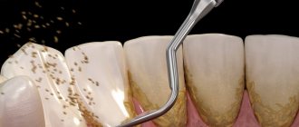
Almost 300 species of microorganisms constantly reside in the oral cavity. Among them there are pathogenic - harmful. Suitable habitats for parasites are braces and plates, carious cavities, crowns, gum pockets, and the back of the tongue. Some teeth have a soft but thick layer of yellow plaque, which over time turns into dark tartar - this is the focus of harmful microorganisms.
Bacteria and microbes form a unique environment that is maintained in the normal state of the body and is capable of self-regulation. In the normal state of the oral microflora, beneficial bacteria outperform pathogenic bacteria.
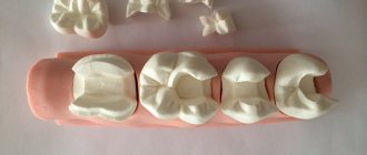
Systematic hygienic teeth cleaning at the dentist is an essential attribute of a healthy and beautiful smile.
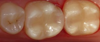
Every time we accept another patient for caries treatment, we are convinced that the price of professional hygiene is disproportionate to the consequences and costs that will have to be borne if it is not done.
It has been proven that even correct, scrupulous and constant home care is ineffective. 90% of adults brush their teeth ineffectively.
Teeth cleaning at Unit using modern technologies: Air Flow and ultrasound gives the maximum effect in maintaining a healthy balance of microflora and local immunity.
Sign up for professional cleaning
Be smart!
The work was added to the website samzan.ru: 2016-03-05
| Ministry of Health and Social Development of the Russian Federation. Volgograd State Medical University. Department of Microbiology, Virology, Immunology with a course in Microbiology. |
| Essay |
| Microflora of the oral cavity |
| Completed by a student of group 7 Faculty of Dentistry Kan.E.G Checked by senior teacher L.A. Blintsova |
2012
INTRODUCTION
The microflora of the oral cavity is normally represented by various types of microorganisms. But they can also be associated with oral diseases such as caries and periodontal disease. They appear when the normal balance between microorganisms, autochthonous and allochthonous microflora is disturbed. The development of modern molecular technologies in the study of oral microflora can help improve diagnostic methods, assess the risk of diseases and their treatment. For example, purulent-inflammatory processes such as dentoalveolar abscess and actinomycosis are usually endogenous infections caused by bacteria, which are part of the permanent microflora, most of them obligate anaerobes. There is a direct relationship between the state of the immune system and the activation of non-pathogenic flora and/or opportunistic microflora of the oral cavity (oportonus-convenient). Opportunistic microorganisms are activated in conditions that are convenient for them—these are immunodeficiency states (6). When local or systemic mechanisms are disrupted, oral candidiasis develops, caused by yeast-like fungi present in the mouth of healthy people (50). The two most common types of oral disease, dental caries and periodontal disease, are associated with microorganisms that form plaque on the surface of the tooth. Dental caries is usually associated with high numbers of S. mutans, Lactobacillus; Counting the level of microorganisms in saliva can be useful for determining the caries risk of patients and for monitoring for preventive purposes. A large number of microorganisms have been identified as potential etiological agents in various manifestations of periodontal disease, and the hypothesis of specific microorganisms involved in plaque formation is still controversial. Their etiology is often complex and may be due to autochthonous microorganisms and external factors in addition to any manifestations of microbial activity present in the oral cavity. (29). It is in dental plaque that the bulk of oral microorganisms are localized; 70% of the volume of dental plaque consists of microbes. 1 mg of its dry mass contains about 250 microbes (1). There are normal microflora of the oral cavity and opportunistic (opportunistic) microflora. However, such a division is difficult to carry out, because any microorganism, even a non-pathogenic microorganism with reduced immunity (AIDS), can acquire pathogenic properties. When characterizing oral microbes, it is most convenient to consider them depending on the localization of pathological changes: caries, periodontitis, periodontal disease, stomatitis, glossitis. In severe diseases such as AIDS, these conditions can occur simultaneously.
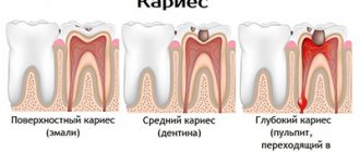
Microbiological aspects of dental caries. Since 1890, it was believed that caries is caused by bacteria that ferment carbon hydrates and sucrose, forming acids that begin to destroy tooth enamel. There were other theories: the presence of “worms”, imbalance of body fluids (disturbance of homeostasis), proteolysis, autoimmune disorders, but these theories have not been confirmed scientifically (51). Theories based on the study of caries in animals turned out to be important for solving questions of the etiology of caries. In the late 1950s, various types of microorganisms and those that caused tooth decay in animals such as rats or hamsters were studied (Table 1). It turned out that caries in the studied animals was caused not by a specific species, but by a large number of different microorganisms. It was believed that not all detected bacteria caused the disease; the leading role belonged to S.mutans and other microbes of the streptococcal group. They caused plaque or caries on the teeth of animals. S. mutans most commonly caused caries localized in fissures and smooth surfaces, where it was associated with other species of microorganisms (Lactobacillus), and in the case of root caries, possibly with Actinomyces species (57). Some researchers have experimentally shown that symbiosis of various cariogenic bacteria, such as S.mutans and Veilonella alcalescens (18,51), is possible. Veilonella species, using lactic acid produced by S. mutans, prevent the demineralization of tooth enamel. This is just one example of the interaction of microorganisms in the complex ecological system that exists on the surface of the tooth. The table lists bacteria that can independently or in associations cause caries.
| Streptococcus | Lactobacillus | Actinomyces |
| S.mutans | A.naeslundii | L.acidophilus |
| S.cricetus | A.vicosus | L.casei |
| S.rattus | A. Israelelli | L.salivarius |
| S.sobrinus | L.fermentum | |
| S.ferus | ||
| S. faecalis` | ||
| S.milleri“ | ||
| S.sanguis | ||
| S.salivarius |
`S.faecalis is called Enterococcus faecalis“The S.milleri group includes the following species: S.anginosus, S.constellatus, S.intermedius
Researchers have paid most attention to the role of mutans streptococci (MS), a term used to describe a group of streptococci that include S.mutans, S.sobrinus, S.cricetus, S.rattus, S.ferrus, S.downei, S.macacae . They were initially thought to be all serovars or biovars of S. mutans, but detailed taxonomic studies have clearly shown that each is a distinct species (55,56). However, these are mainly: S. mutans and S. sobrinus, which are not only isolated from humans, they also cause tooth decay in animals such as hamsters, rats and monkeys. Caries in humans is also caused by bacteria other than MS, these are lactobacilli and actinomycetes. Caries proceeds in stages, the enzymatic activity of plaque bacteria leads to the formation of organic acids, causing a local decrease in pH, and demineralization begins (a reversible process), then microscopic bonds are disrupted and cracks appear along the enamel prisms, a cavity is formed and the underlying tooth tissues are involved. In parallel with these stages, the dominant bacterial flora undergoes changes, demonstrating a phenomenon known as “microbiological succession.” According to modern views, during the stages of caries development, the balance between the autochthonous transient flora on the tooth surface (supragingival plaque) and the obligate flora leads to the beginning of demineralization of the tooth enamel. The number of microorganisms such as MS and Lactobacillus is increasing, which is probably associated with a decrease in pH (to 5.0-4.5), and the number of S.sanguis and A.naeslundii, on the contrary, is decreasing. While the white plaque has already formed, S. mutans predominate, followed by colonization by Lactobacillus species. A large number of other species are also found, such as S.mitis, A.vicosus (57). It is assumed that the isolation of both S.mutans and Lactobacillus indicates the formation of plaque, which is important for the subsequent development of caries. Thus, the detection of different combinations of bacteria can serve as an indicator of the formation of pathological plaque (66). The association between MS in plaque and their involvement in the development of caries has been proven in many studies. Other authors have shown that plaques also form in their absence (28). This suggests that a variety of bacteria can induce dental caries in humans. Some individuals in different countries have been found to have large amounts of MS but no caries. (45). Thus, the disease is probably caused by the activity of the “cariogenic community” of microorganisms rather than by the direct action of any particular one or even several species. A recent study found significant differences between the microbiological composition of dental plaque and dental root plaque. This was revealed in smears of the underlying layer - in carious dentin. Basically, a large number of lactobacilli and pleomorphic Gr (+) rods were detected in the infected dentin. Recently published studies have shown that the bacteria covering the root of the tooth in caries are similar to those associated with gingivitis, with a predominance of Actinomyces species and various other Gr (+) and Gr (-), they subsequently contribute to the formation of dental root caries (30). Further studies of dental root surface plaques revealed large numbers of A. naeslundii and other Actinomyces species (45). Although Gr (+) bacteria were represented in approximately 90% of plaque, Gr (-) anaerobes were also found, such as Prevotella, Selenomonas, Bacteroides (31). Further work is necessary to clarify the bacteriological etiology of the pathology of the tooth root surface.
Determination of caries risk. As noted earlier, caries is the result of the mutual action of microorganisms in dental plaque. The level of cariogenic bacteria in the mouth can be determined by examining all saliva. (63). Lactobacillus counts in saliva have been used for many years to determine caries risk. Many studies have a correlation between MS and Lactobacillus and the prevalence of dental caries; high titers were observed in the presence of dental plaque (7). Other studies have shown that high levels of cariogenic bacteria in saliva were predictive of future caries development (7,40). The ability to count allowed researchers to propose a threshold for the number of microorganisms indicating the development of caries: High and Low risk. There is a clear advantage when it comes to prioritizing the implementation of intensive prevention programs, both for individuals and for the whole group. The level of bacteria that serve as an indicator of a high risk of caries varies among different peoples and depends on many reasons: the age and condition of the teeth, and the environment. Thus, it is difficult to determine the microbial threshold with absolute accuracy for all categories. However, the proposed Table 2 may be useful in clinical studies.
Table 2. Risk of caries development (66)
| Low risk | High risk | |
| Microbes | (number of colonies per ml of saliva) | |
| Mutans streptococci | < 105 | > 105 |
| Lactobacilli | < 104 | > 105 |
One aspect of caries risk is particularly interesting: the relationship between a mother's salivary MS levels and the subsequent colonization of this group of bacteria in her newborns. Studies from Scandinavian countries have shown that children whose mothers had high MS levels >106/ml acquired the same microorganisms at an earlier age than mothers with low risk, and subsequently developed more carious plaque (40,42 ). The correlation between the levels of opportunistic bacteria in the saliva of mothers and their 2- to 5-year-old children was lower in subsequent studies performed in the Netherlands (40–43). However, a highly significant correlation between MS and Lactobacilli levels and caries incidence was seen in children aged 2.5 years and older. Typing of bacteria and studying their morphological characteristics, as well as DNA fragments, showed that the same colonies of MS types were found in children as in their mothers. These technologies are very useful for characterizing intrafamilial caries and its prevention. Another detailed study examined 46 mother-child pairs during the children's first 5 years of life. MS was first detected between 19 and 31 months in 38 children, with a mean age of 26 months. In 8 children (17%) MS was not found until the end of the study. The time between 19 and 31 months has been called the “discrete window of infection.” It may serve as an important factor in planning prevention strategies in children. Despite the recent overall decline in caries rates in the UK and other countries, there are still high levels of disease in certain groups of the population (35). Determination of bacterial titers in saliva is one of the main methods for identifying people at high risk of caries and providing appropriate prevention (39). It is especially useful to screen mothers in maternity clinics with the hope of improving the dental health of their children. (40) Determining whether parents have high or low bacterial titers can be helpful and can be used to monitor diet, oral hygiene, and other preventative procedures. Interpretation of salivary bacterial titers may be promising. An individual with a titer of zero to low levels of MS and Lactobacilli can be confidently said to have a low risk of caries, and conversely, a person with a titer of 106MS/ml and more than 105/ml Lactobacilli can be classified as at high risk. Average data suggest an intermediate or unclear level of risk. Better prediction of caries is possible by taking into account various factors, including determination of bacterial titer in saliva. Now there are special test systems (kits) for lactobacilli and MS and also for Candida (42-48), allowing research to be carried out in any laboratory.
Associations of different types of bacteria in periodontal diseases. Despite advanced research techniques and hypotheses in an attempt to discover the relationship between microorganisms and the etiology of periodontal disease, it is not clear whether any specific microorganisms are involved in the development of the initial stage of periodontitis-gingivitis. Some researchers have suggested that it is caused by Actinomyces israelli, A. viscosus, especially if gingival bleeding occurs (91). Other researchers have shown that a large number of A. naeslundii, A. odontolyticus, Fusobacterium nucleatum, Lactobacillus species, Streptococcus anginosus, Veilonella parvula and Treponema species (166 bacterial species and subtypes identified in 96 samples) may be etiological factors. (92). There have also been several detailed studies on the bacteriology of acute necrotizing ulcerative gingivitis (ANUG), which was thought to be caused by fusobacteria and spirochetes. In one study, microscopy of 22 smears revealed large numbers of Treponema (32%) and Selemonas (6%), Bacteroides (now Prevotella) intermedius (24%) and Fusobacterium spp. (3%) (84, 85). Table 3 provides information on the diversity of microorganisms that can cause periodontitis (51).
Table 3
| Frequently encountered: | Episodic: |
| Porphyromonas gingivalis Prevotella intermedia Actinobacillus actinomycetemcomitans Spirochaetes(Treponema spp.) | Prevotella denticola Prevotella oris Prevotella melaninogenica Bacteroides forsythus Bacteroides gracilis Fusobacterium nucleatum Fusobacterium alocis Fusobacterium periodonticum Eikenella corrodens Wolinella recta Selenomonas spp. Capnocytophaga spp. Campylobacter spp. Treponema denticola Eubacterium brachy Eubacterium nodatum Eubacterium timidum Lactobacillus spp. Peptostreptococcus spp. |
Considerable attention was paid to the frequently detected Porphyromonas gingivalis, Actinobacillus actinomycetemcomitans, and Prevotella intermedia (62). They are currently considered markers of disease activity, but the diversity of subgingival flora makes it difficult to determine the specific microorganism that initiates the disease process (37). But despite the technical difficulties, the results show that in patients with periodontitis, an association was found between P. gingivalis, P. intermedia, A. actinomycetemcomitans and other opportunistic pathogens. Molecular typing methods have shown the presence of these bacteria in spouses and in the family (43,48,64). Such intrafamilial transmission may be an important factor in the subsequent development of the disease, since it has been shown that if one of the spouses has progressive periodontitis, the other also experienced worsening periodontal conditions. Pathogenic microorganisms in periodontitis. The appearance of periodontal disease is mainly associated with the replacement of the predominant Gr (+) bacterial flora of the gingival crevice by Gr (-) anaerobes. A small number of species of Gr (-) flora are associated with specific forms of periodontal disease: Porphyromonas gingivalis in acute and severe periodontitis in adults, Actinobacillus actinomycetemcomitans in localized juvenile periodontitis, Prevotella intermedia and Treponema denticola in acute necrotizing gingivitis. Clinical evidence linking P. gingivalis to periodontal disease is provided by two recent studies (44). Elevated levels of microorganisms were found in periodontal plaque and low levels in healthy areas. The bacteria were eliminated by successful therapy, but were found again in reinfections. This indicates the etiological significance of these pathogens in the development of periodontitis. Of particular interest are data on the microflora of the oral cavity in various immunodeficiency conditions. Immunocompromised conditions. Over the last quarter of the 20th century, there has been a dramatic increase in immunocompromised patients in all countries. Previously, it was associated with the effects of medical treatment during transplantation and the use of corticosteroids and other drugs. Severe immune defects are now associated with HIV infection, which is now the leading cause of immunodeficiency. (53, 76). Thus, the main groups of immunodeficiencies are observed in persons with organ transplants and receiving immunosuppressive therapy, as well as in persons with HIV infection. The group of immunodeficiencies can be characterized as mainly T-lymphocyte immunodeficiencies. Other conditions are rare, but there may be a variety of immune defects, including defects in B cells, neutrophils, and others, which are usually genetically determined. T lymphocytes (T cells) are a heterogeneous population of white cells formed in the thymus. Some T cells (CD4 cells) activate B lymphocytes, which form in the bone marrow and produce antibodies. Other lymphocytes are involved in delayed-type hypersensitivity. These cells also suppress the activity of B lymphocytes (T suppressor cells or CD8). Some of them are capable of damaging or lysing other cells, they are called cytotoxic T lymphocytes. The study of T lymphocytes and their CD4 and CD8 populations is important in clinical practice. Since these cells play an important role in protecting the body from the activation of microbes, including those that are an integral part of the normal microflora of the oral cavity, as well as opportunistic microbes, fungi, and viruses. In HIV infection, a decrease in CD4 T lymphocytes is due to the binding of the virus to cells that have this marker. The CD4 receptor, along with helper T lymphocytes, is found in cells of the nervous system, vascular endothelium and macrophages located in the oral cavity. The totality of damage to these cells determines the pathogenesis of immunological disorders. However, acquired immunodeficiency syndrome (AIDS), the final stage of HIV infection, occurs when the human immunodeficiency virus leads to a sharp decrease in the number of T-helper cells (CD4 lymphocytes). The critical level is a decrease in CD4 lymphocytes below 200 cells per ml of plasma. Most immunocompromised patients are prone to infections caused by microorganisms (Table 4), which are opportunistic in the healthy body of non-immunocompromised people and are activated during immunodeficiency. Such infections are called opportunistic. The first manifestations of these infections can be observed in the oral cavity. Diseases of the oral cavity are prone to relapse, protracted, severe courses are often resistant to treatment and can spread, becoming systemic in nature. In general, the spectrum of infections is wider, and their severity depends on the severity of the immune defect. Plaques on the oral mucosa can be localized to varying degrees, or often cause disseminated disease.
Table 4. Lesions caused by opportunistic microorganisms (29).
| Pathogen | Frequently occurring | Rare |
| Mushrooms | Candidiasis | Aspergillosis Cryptococcosis Histoplasmosis Coccidioidomycosis |
| Bacteria | Devastating gingival and periodontal infections | Atypical mycobacteriosis Tuberculosis Syphilis Epithelioid angiomatosis Gangrenous stomatitis |
Fungal infections in the oral cavity in immunocompromised people. Superficial fungal infections (mycoses) often occur in people with immunodeficiencies (8,60,77). The most common superficial mycosis is oral candidiasis. Table 5 presents data on possible pathogens of mycoses.
Table 5 Fungi associated with superficial mycoses in the oral cavity in patients with HIV infection (82).
| Candida species | Other mushrooms |
| C. albicans C. glabrata C. famata C. kefyr C. krusei C. rugosa C. tropicalis C. guiliermondii | Saccharomyces cerevisiae Rhodotorula rubra Rhodotorula piliminae Fonsecaea pedrosi Prototheca stagnura Trichosporon pullulans |
Oral candidiasis is an early sign of HIV infection and an indicator predicting the development of AIDS (77). Oral candidiasis is also very common in other T-cell defects of the immune system, but it recurs in HIV infection. According to domestic authors (2,4,5), the frequency of mycoses, including candidiasis in HIV, depends on the level of CD4 cells in the blood (Table 6). As can be seen from the table data, the frequency of oral candidiasis when the level of CD4 cells was below 500/μl was 62.1%, and when they decreased below 200/μl it was 94.7%. The authors describe the presence of hyperemic oral mucosa, plaque on the mucous membrane, pinpoint hemorrhages, erosions and other changes. Of the clinical forms of candidiasis, the most commonly observed are erythematous and pseudomembranous forms. The authors describe damage to the tongue: its swelling, smoothness of the papillae and leukoplakia, candidal glossitis.
Table 6. Frequency of mycoses in patients with HIV infection at different levels of CD4 cells in the blood. (O.Kh. Gyaurgieva, 1996)
| Mycoses | CD4 cell count in blood | ||
| CD4 >500/µl | 500/µl>CD4>200/µl | CD4 <200/µl | |
| number of patients (%) | |||
| candidiasis of the oral mucosa and pharynx | 26,7 | 62,1 | 94,7 |
| esophageal candidiasis | 5,6 | 45,0 | 21,1 |
| skin candidiasis | 4,4 | 8,6 | 26,3 |
| vulvovaginal candidiasis | 2,8 | — | 2,6 |
| candidiasis of the larynx, trachea, lungs, intestines, meningitis, disseminated candidiasis | 2,2 | 6,9 | 26,3 |
Bacterial infection in immunocompromised individuals. A wide range of microorganisms may occasionally colonize the oral cavity of immunocompromised patients and may cause oral infections, manifesting as ulcers, which can lead to septicopyemia (26,95). It has been demonstrated that potentially pathogenic bacteria that are part of the normal oral flora (Viridans group streptococci such as Streptococcus mitis, S. sanguis and S. salivarius) (11-16) can cause septicopyemia, including Viridans group streptococci or coagulase-negative staphylococci of the oral cavity. They are increasingly common in patients with leukemia (90–97). Specific oral bacterial infections are less common in immunocompromised individuals. Tuberculosis is a growing problem in immunocompromised individuals, especially those with HIV infection. Infections caused by Mycobacterium tuberculosis and atypical mycobacteria (often M. avium-intracellulare) are more common in HIV infection (13). In this case, damage to the mucous membrane is noted - these are often chronic oral ulcers, accompanied by enlargement of the cervical lymph nodes (20-24). Atypical mybacteria such as M.kansasii, M. avium-intracellulare, cause infections that arise from contaminated water, as well as as a result of medical procedures. The diagnosis of mycobacterial infections can be confirmed histologically or by culture. Treatment of such infections is very difficult due to multidrug resistance (32). In patients with HIV/AIDS, pseudotumor angiomatosis, with lesions of the oral mucosa that clinically resembles Kaposi's sarcoma or pyogenic granuloma, has been described (85).
CONCLUSION
The microflora in diseases does not have a specific specificity; as a rule, various bacteria are isolated, mainly cocci, as well as anaerobes. Lesions of the oral mucosa and tongue can be caused by fungi, bacteria, and viruses. The most pronounced detection is in severe immunodeficiency. In such cases, changes of a mixed viral-bacterial nature are detected in combination with candidiasis of the oral mucosa and leukoplakia of the tongue. Probably, these manifestations can be the cause of incurable periodontal disease and caries. It is possible to highlight further promising areas: - identification of the main microorganisms that cause periodontal lesions - identification of the main cariogenic bacteria for the development of a vaccine against caries - microbiological diagnosis of lesions of the oral mucosa in HIV infection.
Bibliography.
- L.M. Zakarian. Lectures on microbiology with the basics of antiseptics. M.: Almanakh, 2007.
- O. D. Sidorenko, E. G. Borisenko, A. A. Vankova, L. I. Voino. Microbiology and basics of antiseptics. M.: Medicine, 2010.
- A. V. Pinevich. Microbiology of prokaryotes. M.: GOETPR-Media, 2008.
- Antonov V.B., Yarobkova N.D., Chaika N.A. Aspergillosis and AIDS. - St. Petersburg, 2007
- https://dic.academic.ru/dic.nsf/enc_medicine/27121/%D0%A0%D0%BE%D1%82%D0%BE%D0%B2%D0%B0%D1%8F.
13