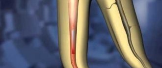
Untimely treatment of caries in children leads to the development of pulpitis - inflammation of tooth tissue, including blood vessels and nerve bundles. The lower molars are most often affected, but the disease occurs even on the anterior elements. Therefore, pulpitis in children occurs even more often than in adults. This is due to a number of factors:
- thin weak enamel;
- large volume of pulp;
- wide root canals;
- less mineralization;
- weak immunity.
In addition to caries, pulpitis can be caused by trauma, including poor treatment.
Symptoms
Inflammation occurs in acute and chronic forms. Acute occurs less frequently, but has more severe symptoms and occurs in two stages:
- serous. The inflamed pulp is filled with serous fluid. There are complaints of severe pain in the tooth, intensifying at night and when chewing. The pain, as a rule, is intermittent, appears once and subsides. Most often, the process occurs in teeth with unformed or absorbable roots. After 5–6 hours, a transition to another stage occurs;
- purulent. Pus forms in the tissues, which begins to melt cells and tissues. The severity of the condition is influenced by immunity, dental condition, and bacterial activity. If the pus has an outlet in the form of a carious cavity or fistula, then the pain is mild. In the absence of outflow, the pain syndrome is bright and long-lasting, intensifying when chewing or eating cold or hot food. The general condition may worsen and fever may appear.
Chronic pulpitis occurs after acute pulpitis or on its own and is characterized by vague symptoms. Pain can only occur under unfavorable factors - cold or hot, too sweet food. The child begins to chew on one side, an unpleasant odor appears, and a bursting and pressing sensation occurs.
The danger of this form is that it is almost asymptomatic. Because of this, inflammation affects more and more tissues. Chronic pulpitis is of three types:
- fibrous. The lightest and most common form. Fibrous tissue grows, the pulp may bleed, and episodic pain appears from mechanical and thermal contact;
- hypertrophic. Develops with severe destruction of the tooth crown. Pathological growth of pulp tissue occurs, which fills the carious cavity;
- gangrenous. The most severe form, accompanied by pulp necrosis. The tooth becomes gray and bad breath appears due to tissue decomposition.
How are baby teeth different from molars?
Baby teeth appear at the age of six months. First, the central incisors erupt on the lower jaw, then on the upper jaw. By about 2.5 years old, the baby already has a full, white-toothed smile. His mouth contains 20 milk teeth, which will serve the child for several years.
After about six years, temporary teeth begin to be replaced by permanent teeth. When the baby brings the first “trophy” to his parents, it causes sincere joy and tenderness. And also the desire to carefully examine what the baby is holding in his fist. And while studying the tooth, parents can draw several wrong conclusions.
pixabay.co/
The first is that baby teeth have absolutely no roots. This impression is created because the root shoot of a fallen tooth is usually located on only one side, and sometimes it does not exist at all. In fact, primary incisors and molars, of course, have roots, but over time they become smaller.
This occurs in order to ensure a natural and painless process of changing the dentition. When the time comes to change the bite, the roots of the baby teeth gradually dissolve. And thanks to this, the indigenous ones “push out” their predecessors from the gums, taking their place.
The second incorrect conclusion may be related to the size of the tooth. It seems incredible that such a “baby” could have the same structure as an “adult” native.
“The structure of baby teeth is almost the same as permanent teeth,” comments Natalya Terentyeva. “They have roots, root and coronal pulp, root canals through which nerve fibers pass. Milk teeth differ from molars in that their enamel is weakly mineralized, which means it is more susceptible to destruction and the development of carious processes.”
How to recognize the problem and is emergency treatment required?
Due to the reduced sensitivity of the pulp, pain during inflammation may be mild and not cause severe discomfort. In this case, the process of tissue destruction occurs rapidly. It is important to visit the dentist regularly and not ignore your child’s complaints of pain when chewing or when eating cold and hot foods.
Treatment for pulpitis should be immediate. The disease is dangerous due to complications - periodontitis or periostitis. It can also spread to a permanent tooth that has not yet erupted and destroy it.
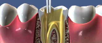
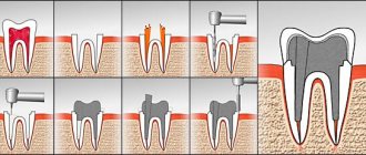
Anatomy of a primary tooth in a child
We are talking about a full-fledged bone formation, the nutrition of which is carried out through numerous microtubules - due to this relationship, hard tissues are strengthened. The structure looks like this:
- crown - the visible part covered with enamel,
- dentine,
- pulp chamber - pulp or neurovascular bundle,
- root system.
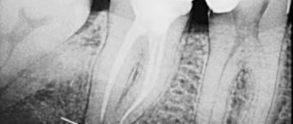
The photo shows the structure of the tooth
. The enamel covering temporary incisors and molars is much more fragile and thinner than in adults. Next comes the dentinal layer with a network of microtubules leading to the pulp - it is this that provides nutrition to all surrounding tissues. It is permeated with blood vessels and nerve endings, and its inflammation leads to severe piercing pain. It should also be recalled that children grow only 20 temporary elements: 8 incisors, 4 canines, 8 molars.
Treatment methods
The treatment of primary teeth in general does not differ from the treatment of permanent elements. If the child’s condition is serious, there is a threat of infection spreading throughout the body, amputation is performed. In most cases, the tooth is preserved to prevent malocclusion. Therapy is carried out in different ways:
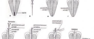
- traditional. Involves treatment in three visits. During the first one, the nerve is opened, a devitalizing paste with arsenic is applied for 24-48 hours or without it for a period of up to 7 days. On the second visit, a pulp mummification mixture based on resorcinol-formalin is placed into the canals. At the last visit, a permanent filling is installed;
- modern. It takes place in 1 – 2 visits. If the child can sit quietly for a long time at the doctor and the roots of the tooth are formed, extraction is performed. The nerve is removed either on the first visit or after applying the paste. Next, the canals are carefully processed, infected tissue is removed, an anti-inflammatory paste (for example, zinc eugenol) is applied and closed with a filling. The composition will gradually dissolve along with the roots when changing teeth;
- partial vital amputation. The doctor removes the upper portion of the nerve and applies an antiseptic and anti-inflammatory medication that seals the remaining living pulp.
When treating teeth with immature roots, a different approach is chosen. This is due to several reasons:
- the apex of the roots has not yet closed and there is a risk of infection of the permanent tooth germ;
- the roots are short and the channels are wide;
- trauma to the upper zone of the root can lead to disturbances in its formation;
- It is impossible to remove the entire pulp and perform a complete canal treatment.
Most often, they choose amputation of the pulp by any method or biological treatment, when the tooth is cleaned of the affected tissue and a paste with calcium hydroxide is applied for several days. After this, a filling is installed.
Methods for depulpation of the dental nerve
The method of removing a nerve in a baby tooth is determined by the stage of pulpitis and the degree of tissue damage. Treatment may involve partial or complete depulpation. The decision on the method of performing the procedure is made by the attending physician.
At the Martinka Children's Dental Clinic, dental nerve removal for children is performed using two methods.
- Chemical. The dentist puts a drug into the tooth cavity that “kills” the nerve. The drug does not affect healthy tissue. While the paste is in effect, a temporary filling is placed on the tooth. After 1–5 days, the filling is removed, and the pulp and nerve are removed. The method is only available for complete depulpation.
- Instrumental. The child is given anesthesia to relieve pain. The tooth is opened and the pulp is removed completely or partially using special instruments. At the end of the procedure, a medicinal paste with anti-inflammatory and antiseptic action is placed into the tooth cavity. A temporary filling is installed. After a few days, the drug is removed and a permanent filling is installed instead of a temporary filling.
If the inflammatory process has started, then upon completion of treatment, the dentist may prescribe a course of antibacterial drugs. Taking painkillers is permissible only as prescribed by a doctor.
Is it possible not to treat pulpitis if the teeth are baby?
There is a common opinion that it is not necessary to treat baby teeth, because they will soon fall out. This is the biggest mistake parents make. Untreated pulpitis leads to irreparable consequences and even affects permanent teeth:
- purulent abscess, periodontitis, life-threatening condition;
- deep infection affecting the germ of a permanent tooth;
- malocclusion due to early tooth extraction due to untimely treatment;
- pain interferes with normal chewing of food, uneven load on the teeth leads to accelerated destruction of other elements.
Features and stages of the procedure
The dentist decides to remove the nerve of a baby tooth.
The procedure begins with a routine examination. It will take the doctor less than half a minute to decide whether to remove the nerve or not. To do this, a temporary onlay is placed on the tooth. Then the removal procedure itself is carried out. Local anesthesia is performed with a solution of ice-caine. The main task of the dentist is to open the tooth enamel using a drill and clean the dental canals so as not to damage the inner row of teeth.
Pulp removal ends with thorough disinfection. After this, the tooth is filled. In difficult situations, a follow-up appointment is scheduled to take an X-ray of the tooth. Sometimes the filling is placed temporarily. A special medicinal composition in the form of a paste is placed under it. And after the time of action of the medicine has expired, the filling is replaced with a permanent one.
Fillings used in dentistry can be of varying quality. There are no uniform standards for doctors. Therefore, parents simply do not know what they are using to glue their children’s mouths shut. But specialized private medical institutions should talk about what materials they work with. Children's fillings, as a rule, have good aseptic characteristics, but their durability is questionable. Dental cement will last about a year and a half.
After the procedure, the tooth is considered dead. It does not react to thermal and acid-base influences. But now it is collapsing at twice the speed. Enamel tubules instead of pulp cannot provide adequate nutrition for the entire tooth.
Prevention
The main method of preventing dental and oral diseases is regular visits to the dentist and treatment of caries in the early stages. It is important to teach a child hygiene and the rules of brushing teeth from the first years of life. Parents should control the diet, which should contain all the necessary substances, limit sweets and allow only water to drink at night.
Doctors at the VIMONTALE clinic treat pulpitis of primary teeth in children of any age. Specialists not only master modern painless techniques, but also know how to find contact with a child, know how to reduce anxiety and win the favor of the youngest patients.
Expert of the article you are reading:
Lozinskaya Alla Nikolaevna
Pediatric dentist, general dentist.
You may also be interested in:
Children's orthodontist Dental treatment for children Correction of bite in children Children's orthodontist Peculiarities of treatment of childhood caries Removal of baby teeth Prevention of childhood caries Silvering of teeth in children
Show more
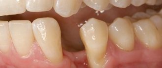
Indications for removal
Indications for the removal of a baby tooth are only two situations: inflammation of the pulp with subsequent death of the nerve or depressurization of dentin.
But the reasons for this can be a variety of situations, including injuries:
- Caries
- Pulpitis
- Periodontitis
- Traumatic brain injuries, strokes
Caries on children's enamel develops in a matter of days. If harmful microorganisms manage to get into the pulp, the baby tooth will turn black after a couple of weeks. And at the site of infection, a hole is formed through which infection of the entire body, abscess of the gums and bone tissue can occur.
Pulpitis is a consequence of advanced caries. The soft tissues of the pulp chamber cease to perform their functions. The tooth is attacked by microbes from the inside. They gradually destroy blood vessels and nerve endings in the pulp, turning them into a source of infection.
Periodontitis poses the greatest danger, since the infection penetrates into the area of the neck of the tooth, which is adjacent to the gums and roots. In advanced situations, this can cause complications when the lower teeth erupt. For example, excessively compacted tissue can cause the growth of a molar to grow abnormally.
If there are any injuries or chips, the child should be seen by a doctor. Every meal you eat will promote the growth of bacteria. In such cases, the teeth are either removed completely or the broken part is built up.
Our patients
Recommendations from patient Ekaterina
Sokhov Valery Borisovich
Dentist surgeon, general dentist, implantologist, dental therapist, orthopedic dentist
Make an appointment 8 (499) 520-98-70
Make an appointment
Patient recommendations Semenov Yu.
Sokhov Valery Borisovich
Dentist surgeon, general dentist, implantologist, dental therapist, orthopedic dentist
Make an appointment 8 (499) 520-98-70
Make an appointment
Recommendations from patient Alekseeva O.V.
Akhmedkhanov Said Rashidovich
Dental surgeon, general dentist, implantologist, orthopedic dentist, dental therapist.
Make an appointment 8 (499) 520-98-70
Make an appointment
Recommendations from patient Zolotareva S.V.
Akhmedkhanov Said Rashidovich
Dental surgeon, general dentist, implantologist, orthopedic dentist, dental therapist.
Make an appointment 8 (499) 520-98-70
Make an appointment
Recommendations from patient Vera
Akhmedkhanov Said Rashidovich
Dental surgeon, general dentist, implantologist, orthopedic dentist, dental therapist.
Make an appointment 8 (499) 520-98-70
Make an appointment
Recommendations from patient Mikhail Ivanovich
Akhmedkhanov Said Rashidovich
Dental surgeon, general dentist, implantologist, orthopedic dentist, dental therapist.
Make an appointment 8 (499) 520-98-70
Make an appointment
Collapse
Tooth preservation
In our clinic, doctors follow the most important principle when treating small patients: do everything possible to keep the pulp alive! Only thanks to it will the tooth be completely formed, its roots will become stronger, and the enamel will increase to the required thickness.
Therefore, pediatric dentists at the Dentville clinic use materials that are biologically compatible with the child’s body - constant monitoring of pulp regeneration processes allows it to be restored even in cases of deep nerve damage by caries.
Features of the root system of children's teeth
The structure of all dental canals includes coronal, middle and apical (apical) parts. The first, represented directly adjacent to the mouths, is the largest and widest. The middle part connects the coronal region with the apical zone of the root. The apical region is located below and often has a number of anatomical features: pathological or physiological resorption, fusion of several canals, lateral placement or open structure of the hole, strong narrowing or bending, ramification (branching), etc. The narrowest part of the root is located at the dentinocemental border. At a distance of 0.5-1 mm from the radiological apex there is a physiological opening. At an early age, root canals do not have a perfectly round cross-section. Only with age, due to the deposition of replacement dentin, its internal space takes on the shape of a circle.
Preventive measures
Obviously, it is better to prevent a dental problem than to subject the child to treatment and, accordingly, stress. But for this it is necessary to take care of prevention, and for this purpose, parents should take note of the following recommendations:
- The sooner you start taking care of the condition of your child’s teeth and oral cavity, the better. Even during pregnancy, the expectant mother should eat properly and also enrich her diet with foods high in calcium and fluoride. At the same time, you should start carrying out daily hygiene procedures even before the first teeth appear - you can clean your gums from bacterial plaque using special napkins or fingertips,
The photo shows oral hygiene in a baby using a special napkin. - As soon as the child reaches the age when he can brush his teeth on his own, he needs to be taught daily hygiene. At the same time, he must learn how to clean correctly, and a pediatric dentist will help you with this,
The future smile depends on the health of baby teeth - You should limit your child’s consumption of sweets, including sweet mixtures for little ones. Excess sweets in the diet is one of the most common causes of dental caries in childhood.
- Regular preventive examinations at the dentist’s office will allow you to detect the problem in time, and therefore quickly deal with it. At the same time, it is better to show the baby to a specialist as soon as his first teeth appear.
Parents need to understand that treatment of baby teeth is an extremely important condition for the health of permanent teeth. Therefore, if caries or any other dental diseases develop, the child should be immediately shown to a specialist. And in order to prevent problems from arising, it is necessary to take your child to the dentist for preventive examinations 3-4 times a year.
Instead of empty spaces - tabs
The Dentville Clinic pays close attention to young patients! If our doctors are faced with the impossibility of saving a tooth, then we offer unique solutions that will help avoid bite problems that may arise due to voids:
- microprosthetics;
- lightweight crowns suitable for children's teeth;
- installation of lightweight tabs.
In the future, microprostheses, inlays and crowns can be replaced with ceramic structures, for example, crowns - and then the “adult” dentists of the Dentville clinic will be waiting for you!
What materials are used
Many parents are interested in what is currently used to fill children’s teeth. Not so long ago, the choice of materials used to fill children's teeth was not as diverse as it is today. At one time, dentists more often resorted to using amalgam, but this composition cannot be called completely safe and harmless to the body, especially when it comes to a child. Later they began to use glass ionomer cement, but it also has its disadvantages. For example, it takes quite a long time for the material to harden, which is not always convenient if there is a baby in the dental chair.
On a note! Often, special pastes are used to fill the root canals of baby teeth. Such compositions are not able to provide perfect tightness if used to fill a vertical cavity. In addition, after some time they dissolve, and therefore are not intended for permanent filling.
This is how a child’s teeth are filled with reflective composites.
To date, the situation has changed a little, and today pediatric dentists increasingly prefer fillings made of light-curing composite, as well as compositions that are a combination of a hybrid composite and glass ionomer cement. These materials are characterized by increased strength, are easy to polish and have a high degree of adhesion.
Is it possible to restore a dead nerve?
If the nerve does die, our dentists can help restore it. We have a unique revascularization technique that helps “bring back to life” pulp tissue. In this case, the doctor carries out the work, dividing it into several stages:
- The dental canals are sterilized and cleaned.
- Filling the channels with medicine and the patient's blood cells.
- Sealing the canal.
- Over the course of several months, a special tissue appears that replaces the functionality of the nerve and allows the tooth to develop and grow, and the roots and enamel to become stronger.
During the treatment process, doctors will monitor the process of pulp restoration using computed tomography.