Author of the article:
Soldatova Lyudmila Nikolaevna
Candidate of Medical Sciences, Professor of the Department of Clinical Dentistry of the St. Petersburg Medical and Social Institute, Chief Physician of the Alfa-Dent Dental Clinic, St. Petersburg
Endodontic treatment, affecting the internal parts of the tooth and, first of all, the roots, is one of the most important sections of modern dentistry. The effectiveness of all treatment procedures for such serious and dangerous diseases as pulpitis, periodontitis, purulent abscess, etc. often depends on the quality of tooth root canal treatment.
Being a very important part of the tooth, the roots provide its fixation in the jaw, nutrition and blood supply. However, the root can also easily cause pain; Many diseases associated with untimely or incorrect root canal treatment lead to severe consequences, including tooth loss. Infection can penetrate into the root canal either as a result of a pathological process (for example, caries) or as a result of errors made when performing certain dental procedures. There are cases when pathogenic microflora penetrates inside the canal as a result of a tooth injury that violates the integrity of its structure, the outer shell.
Root canal filling takes a central place in the treatment of pulpitis, periodontitis and similar diseases. Effective root canal treatment is impossible without solving this problem. If the canal is sealed in violation of the technology, or not tightly enough, then a focus of infection and inflammation is likely to form in its cavities. Sometimes pain occurs after root canal filling, caused by the same infection, mechanical, thermal or chemical damage to the tooth tissue during treatment. To avoid these and other adverse consequences, the dentist must follow technology at every stage of root canal filling, starting with the preparatory stage.
Preparation for filling
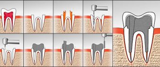
Before filling the canals with filling material, the doctor must thoroughly clean them and prepare the cavity in a special way. This process takes place in several stages:
- Removal of affected tissue
Most often, the need for root canal treatment arises as a consequence of another pathological process, in particular caries. To destroy the source of infection and open access to the mouths of the canals, the dentist removes dead and diseased tissue using a bur. - removal
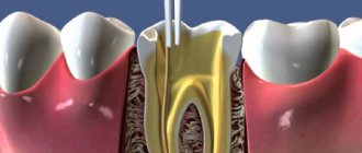
Pulp is the sensitive tissue of the tooth, a plexus of blood vessels and nerves located in the coronal part and inside the roots. The method of filling root canals involves removing the pulp using a special tool. Most often, this procedure, like the previous one, is performed under local anesthesia to eliminate pain and discomfort when removing the pulp. - Measuring canals
In order to perform a quality filling, the doctor needs to obtain as accurate information as possible about the properties and current condition of the canals. For this purpose, before treatment, as a rule, a targeted radiograph is performed. An important procedure is also measuring the length of the channels. The length is individual and depends not only on the size of the root, but also on the degree of curvature of the canal. - Mechanical processing
Before filling, the channels are cleaned and expanded using special tools. This is necessary to completely remove the affected tissue, as well as to more densely and evenly fill the canal cavity with filling material. Mechanical processing is performed using special thin tools - files. With the help of a file, the canal is passed completely, from the mouth to the apex.
Only after completing all the preparatory steps can the doctor proceed to filling.
Let's look at the root: how are the root canals of teeth filled?
The more a person is afraid of dentists, the more closely he eventually has to interact with them and the longer the dental treatment will be for him - a simple and banal truth, but, unfortunately, not all people take note of it. If caries treatment is not carried out in a timely manner, the infection gradually spreads “inward”, capturing more and more new territories and reaching the pulp chamber. Pulpitis develops - an extremely painful and unpleasant disease, which is popularly called “inflammation of the dental nerve.” This is where most people don’t just go to the dentist, but run – because the pain prevents them from sleeping, eating, or working normally. And if the patient has already begun to have inflammation of the tooth canals, treatment will require quite serious and lengthy treatment: cleaning the tooth canals from infected and dead tissue, their special antibacterial treatment, careful filling of both the canals themselves and the dental crown with the cavity.
Almost everyone knows about such a dental procedure as treatment and filling of dental canals, and many people have encountered it from personal experience. But most people usually don’t know the intricacies of the process, how exactly such treatment takes place, why it is needed and what it gives in the end. So what are the standards for filling canals, why and how exactly is this procedure carried out, and what can a poorly filled canal threaten a patient with? Dentist-therapist at the 32 Dent clinic Larisa Aleksandrovna Kravtsova will help you get reliable answers to all these questions.
What determines the quality of filling?
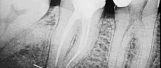
The technique of filling root canals requires considerable skill from the dentist. Errors that negatively affect the effectiveness of treatment can occur at any stage. Inaccurate measurement of the length of the canals can lead to under- or over-filling of the canals, which almost always results in the development of inflammation of the tooth and surrounding tissues. If the length of the channels is determined incorrectly (not to the full length), they are also processed. The consequences of errors in measuring length include pain, neuralgia, swelling of the gum tissue, the development of periodontitis and other inflammatory diseases, and tooth extraction. Only in some cases can the canal be resealed without serious consequences. A control radiograph, which is required to be performed after filling, usually helps to detect an error in a timely manner.
In order to avoid errors when measuring length, clinics today use a special device - an apex locator. The device is connected to a file that is placed into the root canal. Using an apex locator allows you to record the moment the file reaches the root apex. In some cases, a radiograph with files installed inside the canals is used for more accurate control.
Doctors also make a large number of mistakes at the mechanical processing stage. Modern methods of filling root canals necessarily require preliminary expansion of the canals. The untreated channel, as a rule, has a too small and, moreover, variable diameter; inside the canals there are usually many narrowings and expansions that prevent high-quality filling. To eliminate them, mechanical treatment is used.
Cleaning and widening the canal can be done either with simple hand instruments or with a special endodontic handpiece. In the first case, the doctor literally performs all operations with his hands - independently determining the depth of immersion of the file, the force of pressure and the speed of rotation. You have to rotate the tool with your fingertips. Naturally, such a procedure does not always allow high-quality cleaning of the entire surface of narrow canals and sometimes leads to the tip of the instrument breaking off, which can cause poor-quality filling or pain after filling the root canal.
Mechanical processing using an endodontic handpiece is much more reliable and precise. The tip is connected to a microdrive, which ensures its rotation and also automatically controls the load and rotation speed to prevent breakage. The use of such technological tools makes it possible to achieve the highest quality of treatment and significantly reduce the risk of complications, but does not guarantee 100% effectiveness of treatment.
When is tooth canal cleaning performed?
It is necessary to treat and fill root canals in the following cases:
- If caries has progressed to pulpitis (infection has spread to the dental pulp, or colloquially “dental nerve”). The pulp will have to be removed (the tooth depulped), and the root canals in which it was located will have to be thoroughly cleaned of tissue debris and sealed.
- If it is necessary to prepare a dental unit for the installation of a crown and the doctor believes that depulpation is necessary. Previously, when installing single crowns or supporting crowns of a bridge, the teeth were necessarily subject to depulpation; now this procedure is not performed in all situations and only when installing certain types of dentures.
- If there is a need for re-treatment of the tooth after the occurrence of a secondary source of infection or with the development of periodontitis, inflammation of the tissues around the tooth root (develops with poor-quality canal filling).
In all these situations, endodontic treatment is carried out (the name comes from the Latin words “inside” and “tooth”, that is, it can be literally translated as intradental treatment) with mandatory filling of the dental canals.
If you have a problem similar to that described in this article, be sure to contact our specialists. Don't diagnose yourself!
Why you should call us now:
- We will answer all your questions in 3 minutes
- Free consultation
- The average work experience of doctors is 12 years
- Convenient location of clinics
Methods for filling root canals
There are several basic techniques used in the treatment of pulpitis and periodontitis:
- One-paste method
The channel is filled with a plastic, subsequently hardening material. The method is outdated and gives a large number of complications. - Single pin method
First, the root canal is filled with a special paste, and then a gutta-percha pin is inserted into it. The percentage of complications is lower, but this method is also gradually being phased out. - Filling with “thermophile”
A more modern method, also based on the use of an initially plastic material - heated gutta-percha. The fluid state allows the material to densely fill all cavities inside the canal, which significantly reduces the risk of complications. The disadvantages of the method include high requirements for the doctor’s qualifications and relatively high cost. - Gutta-percha condensation method
This method ensures even more dense filling of the canal lumen with filling material. Currently, the method is one of the most common. The procedure is performed in several stages.
Root canal filling using lateral condensation of gutta-percha
- Selection of the main pin
The selection of the appropriate pin depends on the diameter of the canal after machining and expansion. - Filling the canal
Before installing the pin, the canal is filled with a special paste - sealer. It provides the necessary seal. - Compaction of the main pin
To compact the filling and free up space for new pins, a special tool is inserted into the canal cavity - a spreader. The reciprocating movements of the spreader push the pin toward the canal wall. - Insertion of additional pins
Depending on the diameter of the canal, at this stage 8 to 12 additional pins of small diameter are inserted and sealed. - X-ray examination
The filling density of the canals is checked using x-rays. If the canal is not sealed to the very top or, on the contrary, the filling material extends beyond the root, the pins are removed and the process is repeated again. - Removing excess sealer and gutta-percha
After the desired result has been achieved, remove excess parts of the filling materials protruding from the mouths of the root canals. For this, a special hot tool is used. - Installation of a temporary filling
The final stage of the procedure is the installation of a temporary filling. During the next visit, the doctor re-checks the quality of the filling and restores the crown of the tooth using a permanent filling. Simultaneous filling of canals and crowns is unacceptable.
Complications after root canal filling
If a filled tooth hurts after visiting the dentist, there is a high probability of complications. Filling is most often used in the treatment of caries and pulpitis, poor treatment of which can stimulate the progression of the disease and the occurrence of pain.
If after filling there is pain in the problem tooth, treatment will be carried out depending on the cause of the complication. One of the reasons for the complication is insufficient treatment of the tooth before filling. Treatment for such a complication consists of removing the filling, cleaning the problem areas and re-filling.
After treatment of caries, if the doctor is insufficiently qualified, the following unpleasant consequences are possible:
- enamel damage;
- pulp burn during tooth preparation;
- acid contacting dentin.
After canal filling, the following complications may occur:
- Perforation of the root wall. Symptoms of perforation upon examination include instrument failure, pain, and bleeding. Treatment in this case begins with studying the situation in the root lumen on an X-ray image. Then the perforation is closed with cement or other filling material.
- Insufficient antiseptic treatment can cause infection. Needs to be re-treated.
- The development of infection in the tooth as a result of incomplete filling of the canal with filling material. Treatment is repeated cleaning and refilling.
- Extension of filling material beyond the root apex. Depending on the intensity of pain and sensitivity of the patient, there are two options: at the stage of painful sensations, take painkillers or re-treat the tooth.
- Reaction to temperature changes in a canal missed during filling.
- Pain due to a broken instrument in the canal. The fragment is easily detected on x-ray. This tooth often needs to be removed.
- The appearance of a cyst or granuloma after improper canal treatment.
- Periodic inflammation of the gums, formation of gumboil or fistula due to dental canal treatment performed incorrectly.
- Change in tooth color due to improper treatment.
- Due to a poorly treated canal and an inflamed abutment tooth, replacement of a high-quality denture is often required.
- Loss of a filling due to an inaccurate procedure. Refilling required.
- An allergic reaction to filling materials can occur due to the characteristics of the body.
Interesting fact
The widespread use of implants in modern dentistry became possible thanks to Professor Ingvar Brånemark from Sweden, who in 1965 discovered osseointegration - the process of healing and fusion of bone tissue with a titanium implant. The bioinertness of titanium practically eliminated its rejection by the body.
The first “dentists” were the Etruscans. They carved artificial teeth from the teeth of various mammals as early as the 7th century BC, and also knew how to make bridges strong enough for chewing.
How to check the quality of canal filling?
Quality control of fillings is an integral part of the treatment process and is performed at every stage. X-ray control is especially important after filling. It allows you to identify areas of insufficiently dense obturation, detect pins or sealer residues protruding beyond the root apex, identify fragments of files and other defects. All this can lead to severe pain after treatment, as well as to the development of complications.
The x-ray image should clearly show the canal cavities, densely filled with filling material. There should be no enlightenment. The filling material should reach the very top of the canal.
Following simple rules of prevention also helps to avoid complications. Pain after treatment can last from several days to a month and does not always indicate ineffectiveness or low quality of treatment. However, if they appear, you should consult your dentist. Also, after treatment, it is recommended to abstain for some time from physical activity, drinking alcohol, and eating hot and spicy foods.
To prevent pulpitis, carefully monitor your oral hygiene. Use only professional products. If during the day you do not have the opportunity to brush your teeth after eating, use Asepta Fresh mouthwash. It disinfects the oral cavity, effectively fights caries, and normalizes acidity.
Clinical researches
As a result of clinical experiments using the Asepta series of products, conducted at the Kazan State Medical Academy, the complex use of anti-inflammatory drugs from the Asepta line contributed to faster relief of inflammation, the combined use of balm, gel, rinse, toothpaste and vitamin-mineral complex mutually enhanced the therapeutic effect did not require daily visits to the dentist.
Sources:
- The use of drugs from the Asepta line in the complex treatment of inflammatory periodontal diseases (N.V. Berezina E.N. Silantyeva S.M. Krivonos, Kazan State Medical Academy. Kazan.) N.V. BEREZINA, E.N. SILANTIEVA, S.M. KRIVONOS Kazan State Medical Academy
- The role of anti-inflammatory rinse in the treatment of periodontal diseases (L.Yu. Orekhova, A.A. Leontyev, S.B. Ulitovsky) L.Yu. OREKHOVA, Doctor of Medical Sciences, Prof., Head of Department; A.A. LEONTIEV, dentist; S.B. ULITOVSKY, Doctor of Medical Sciences, Prof. Department of Therapeutic Dentistry of St. Petersburg State Medical University named after. acad. I. P. Pavlova
- Report on determining/confirming the preventive properties of commercially produced personal oral hygiene products: Asepta toothpaste used in combination with Asepta mouthwash and Asepta gum balm Head. Department of PFS Doctor of Medical Sciences Professor S.B. Ulitovsky St. Petersburg State Medical University named after Academician I.P. Pavlova. Faculty of Dentistry. Department of Preventive Dentistry.