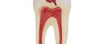
Such a chronic form of pulpitis as hypertrophic pulpitis is rare. It develops when timely treatment of fibrous pulpitis has not been carried out and is accompanied by inflammation of the pulp and its proliferation. Chronic hypertrophic pulpitis does not lead to severe pain, which significantly complicates its identification and timely provision of professional dental care. This is very bad, since the pathology can lead to the development of flux or sepsis. Abnormally overgrown pulp tissue protrudes outward and can become easily infected.
Clinical manifestations:
- Mildly manifested pain symptoms arising from exposure to irritants of various natures;
- Pulp bleeding;
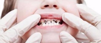
- An almost completely destroyed dental crown and a deep carious cavity from which the pulp protrudes;
- Halitosis, which occurs due to the inability to carry out full hygienic procedures for the oral cavity.
In our clinic you can get a free dental consultation!
CAUSES AND SYMPTOMS
In most cases, the precursor to this form is acute inflammation of the “nerve” of the tooth, which is accompanied by severe pain. This happens after opening the canal in patients with high body resistance, when pathways appear for the normal outflow of inflammatory exudate.
In young children and patients with low body reactivity, this disease may initially become chronic, without clinical manifestations of the acute stage.
TYPICAL SYMPTOMS:
- aching pain, worse in the evening and at night;
- sharp prolonged reaction to a temperature stimulus;
- feeling of fullness and heaviness in the tooth;
- acute pain when probing the canal.
ACCURATE DIAGNOSTICS
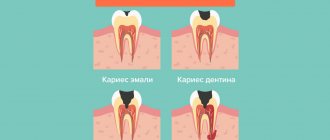
The most important thing is to clearly differentiate fibrous pulpitis from other diseases. In particular, it is distinguished from deep caries by a prolonged pain reaction to external irritation, as well as the absence of spontaneous acute pain radiating to the temple or jaw.
The gangrenous form is characterized by a grayish color of the enamel, a putrid odor, and pain when probing the mouth of the canals. With pathological hypertrophy of the soft tissues of the tooth (another disease in the classification of pulpitis), the patient complains of prolonged pain not associated with irritating factors, and during the examination the doctor sees an overgrown bleeding pulp.
If you use the services of our dentistry, we will perform an accurate differential diagnosis using special research methods. After this, we will select the optimal method of therapy.
Our doctors
18 years of experience
Baghdasaryan
Armen Evgenievich
Chief physician, dentist-orthopedist-therapist
Graduated from VSMA named after. N.N. Burdenko. Internship on the basis of MGMSU named after. A.E. Evdokimov in “General Dentistry”.
Clinical residency at the Moscow State Medical University named after. A.E. Evdokimov in “Orthopedics”.
More about the doctor...
5 years experience
Sadina
Ekaterina Vladislavovna
Dental therapist, surgeon
Penza State University Medical Institute, specialty “Dentistry”.
In 2021, she underwent professional retraining in the specialty “Therapeutic Dentistry” at the Moscow State Medical and Dental University named after A.I. Evdokimov.
More about the doctor...
8 years of experience
Arzumanov
Andranik Arkadievich
Dentist-orthodontist
Graduated from Moscow State Medical University. Internship - Moscow State Medical University at the Department of Orthodontics and Children's Prosthetics.
Residency at Moscow State Medical University at the Department of Orthodontics and Children's Prosthetics. Member of the Professional Society of Orthodontists of Russia since 2010.
More about the doctor...
Etiology
The causes of fibrous pulpitis can be different. As already mentioned, it can be the outcome of an acute form of inflammatory processes, in which the crown of the tooth is opened and exudate flows out, or it can occur primarily. The causes of pathological processes are:
- medical errors during caries treatment;
- advanced caries;
- dental injuries.
In our clinic you can get a free dental consultation!
Diagnostics
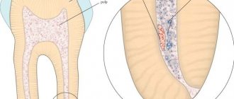
Diagnosis of hypertrophic pulpitis, first of all, involves examination by a dentist. He collects anamnesis and analyzes the nature of pain, asking the patient about them. As a result, it is possible to establish that some time ago the patient experienced intense pain symptoms, which over time almost completely disappeared. During the examination, the dentist reveals a deep carious cavity that contains bleeding granulations, the probing of which is not painful. When probing the pulp, intense pain occurs. When studying a polyp, it can be determined that it originates in the pulp chamber. If pulpitis is at the initial stage of development, the sprouted tissues have a bright red color, while in advanced ones it is light pink. Diagnostics also includes:
- thermal test (the result of which is negative);
- radiography (the image of which clearly shows the absence of a septum between the pulp and the carious cavity);
- electroodontic diagnostics (which shows reduced excitability of the pulp).
Chronic pulpitis
The age category of people suffering from this disease cannot be precisely identified. It is observed in both young and mature patients. In children, chronic pulpitis of the primary form can also be found when the roots are not formed. The chronic stage of pulpitis is much more common than the acute stage, accounting for approximately 75% of 100.
Classification of chronic pulpitis
In dentistry, there are three main forms of this disease. This is a fibrous, gangrenous and hypertrophic variety. Special attention is paid to exacerbation of chronic pulpitis.
Chronic fibrous pulpitis
The most common type is fibrous, accounting for about 69% of the total number identified. In this case, the pulp turns into a fairly dense scar formation of a grayish color. The change occurs due to the active proliferation of coarse fibrous connective tissue. Foci of petrification and hyalinosis are also observed. If left untreated, gangrene, cellulitis, or microabscesses may occur.
Chronic hypertrophic pulpitis
The hypertrophic type of chronic pulpitis is observed extremely rarely (0.5 out of 100%). It is characterized by division into granulating and polypous groups. In the first case, dentin is replaced with osteodentin. Granulation tissue is also formed, which affects the carious cavity. In the second case, a polyp is formed; it has a mushroom-like appearance with an ulcerative surface.
Chronic gangrenous pulpitis
Only 2 out of 100 patients suffer from this disease. It represents an ulcerative lesion or necrosis of the pulp. When dissecting the tooth cavity, dark tissue detritus can be found. In many cases it is possible to save the healthy part of the pulp.
Exacerbation of chronic pulpitis threatens gangrene, so it is important to take all possible measures to prevent it.
Prevention and prognosis of chronic pulpitis
Only timely and competent treatment can guarantee a positive prognosis, namely the preservation of the “native” tooth and the elimination of inflammation. If you start the disease, it will not be easy to avoid complications. The most dangerous are:
- purulent periostitis;
- periodontitis;
- osteomyelitis.
It should be borne in mind that each of the listed diseases has its own complications that may arise in the future if the patient does not act. To prevent them, at the first symptoms, it is recommended to immediately visit a dental clinic.
Prevention of chronic pulpitis involves maintaining daily oral hygiene. Regular “control” examinations will help avoid unpleasant consequences.
Slowly developing chronic pulpitis is acute pain, the risk of flux (periostitis), progressive inflammation, the formation of cysts, phlegmon and fistulous tract. Any painful sensation that comes from the teeth or gums should be diagnosed immediately. This will allow you to maintain a healthy smile, as well as avoid the need for long-term and painful treatment for complications of chronic pulpitis.
Treatment
Treatment of hypertrophic pulpitis is carried out only by partial or complete removal of the pulp, i.e. using surgical techniques. Their choice is made individually, depending on the degree of soft tissue damage.
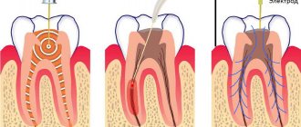
Vital extirpation
This technique involves partial removal of the pulp from the orifice and coronal part. The procedure is performed under local anesthesia and does not cause pain. Partial removal of the pulp allows you to preserve its functionality, and therefore the tooth itself remains alive. An important role is played by how tightly and correctly the medical pad was applied after removing the pulp. Thanks to it, you can eliminate the risk of pulp infection during the filling process and after it.
Devital extirpation
This technique is aimed at completely removing the pulp from the crown, mouth and root of the tooth. Removal is carried out in two stages using devital paste, which the dentist applies to the pulp during the first visit. During the second visit, dead pulp is removed, dental canals are filled and the crown of the tooth is restored.
Forms of the disease
This pathological process develops in two directions:
- Formation of granulations. Its essence lies in the growth of granulation tissue from the pulp chamber into the carious defect. This is a compensatory reaction aimed at filling the existing void.
- Polyp of the neurovascular bundle. It occurs in the later stages of the disease. This type is characterized by an adhesive process. Gingival epithelial cells grow into granulations that have grown from the dental cavity and firmly merge with it, forming one whole.
Where to contact?
The “Good Hands” dental clinic always means high-quality treatment, attentiveness and helpfulness of the staff, and professionalism of the dentists. By contacting us, you can be sure that you will be treated by experienced specialists who have successfully treated hundreds of patients. They use gentle techniques and have modern equipment and high-quality anesthetics, making the treatment as comfortable as possible for the patient, and the results invariably please him! You can schedule a free consultation with us and find out all the necessary information right now by filling out an application on our website or calling us by phone.
Why does pulpitis appear: a review of the main reasons
The main causes of pulpitis are:
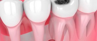
1. Untreated caries. Carious bacteria little by little destroy first the enamel of the tooth, and then its hard tissues, and over time this process reaches the pulp chamber, where it affects the nerve of the tooth.
2. Low quality of dental treatment. If, when treating caries, the doctor does not properly treat the tooth and its canals, a recurrence of pulpitis is possible. It is for this reason that the choice of dentistry should always be treated with maximum attention!
3. Mistakes made by the doctor when preparing the tooth for prosthetics.
Before prosthetics, the teeth that should support the denture are ground down to the thickness of the future crown. If, when grinding teeth with not removed pulp, the doctor overheats the tissue, pulpitis may develop in the future.
4. Some gum diseases, for example, periodontal disease, can lead to the development of pulpitis. This disease causes tooth roots to become exposed and deep pockets to form where the tooth meets the gum. In such pockets, bacteria actively develop, causing caries and, if left untreated, pulpitis will appear over time.
Another cause of tooth pulpitis can be trauma, which leads to the opening of the pulp and infection.
What is periodontitis?
| Periodontitis, like pulpitis, is a complication that accompanies the development of caries. The disease may appear due to injury or improper dental treatment. With periodontitis, the inflammatory process moves to the periodontal area, which is a narrow space of connective tissue between the bone socket and the tooth itself. According to the clinical course, the disease can be acute or chronic. Acute periodontitis is characterized by sharp pain, sensitivity to cold or hot, and swelling of the gums. Flux forms in the area where gum tissue is damaged. The chronic form is often asymptomatic, but this is where the danger lies, since the inflammatory process begins to actively develop deep in the tissues. |
There are the following forms of chronic periodontitis:
- Granulomatous periodontitis;
- Fibrous;
- Granulating.
It is important to note that periodontitis is a dangerous disease for health and cannot be cured on its own. You should definitely contact your dentist in order to diagnose and eliminate it on time.