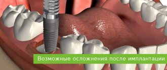
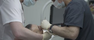
Carrying out any surgical interventions in the area of the facial part of the skull, including dental procedures, is extremely dangerous due to the close location of important anatomical structures. When performing seemingly simple tasks during surgery, damage to atypically located nerves, vessels, and ducts of the salivary glands is possible, which leads to significant complications. Numbness after sinus lift and bone grafting may appear at different stages of the operation and postoperative period; its causes can be very diverse and closely intertwined with each other.
A little about the anatomy and course of the branches of the trigeminal nerve
The teeth of the upper and lower jaw are innervated by the branches of the trigeminal nerve, which provide all types of sensitivity directly to the teeth, skin and mucous membrane of the lips and cheeks. The lower jaw - by the mandibular nerve, the upper - by the alveolar branches of the maxillary nerve, forming the dental plexus.
The alveolar branches of the maxillary nerve pass along the wall of the maxillary sinus to the apexes of the roots of the upper teeth, where they form a nerve plexus responsible for the sensitivity of the upper teeth and gums. The mandibular nerve passes through the bone canal of the same name, innervating the lower teeth and gums, lower lip, and part of the cheek.
Main causes of nerve damage during implant placement
Damage to the maxillary or mandibular processes of the trigeminal nerve, which is the main cause of long-term numbness of the lips, tongue, and chin after dental implantation, can be caused by an unintentional mistake by the dentist or negligence. Damage to the mandibular nerve, which provokes paresthesia, can occur in the following cases:
- Incorrect selection of implants (chosen too long).
- The specialist has not studied the anatomical features of the jaw and the implanted implant touches the nerve ending.
- Injury to the nerve ending by a needle during the administration of anesthesia or dental instruments during bone preparation.
If numbness in the lower part of the face is caused by injury to the nerve endings during the administration of anesthesia, the discomfort will go away on its own after the nerve fibers heal. Typically, the healing period ranges from 3 months to six months. You can understand that numbness of the lip and chin after implantation is associated with damage to the trigeminal nerve by the presence of the following signs after surgery:
- salivation increases;
- difficulty eating;
- facial expressions and articulation are impaired;
- the sensitivity of the tongue and lips decreases;
- there is pain and discomfort in the implant area;
- numbness is felt on the side where the titanium root is implanted.
Chief physician, Salatsky Dmitry Nikolaevich
Sign up for a free consultation
+7
If the feeling of numbness in the lower jaw does not decrease within several hours after implantation, then an urgent consultation with the doctor who performed the operation is necessary. Only after conducting diagnostic studies will a specialist be able to identify the extent of nerve damage and, based on this, prescribe appropriate treatment.
Characteristics of sinus lifting and bone grafting
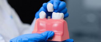
Bone grafting in dentistry is designed to increase the volume of bone tissue to create suitable conditions for placing a dental implant. An increase in the volume of hard tissues can be carried out both through the use of one’s own bone material and with the use of synthetic substitutes. Bone grafting is performed on both the lower jaw and the upper jaw.
Sinus lifting is an operation performed exclusively on the upper jaw and, in essence, is a type of bone grafting. Its task is to raise the bottom of the maxillary sinus in order to increase the volume of bone mass. Additional bone material can be either your own or artificial. There are two ways to perform a sinus lift: open and closed. They differ in the complexity of execution and the ability to add different amounts of bone tissue.
Dental implant placement should not be confused with bone grafting or sinus lifting. These are two different operations, although they can be performed simultaneously. If there is initially a small amount of bone tissue, it may not be possible to place a dental implant immediately. Then, first, bone tissue is built up, and only a few months later, an artificial tooth is implanted.
How to distinguish the effects of anesthesia from numbness
Numbness of the jaw after removal (paresthesia) and loss of sensitivity due to a “freezing” injection given before surgery are completely different things. The anesthetic contained in the injection temporarily blocks the transmission of nerve impulses along the nerve trunk. Otherwise, removal will be very painful - and this is a lot of stress for the body. After an injection of anesthesia, the patient begins to understand that the cheek, gums, tongue, and part of the lip are gradually becoming numb. Some people notice that goosebumps “run” along the neck, near the nose, under the eye. The effect of the injection is maximally manifested after 10-30 minutes, and numbness may persist for another 1.5-5.5 hours. The duration of “freezing” depends on the concentration of the substance and the characteristics of the patient’s body.
How long does numbness last after tooth extraction? When the anesthesia wears off, sensitivity gradually returns to the tissues. This usually occurs several hours (on average 2 hours) after treatment or removal. But not always. Sometimes the numbness does not go away the next day, a week later, or even longer. Here it is not anesthesia that is “to blame”, but some features of the body’s recovery after the intervention or other reasons, which will be discussed below.
Causes of numbness after surgery
It should be immediately mentioned that numbness associated with anesthesia will not be considered, since it is a normal reaction to the administration of the drug. Anesthesia can be maintained to one degree or another from several hours to a day, after which sensitivity returns in full and sometimes painful sensations of varying severity are observed. Also, a slight decrease in sensitivity may persist due to existing edema, which is a variant of the normal condition.
Severe loss of sensation occurs when the branches of the trigeminal nerve are damaged or compressed.
Treatment
Numbness after anesthesia goes away on its own if no damage or disturbance has occurred. In case of complications after surgery, the help of a specialist is necessary if:
- numbness after treatment or removal does not go away after 24 hours;
- when removing a wisdom tooth - after 1-5 days.
The nerve is completely restored after 2-3 weeks. Associated symptoms should be taken into account. Shooting pain in the tooth, burning sensation in the gums, sagging tissue indicate disturbances that occurred as a result of the operation. Nausea or vomiting is a sign of poisoning in the body. In difficult cases, treatment is required.
Complications after the use of anesthesia are quite common. If the damage is not too complex, and the doctor’s recommendations are fully followed, then surgery can be avoided.
Medicines
To restore damaged nerves and normalize blood circulation, dentists prescribe:
- vitamin complexes containing vitamins B and C;
- Actovegin, Piracetam and other medications.
Folk remedies
At home, rinsing with oak bark and chamomile decoction is used to relieve inflammation. Valerian root is used in an infusion, as it has a sedative effect and relaxes muscles. Furacilin solution has proven itself well; it is used for rinsing.
It is not recommended to use heating or cooling yourself. Despite the feeling of relief in the first minutes, such procedures can cause harm.
Physiotherapy
Dentists resort to prescribing ultrasound therapy to warm up the designated areas. This promotes tissue restoration.
Problems in the lower jaw
Loss of sensitivity after bone grafting, like any other surgical interventions, is much more common in the lower jaw. This is due to the fact that during surgery, with an atypically high location of the mandibular canal or a small amount of bone tissue, nerve damage or compression may occur.
Nerve damage during surgery
Thanks to the widespread use of radiography and computed tomography, such a complication is now extremely rare. It may be associated with an atypical location of the mandibular canal, fragility of bone tissue, or medical error.
During the process of access, removal of tooth root debris, and formation of new bone tissue, the nerve may be damaged by the instrument or compressed by excessive amounts of new bone mass. In this case, after the end of anesthesia, numbness of the teeth anterior to the site of bone grafting, the lower lip, chin, and part of the cheek may persist.
Nerve compression due to implant displacement
If a dental implant changes its location due to injury, inflammation, or violation of medical instructions, compression of the mandibular nerve may occur. A similar situation can develop both in the first days and weeks after surgery, and after a long time.
With such a nerve trunk injury, extremely severe pain may initially be felt, and only then numbness may appear.
Nerve compression due to abnormal healing
After bone grafting, the slow process of new bone formation begins. Sometimes regenerative processes follow a pathological path, when newly formed tissues compress surrounding structures, blood vessels, and nerves.
This condition develops gradually, over 1-3 months after surgery. Numbness also does not appear immediately. Initially, it may be almost invisible or appear occasionally, passing after some time. Over time, numbness may increase or may remain at the same level.
Nerve damage during implantation: diagnosis, treatment, prevention
Damage to branches of the trigeminal nerve (eg, inferior alveolar, lingual, mental, or infraorbital) is a potential complication that can develop during the dental implant procedure.
Direct damage to the nerve fiber can be caused by injury, inflammation, or the result of an infectious factor. Most often, the branches of the trigeminal nerve are affected during anesthesia, flap separation, bone augmentation, osteotomy and direct installation of a titanium intraosseous support. Since the restoration of damaged nerve fibers is quite problematic, the best tactics for treating such complications is prevention. Therefore, it is extremely important for the doctor to understand the features of the histology and anatomy of the nerves of the maxillofacial region, and to be informed about the symptoms that most often accompany their lesions. Also, the clinician must take into account aspects of differential diagnosis in order to correctly establish the cause of the development of certain symptoms, based on which in the future he will have to carry out appropriate treatment. Treatment options for trigeminal nerve branch lesions include the use of various pharmacological agents, monitoring with physical therapy, or even removal of the problematic dental implant.
In this article we will discuss approaches to the treatment of dental patients with damage to the nerves of the maxillofacial area associated with the dental implantation procedure, as well as the main aspects of the etiology and pathogenesis of such pathologies in general.
Anatomy and histology of the trigeminal nerve
The trigeminal nerve is the fifth and largest pair of cranial nerves, which consists of the following branches: the ophthalmic nerve (V1), the maxillary nerve (V2), and the mandibular nerve (V3). The mandibular nerve is the largest branch and innervates the lower lip, chin area, teeth, adjacent soft tissues, lower jaw and part of the external ear. The motor fibers of the mandibular nerve are not damaged during the implantation procedure because they arise from the main branch of V3 before exiting the mental foramen. The main structural unit of a nerve is the nerve fiber. The structure of V3 is dominated by myelinated nerve fibers. Each axon and Schwann cell is covered in connective tissue called the endoneurium. Groups of nerve fibers form bundles that are surrounded by epineurium. Damage to any part of the nerve bundle can lead to neurosensory impairment. The trigeminal nerve consists of 7000-12000 axons, and the number of bundles varies in different parts of the maxillofacial region. The inferior alveolar nerve (IAN) is polyfascicular (consisting of more than 10 fascicles), while the lingual nerve contains only a few similar nerve structures. Since the NAN is characterized by a large number of nerve bundles, its regenerative abilities are also significantly higher compared to the lingual nerve.
Types of nerve lesions
Lesions of the trigeminal nerve can be caused by compression, stretching, complete or partial disruption of the integrity of the nerve fiber. Damage may result in neurosensory changes in touch, pressure, temperature, and pain. Such pathologies significantly affect the patient’s comfort and ability to talk, eat, kiss, shave, apply makeup, brush teeth and drink normally. In addition, neurosensory disorders also affect the patient's ability to interact normally in society. Signs of these pathologies can be identified directly during surgery (if there is a pain symptom), or during long-term monitoring of the patient’s condition. To describe traumatic lesions of axons of varying degrees of complexity, the following terms are used:
- neurapraxia - a lesion in which the integrity of the nerve fiber is preserved, and the mechanism of injury is associated with stretching or impact such as blunt trauma; Sensitivity usually returns to normal within a few days or weeks.
- axonotmesis - damage to the nerve, in which the processes of its degeneration and regeneration develop, but the axon itself does not lose its integrity, and sensitivity is normalized over 2-4 months; however, sensitivity after recovery may be slightly less than before the intervention, and in some clinical cases it is characterized by accompanying dysesthesia.
- neurotmesis - damage to the nerve, in which there is a violation of its integrity, and the prognosis for the restoration of normal sensitivity is unfavorable.
The International Association for the Study of Pain has standardized nomenclature regarding traumatic nerve injuries. In particular, the definition of the term paresthesia, which was previously used to refer to loss of sensation, was changed. Current terminology provides the following definitions:
- paresthesia - a change in sensitivity without accompanying discomfort;
- dysesthesia - a change in sensitivity, which is accompanied by unpleasant sensations;
- anesthesia - loss of sensitivity.
To describe changes in neurosensory functions, terms such as allodynia (the occurrence of pain to stimuli that normally do not provoke pain), causalgia (the presence of persistent burning pain), hypoesthesia (decreased sensitivity to the action of stimuli), hyperesthesia (increased sensitivity to action) are also used. irritants).
When nerves are stretched or compressed, the perineurium protects the bundles from damage. However, lengthening the nerve by more than 30% can provoke structural damage. When the integrity of the nerve is completely disrupted, symptoms of anesthesia and a decrease in certain sensory functions develop. When the integrity of the nerve fiber is partially disrupted, various symptoms of damage, including dysesthesia, may be observed. It should be noted that the presence of persistent pain after surgery is not a criterion for determining the potential for complete restoration of the function of the affected fiber.
After damage to the peripheral nerve, Wallerian degeneration begins to develop, which continues for several weeks and even months. Axonal necrosis develops distal to the site of traumatic intersection. Degeneration in such cases is progressive and irreversible and lasts for up to 18 months. The ability of the affected nerve area to heal is influenced by factors such as the patient's general health, age, and type of injury. A key point in the process of nerve recovery after damage is the formation of scar tissue in the area of endoneurial tubules.
Evaluation of traumatic lesions of the trigeminal nerve
NAN is most often affected during the installation of dental implants. Signs of inferior alveolar nerve involvement include anesthesia, paresthesia, or dysesthesia in the skin, lower lip, cheek, and gums up to the second molar site. Patients with damage to the lingual nerve are characterized by uncontrolled salivation, biting the tongue, a feeling of heartburn, loss of taste, changes in speech and swallowing function, numbness of the mucous membrane and tongue. Both during and after surgery, all potential symptoms of sensorineural impairment should be documented. Areas of altered sensitivity are mapped (both by location and area of the affected area). Thus, it is possible to monitor changes in all parameters in the future, and determine whether the patient needs microsurgical intervention or not. To identify and determine the extent of disorders, both objective and subjective diagnostic tests are used, which are conventionally divided into mechanoceptive (response to mechanical stimuli and compression) and nociceptive (sensation of pain).
Mechanoceptive tests include static touch with a soft brush, two-point discrimination, and determination of the direction of brush movement. The sensation of a needle prick and the recognition of thermal stimuli are classified as nociceptive diagnostic procedures. To compare indicators, not only the affected area is always diagnosed, but also a symmetrical area, thus accurately identifying the fact and degree of neurosensory impairment. If the patient complains of loss of taste, a cotton swab moistened with salt or sugar is used for diagnosis.
Prevalence of traumatic nerve injuries
After implantation, permanent loss of sensitivity in the lip area due to traumatic damage to nerve fibers is observed in 0-36% of clinical cases. However, these data can be considered somewhat outdated and do not correspond to the approaches of modern implantological practice. After all, earlier during operations, dental surgeons more often used vestibular incisions, which caused sensitivity disorders to develop. Today, during the installation of dental implants, midline mucosal incisions are made along the top of the residual ridge, and the entire procedure is pre-planned, taking into account the data obtained after a computed tomographic examination. Thus, it can be assumed that the prevalence of nerve fiber damage due to implantation is significantly less than 36%.
Dannan et al reported that the incidence of nerve damage with implantation was as high as 2.95% (5 of 169 patients treated) in cases of temporary neurosensory changes, and 1.7% in cases of irreversible implant-associated neuropathies. Another study found that the incidence of nerve damage after maxillofacial surgery was 2.69% (42 of 1559 patients), with an even lower percentage of irreversible neurosensory damage, but the exact number was not specified in the study. . In the author's opinion, however, even such rates of implant-associated damage to neural structures are too high for clinical practice. Transient loss of sensation in the lip can often be associated with swelling, which is observed during the first two weeks after surgery.
Traumatic damage to the lingual nerve during surgical procedures
The lingual nerve in the region of the mandibular molars passes through the soft tissues on the lingual side of the jaw. Sometimes the nerve is located coronal to the surface of the bone tissue and is tightly adjacent to the cortical bone plate on the lingual side. Therefore, any surgical interventions must be carried out very carefully in this area. After removal of the third molars of the mandible, lesions of the lingual nerve are observed in 0.5-2.1% of clinical cases. Traumatic disorders of the lingual nerve during dental implantation are not a common phenomenon and are recorded quite rarely. To prevent such complications when installing dental implants, the following rules should be followed: only intrasulcular incisions can be made without releasing incisions and flap separation from the lingual side; During flap separation, it is necessary to avoid overstretching it and maintain a safe distance when performing osteotomy. 90% of all recorded cases of neurosensory changes associated with lesions of the lingual nerve resolve within 8-10 weeks after surgery.
Preoperative planning: prevention of traumatic nerve injuries
To prevent most complications associated with the installation of dental implants, it is necessary to ensure careful planning of the surgical intervention. Using the capabilities of computed tomography and surgical templates allows you to avoid unexpected outcomes of iatrogenic intervention. When installing a dental implant, a minimum of 2 mm of bone thickness must be left between the apical part and the coronal part of the mandibular nerve canal. In addition, it is important to adhere to the specified osteotomy length and strictly follow the bone preparation protocol. The presence of 2 mm of bone thickness also avoids excessive bone compression in the area of the nerve after installation of a titanium intraosseous support (photos 1 - 2).
Photo 1. The implant was installed in the area of the 30th tooth. After the anesthesia wore off, the patient began to complain of parasthesia in the area of the right lip and chin. On an x-ray taken immediately after implantation, there are no signs of implant penetration into the mandibular nerve canal.
Photo 2. The implant was installed 10 years ago, and during this time the patient was able to adapt to changes in sensitivity. The CBCT image shows that the implant in the area of the 30th tooth is much closer to the nerve canal than previously thought.
If necessary, to ensure the safety of the intervention, short dental implants can be used. It is also important for the doctor to be familiar with the absolute length of all drills that are used during the manipulation, since failure to take these parameters into account can provoke excessive deepening by more than 0.4-1.5 mm relative to the selected safe boundary. To control the deepening into the bone tissue, it is also recommended to use special stoppers. However, the physician must understand that neither the thickness nor the density of the bone tissue over the area of the nerve ensures the safety of its condition during the osteotomy procedure, therefore applying too much force and pressure during the preparation of the bone tissue is strictly prohibited. Finally, it should be noted that up to 50% of lawsuits related to nerve damage after implantation are caused by the lack of informed consent on the part of the patient, which the doctor must obtain before surgery. It is also a good idea to assess the patient’s neurosensory parameters before the intervention in order to compare them with the data that will be obtained after implantation.
Local anesthesia: a potential cause of nerve damage
Traumatic lesions of the mandibular and lingual nerves can occur during anesthesia due to needle trauma, hematoma, and exposure to the components of the anesthetic solution. The mechanisms of such lesions are still quite unexplored. One retrospective study estimated the incidence of nerve injury during anesthesia to be between 1/26,762 and 1/160,5716 cases, while Haas and Lennon predicted the incidence of such complications to be 1/785,000. Other data suggest that the prevalence of short-term transient lesions of the mandibular and lingual nerves as a result of anesthesia ranges from 0.15% to 0.54%. Whereas cases of the development of permanent changes in sensitivity of the same etiology are quite rare, with a prevalence of 0.0001-0.01%. After performing mandibular anesthesia, 3-7% of patients experience electrical sensations that resolve spontaneously over time. However, when the clinician notes that the patient has overreacted to the needle insertion, the needle may need to be withdrawn and repositioned slightly. Methods for the treatment or prevention of nervous complications associated with the anesthesia procedure have not yet been developed. Between 70% and 89% of anesthesia-related neurosensory lesions develop in the region of the lingual nerve. This trend can be explained by the fact that the lingual nerve consists of only a few bundles, while the lower alveolar nerve consists of a huge number of them, which, in turn, increases its potential for regeneration. From a geometric point of view, everything is explained much more simply: the size of the needle is on average 0.45 mm, while the diameter of the lingual nerve is 1.86 mm, and the diameter of the inferior alveolar nerve is 3 mm.
Anesthesia-associated nephropathy most often develops after anesthesia with 4% solutions of articaine or pilocarpine. Compared to lidocaine, pilocarpine and articaine cause 7.3 and 3.6 times more neurosensory impairment. Garisto et al reported that in 4 of 9 studies, the complication rate was higher with prilocaine or articaine 4% solutions than with lower concentration anesthetic injections. According to the authors, local anesthesia with these drugs should be avoided in order to reduce the incidence of associated neuropathies after iatrogenic interventions. However, according to Malamed, the cases in which Arcticaine has demonstrated a greater association with neuropathies than lidocaine are anecdotal, and do not have sufficient evidence-based argumentation. Similarly, in 2013, after an extensive review of the literature, Toma and colleagues concluded that studies suggesting extreme neurotoxicity of articaine were retrospective in design, the data presented were at high risk of bias, and the findings should not be regarded as sufficient evidence. . The authors concluded that direct trauma to the nerve fiber is the predominant cause of neurosensory impairment, and the latter has little to do with the chemical toxicity of the anesthetics used. In general, there is disagreement in the literature on this issue; Therefore, clinicians should make decisions regarding the use of higher concentrations of anesthetics based on the conditions of each individual clinical case, while interpreting prior information and taking into account the recommendations of the drug manufacturers.
Osteotomy procedure for implantation
The osteotomy procedure should be performed using well-sharpened drills and abundant irrigation. Hypothetically, overheating of the intervention area during osteotomy can provoke traumatic nerve damage. The extent of bone necrosis caused by overheating is directly proportional to the preparation temperature under which iatrogenic intervention was performed.
Eriksson and Albrektsson summarized that performing an osteotomy at 47°C for 1 minute may provoke subsequent bone resorption. In cases of progressive resorption, given the shift in the position of the mental foramen, a transcrete incision is contraindicated; instead, it should be shifted to the lingual side. When placing implants anterior to the mental foramen, careful analysis of CT images should be performed, which may help detect the presence of a loop of mental nerve.
Flap separation procedures
As a rule, flap separation does not provoke any neurosensory disturbances, however, the doctor should still pay great attention when performing this iatrogenic intervention in the chin area. The doctor must clearly understand where the mental nerve exits the mental foramen, so that when separating the flap it does not cause damage to the nerve fiber.
Tooth extraction
Before extracting molars and premolars in the lower jaw for subsequent installation of dental implants, the relationship between the position of their roots relative to the course of the inferior alveolar and mental nerves should be carefully studied. Care should also be taken to curettage the sockets after resection, since periapical lesions may often be close to neural structures (Figures 3–4).
Photo 3. The patient sought help for pain in the area of the 31st tooth. The X-ray image shows signs of acute apical periodontitis.
Photo 4. The results of CBCT diagnostics indicate that the pathological focus is located near the nerve canal. The tooth was carefully removed, followed by careful curettage of the defect area.
Pharmacological therapy of neuropathies associated with the installation of dental implants
There is no clear opinion regarding which drugs are best to use in diagnosing traumatic lesions of the nerves of the maxillofacial area. Some authors prefer corticosteroids and nonsteroidal anti-inflammatory drugs (NSAIDs). It is worth remembering that the use of various pharmacological agents is relevant only if the integrity of the nerve fiber as a whole is preserved. In cases of sensory impairment after injection, the patient can be given dexamethasone 4 g/ml directly to the area of the injury, and after 3 days the dose of steroids can be increased. If a nerve is compressed or a fiber is injured during surgery, 1-2 ml of dexamethasone is administered intravenously, after which dexamethasone is prescribed orally for 6 days (4 mg, 2 tablets after meals for three days, then 1 tablet after meals for another three days). If the integrity of the nerve is compromised, steroids may be prescribed for a period of 5 to 7 days. Impaired sensitivity can be relieved by 800 mg of ibuprofen, which should be taken three times a day for 3 weeks. If neuropathy develops after removal of the implant, patients can also be prescribed 800 mg of ibuprofen three times a day and with the same frequency 500 mg of amoxicillin for 5-7 days. In parallel, 50 g of prednisolone is prescribed, reducing the dose by 10 mg every day (for 5 days). Post-traumatic neuropathy can be treated with low doses of antidepressants and antiepileptic drugs.
In combination with a pharmaceutical approach, Renton and Yilma recommend physical therapy interventions for patients with chronic neuropathy. The goal of such a treatment algorithm is to reduce the patient’s discomfort and help him get rid of pain. Bagheri and Meyer generally question the effectiveness of corticosteroids in the treatment of traumatic lesions of the inferior alveolar nerve, since the density of the bone tissue forming the nerve canal is so high that it is simply impossible for medications to reach their space. At this time, there are no results from clinical studies that confirm the feasibility and success of using corticosteroids or NSAIDs for traumatic neuropathies associated with the installation of dental implants.
When to refer a patient to a microsurgeon
Universal recommendations regarding when to refer patients with traumatic trigeminal neuropathies to a microsurgeon have not yet been developed. Some authors argue that the patient should be referred to a specialist immediately after noticing sensory disturbances after the implantation procedure. Other researchers recommend referral to a microsurgeon after 2, 3, 4, or even 6 months of monitoring if signs of traumatic neuropathy are observed during these months. Ziccardi and Zuniga recommend microsurgical intervention for up to 1 year after the implantation procedure, since the effectiveness of microsurgical correction noticeably decreases one year after the intervention. When determining the fact of traumatic neuropathy after implantation, the physician must determine whether the patient needs to be referred to a microsurgeon, or whether the situation can be improved with the use of various pharmacological drugs.
Sometimes explantation, or slight “unscrewing” of the implant from the bone, can help resolve the symptoms of traumatic sensory dysfunction. The results of individual studies indicate that the advisability of referral to a microsurgeon is justified only by stating the fact of a complete violation of the integrity of the nerve. When the doctor is sure that he did not violate the integrity of the bone canal of the nerve, then the disturbance in sensitivity can be caused by compression of the nerve fiber or an inflammatory effect. In such cases, a pharmacological approach to treatment can also help. Meanwhile, the doctor must understand that even if he did not perforate the main nerve canal, the nerve often has additional branches, the course of which is quite difficult to predict. The latter is possible only with a careful analysis of the data obtained after CBCT scanning, because two-dimensional radiographs provoke superimposition of individual anatomical structures, therefore, the prediction of possible traumatic nerve damage with their help is ineffective (photo 1-2).
In cases of discomfort after implant installation, if it can be confirmed that the intraosseous support is not located near the nerve canal, treatment algorithms are variable. Bagheri and Meyer suggest monitoring for 3 to 4 months to see if sensory improvements are observed before referring the patient to a microsurgeon. The authors also claim that if the position of the implant is too close to the nerve, the level of compression of the latter can be reduced by a slight reverse movement of the implant. Other researchers suggest explanting the intraosseous construct within 36 hours and immediately prescribing steroids if the patient exhibits signs of neurosensory impairment after surgery.
Khawaja and Renton described four clinical cases of the development of sensory symptoms after the installation of dental implants, in which the intraosseous structure was not located near the nerve. However, upon explantation, the symptoms of neurosensory disorders resolved quite quickly in two of the four patients. Interestingly, positive dynamics were observed in patients whose implants were removed within 18 and 36 hours, and residual symptoms were observed in patients whose explantation was performed 3 and 4 days after surgery.
Renton, Dawood and colleagues suggest that patients with such symptoms should be monitored for no more than 3 months, since after this period the risk of developing neural changes outweighs the potential success of microsurgery. Ziccardi and Steinberg in their review article recommend initial monitoring for 1 month, and if symptoms improve, continue to monitor the patient. If the symptoms do not improve or worsen, it is necessary to refer the patient to a microsurgeon. Research results indicate that patients operated on for neurosensory impairment 6-8 months after implantation have the same level of success as patients operated on in a shorter period after iatrogenic intervention. According to the authors, earlier intervention is possible and more effective, but there is no evidence to support this assumption. In addition, microsurgery is not indicated in cases of only minor neurosensory changes, since the risk from the intervention itself in such cases exceeds the predicted success rate. There is also a 12-week waiting period - on average, this is the amount of time a doctor needs to decide whether to refer a patient for microsurgical surgery.
It is logical that in cases where patients have acute pain, it is not necessary to wait 12 weeks. Before arguing for certain approaches to treatment, the doctor must objectively compare X-ray data and clinical symptoms, and then take into account all possible medico-legal aspects of the violations.
Surgical restoration of damaged trigeminal nerve
There are specific reasons that justify the need for microsurgical restoration of the damaged trigeminal nerve, and contractual factors that determine the prognosis of this manipulation. Ziccardi and Zuniga formulated the following list of reasons that justify referral of a patient to a microsurgeon: sensory impairment that persists for more than 3 months and interferes with the patient’s normal life activities, confirmation of the fact of nerve transection, lack of improvement in signs of hypoesthesia, development of pain after implantation. After microsurgical correction is performed, a number of factors influence the success of it: the waiting time after implantation, the type and volume of the lesion, the degree of vascularization of the affected area, the experience of the surgeon, the method of harvesting and preparing the graft, the presence of tension in the repair area, the age and general health of the patient.
In fact, microsurgical restoration of the branches of the lingual and inferior alveolar nerves is possible. However, the success rate of such manipulations is very variable - on average, researchers indicate only 50-59.4% effectiveness of such treatment, and in only two studies the results of the intervention reached levels of 81.7% and 63.1%. In all the studies conducted, the number of subjects is quite small, so it is methodologically impossible to compare the final results of such studies. However, 50-60% of patients after microsurgical correction show signs of improvement in neurological impairment. Ziccardi and Zuniga warn that patients with severe sensorineural impairment should be informed that full rehabilitation is virtually impossible. Also, many researchers indicate that the effectiveness of microsurgical procedures in cases of treatment of anesthesia, dysesthesia and spontaneous pain is exaggerated. To summarize, microsurgical correction can help individual patients, but the level of predictability of the results of such treatment is ambiguous. Thus, the best treatment for neurosensory disorders associated with implantation is their prevention.
Conclusion
A common dilemma in clinical practice is: if an implant is successfully osseointegrated but causes mild paresthesia without pain, what should be done with it? After all, explantation may not always help resolve symptoms, and retention of the implant in the bone with actual nerve damage can trigger the development of neuroma. The latter is formed as a result of excessive healing of the area of the damaged nerve and hyperplasia of adjacent tissues, and very often requires subsequent surgical removal. The decision to choose a possible treatment method should be made together with the patient after a thorough discussion of all possible options, and before starting rehabilitation, the patient must formally confirm his consent by filling out a special written form.
Authors: Gary Greenstein, DDS, MS Joseph R. Carpentieri, DDS John Cavallaro, DDS
Problems in the upper jaw
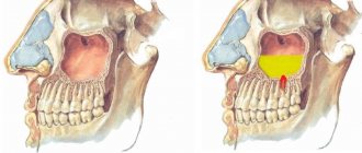
Numbness after sinus lifting and bone grafting in the area of the upper teeth and gums is less common than in the lower jaw. This is due to the fact that the innervation of these structures is carried out by three groups of branches of the maxillary nerve, intertwined with each other and forming the dental nerve plexus. Such a structure is quite difficult to damage or completely disable.
The loss of sensitivity in the area of the upper lip and upper half of the cheek cannot in any way be associated with dental interventions due to the fact that completely different branches of the maxillary nerve go to them.
Damage to the alveolar branches or dental plexus
This complication can only develop in conjunction with penetration into the maxillary sinus during surgery, which should not occur if the operation is performed correctly.
Local, mild numbness develops. One or two teeth may be involved with incomplete loss of sensitivity.
Improper placement of the implant or complications in the postoperative period
Dislocation of the skeleton of a prosthetic tooth can occur due to injury, inflammatory processes, or neglect of the dentist’s recommendations. Diagnosis of this condition is not particularly difficult due to the obvious pathological state of the implant.
There may be loss of sensitivity due to chronic odontogenic sinusitis. The condition develops over a long period of time, occurs with severe symptoms of sinusitis, and numbness develops in extremely severe and advanced cases.
How long does the anesthesia wear off?
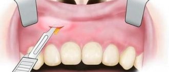
Dentists use special types of anesthesia:
- using applications (in this case, the anesthetic is applied to the area treated with antiseptic substances in the form of a gel, ointment, solution);
- due to the conductor, that is, through injections (carried out for a long-term type of dental intervention, Novocain is used);
- infiltration (three-stage type, indicated for complex manipulations).
After surgery to remove or treat, depending on the type of anesthesia, a feeling of numbness remains. If this is not due to disturbances in pain relief, then the numbness goes away after a certain period of time.
Experts have established a maximum threshold for each type of anesthesia and recommend closely monitoring how much time has passed since dental procedures were performed:
- numbness after application of anesthesia can last up to 30 minutes;
- after conduction – up to 5-6 hours;
- after infiltration – up to 3-4 hours.
The characteristics of the patient should be taken into account, such as age, weight, general health.
Diagnostics
If partial or complete numbness occurs after sinus lifting and bone grafting, you should undergo a number of diagnostic procedures:
- Examination of the oral cavity by a dentist. Allows you to assess the general situation and in some cases even make a diagnosis immediately. Must go through first. If there is displacement of the implant or nerve injury during surgery, then further examination methods can be of a purely clarifying nature.
- Assessment of neurological status based on damaged branches of the trigeminal nerve. This task is more likely for a neurologist, rather than for a dentist. By testing reflexes and using special methods to determine sensitivity, it is possible to assess the presence/absence of nerve damage.
- X-ray or computed tomography. These examination methods make it possible to establish in detail and accurately the cause of loss of sensitivity. With the use of modern technologies, it becomes possible to calculate down to the millimeter the direction and degree of displacement of the installed structure, existing pathological foci of regeneration, and determine the presence of an inflammatory process both at the site of bone grafting and directly in the maxillary sinus.
Dangerous complications and natural consequences of surgery
Below is a table with natural symptoms and dangerous complications that occur immediately after surgery (manifest within 7 days). The most important thing is that most of these can be solved quite quickly and easily. The main thing is to see a doctor in time.
| Symptom | Normal condition | Complication |
| Jaw pain | Appears after the anesthesia wears off. Weakens and disappears within 2-7 days. | The pain does not stop and intensifies. Painkillers do not help or help little. There is a high risk of soft tissue inflammation or nerve damage. |
| Swelling | After implantation, swelling may appear within a week and not subside. | The swelling does not go away. It hurts when touched. Increases in size. |
| Bleeding gums | Minor blood loss for 2-7 days from the incision site (where the gum was sutured or a bone graft was performed). | Significant bleeding after surgery that does not stop. Most often indicates damage to blood vessels. |
| Temperature increase | It can rise to 37.5-38.0⁰С (within 2-7 days). This is a normal reaction of the body to the introduction of a foreign body. | The temperature does not subside and increases after 7 days. Swelling forms. All signs of inflammation. |
| Numbness | The result of anesthesia, which wears off within 24 hours. | The numbness does not go away. Perhaps a nerve was damaged during the operation. |
Now let's talk about later complications that appear several years after the procedure for installing an artificial root.
Peri-implantitis
. An inflammatory process affecting the tissue around the implant. If measures are not taken in time, it can lead to resorption of gingival bone tissue.
| How to treat peri-implantitis? At the initial stage, it is medicated with the use of laser/ultrasonic cleaning of the implant (for this it is removed). |
Implant failure
. The rarest complication that develops over a long period of time and not instantly. Accompanied by the following symptoms: inflammation, bleeding, pus, implant mobility, pocket enlargement and bone thinning.
| It is important to know! Complications arise both with implantation using the classical method and when using a protocol with delayed loading. However, in practice, it is the classical method that is more dangerous due to the method of installing the implant, a large number of additional procedures and an impressive list of contraindications. The Dr. Fedorov Implantation Center uses advanced implantation techniques |
Treatment options
If the nerve trunk, plexus or branches of the nerve are damaged or compressed, there are two ways to solve the problem.
The first is to leave everything as is, without performing surgery. Such a solution to the problem is acceptable in case of incomplete loss of sensitivity or a small area of focus, with the exception of cases with displacement of the denture. Compression of the nerve due to regenerative processes will stop progressing after a certain point, “frozen” in one place. Odontogenic sinusitis can be treated with medication using antibiotics.
The second method is to perform surgery. The operation is indicated for injuries during sinus lifting and bone grafting, displacement of dentures or bone mass, extensive or complete loss of sensation. During the operation, the cause of the numbness is eliminated and, if necessary, the damaged nerve is sutured. Restoration of sensitivity takes a long time, sometimes never returning to its original state.
Diagnosis and treatment
As a rule, it is possible to determine the presence of numbness and find the cause of this ailment already at the stage of conversation with the patient. The dentist must conduct an in-person examination, on the basis of which he may prescribe the following studies: radiographic, tomographic, and Dopplerography of the vessels of the jaw. Unfortunately, without a thorough preliminary diagnosis, making a correct diagnosis and prescribing a treatment regimen is impossible. That is why, if necessary, it is not recommended to refuse to do some tests.
Most often, therapy is limited to the use of certain medications without the need for surgery. Sometimes it is necessary to perform an operation using one of the methods of jaw microsurgery. In both situations, the doctor’s main goal is to relieve the patient of discomfort and return the tissues to their former sensitivity.
It is important to understand that timely seeking help guarantees the fastest possible cure. A disease whose duration exceeds 3 months will be much more difficult to overcome. If numbness persists for a year, then it will be almost impossible to regain sensitivity. After treatment, the following therapeutic procedures may be prescribed: UHF, electrophoresis, laser therapy, exposure to diadynamic currents, etc.
When do you need a doctor’s help and who to contact?
How long does numbness last after tooth extraction and when is it time to sound the alarm? It was already mentioned above that sensitivity should be restored a few hours after the anesthetic injection was given. But situations are different - for example, after a complex removal of the “eight”, after osteoplastic surgery or installation of an implant, etc. numbness can last 1-2 days, less often up to 5-7 or even up to 10-14 days. But, as practicing dentists note, sensitivity is restored after 2 weeks in almost all patients. If after 14 days there is no improvement, then you need to go to the doctor. To begin with, you can contact the dentist who performed the previous treatment, then you can contact a neurologist or a neurophysiologist.
Read on the topic: is it necessary to take an x-ray after a tooth has been removed?