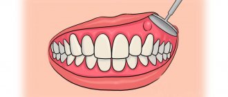
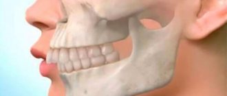
Exostosis of the jaw is a benign formation that manifests itself in the form of an osteochondral growth (osteophyte). The protrusions can be single or multiple and are located on the jaw bone. Maxillary osteophytes most often form on the outer (buccal) surface of the alveolar ridge, and exostosis on the lower jaw - from the inside, on the lingual side. A palatal bone growth (palatal torus) is also found.
Exostoses are painless and do not cause any discomfort at an early stage. But as it increases, the formation begins to cause a lot of discomfort - it complicates eating, affects sound pronunciation, interferes with prosthetics, etc. Treatment of osteophytes is only surgical. Removing exostosis on the gum is a low-traumatic operation that takes about an hour. The operation is performed under anesthesia, so the patient does not experience any discomfort. The method of surgical intervention depends on the location of the osteophyte.
Reasons for appearance
Their formation is associated with improper healing of post-extraction losses or improper preparation of wounds after surgery. This may result in sharp bone fragments being left behind. They cause mechanical irritation of the mucous membrane and pain. In addition, they can interfere with the proper fabrication, installation and use of removable dentures.
After tooth extraction, accumulations of macrophages, lymphocytes and neutrophils form around the smallest bone fragments. Later, mesenchymal cells penetrate the clot and create new connective tissue, creating new vessels. Callus is formed from it, which as a result of subsequent processes is transformed into new bone tissue. However, sometimes various local or general factors can prevent proper remodeling of the extraction socket. These may be local inflammatory processes, stimulants, vitamin deficiencies or immunological disorders in various chronic diseases.
Treatment and prevention of exostosis of the jaw
Surgical excision of the bone growth is the only effective treatment for exostosis. The intervention is performed under local anesthesia, so the patient does not experience discomfort. It is worth saying that there are two techniques for performing the operation. The choice of a specific technique depends on the location of the osteophyte:
- Removal of the palatine torus. In this case, the doctor makes a small linear incision, as well as two releasing incisions - in front and behind. After this, the dentist peels off the mucous membrane and removes the osteophyte. Extraction can be carried out either as a single block or in fragments. Next, the bone tissue is smoothed, and then interrupted sutures are applied.
- Removal of alveolar osteophytes. The procedure for performing the manipulations is no different from the previously discussed technique. The main difference is the configuration of the cut - in this case it has a trapezoidal shape. Otherwise, surgical treatment of maxillary and mandibular osteophytes is the same as removal of the palatine torus.
As for prevention, there are no specific measures to prevent this disease. The only thing that can be recommended is to visit the dentist regularly, at least twice a year, for examinations.
Still have questions? Sign up for a consultation at our clinic or call:
Symptoms
Symptoms of jaw exostosis in the early stages are usually not pronounced. Until osteophytes reach 3 mm in size, they are difficult to feel with the tongue or see with the naked eye. However, larger bone processes make themselves felt:
- When touched with the tongue, they are felt as a hard, smooth, painless bulge.
- Visually distinguishable as tubercles, while their shade is lighter than the surrounding mucosa.
- May cause discomfort or even pain. If they are localized, for example, in the area of wisdom teeth, then pain may occur when the mouth is pulled wide open.
Causes, symptoms, diagnosis
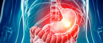
The reliable reasons for the formation of exostosis have not yet been established. However, among the most common factors leading to the development of the disease are injuries, inflammatory processes occurring in the bones, and congenital jaw anomalies. In addition, the appearance of osteophytes may be a consequence of general diseases, for example, insufficient function of the endocrine system.
Small osteophytes do not cause complaints from the patient - there is no pain, the mouth opens freely and fully, the mucous membrane in the area of the growth does not have pronounced pathological changes. Therefore, most often exostosis is detected by a dentist - during a preventive examination or in preparation for prosthetics.
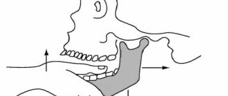
Diagnosis of the disease is carried out on the basis of X-ray data. The image clearly shows a bone growth that is not fused with the surrounding tissues and has clear boundaries.
Mandibular exostosis with the formation of osteophyte on the inner surface of the alveolar process.
Diagnostics
Sometimes bone spurs can reach significant sizes and take on unusual shapes with no or minimal symptoms of pain. They must be differentiated histopathologically from, for example, tumors of mesenchymal origin such as osteochondroma or osteochondroma. For this purpose, the dentist, after visual examination and palpation of the exostosis, refers the patient for examination:
- X-ray – accurately shows the size and location of the formation, its position in relation to the roots of the teeth and other elements of the dental system.
- Biopsy - cytological examination is necessary for the differential diagnosis of benign formation.
Causes
Reasons for the formation of growths:
- inflammatory, infectious processes, purulent accumulations;
- mechanical impacts, injuries causing bone destruction, jaw displacement and other damage;
- deviations of row segments, formation of nodular hardenings;
- disorders of the endocrine system, hormonal levels.
One of the reasons for the development of pathology is complex tooth extraction. Often, when a wound heals, the tissue around it becomes overgrown with moving areas. If done incorrectly, compactions may appear and exostosis will gradually develop.
How is exostosis removed?
The only way to cure exostoses is to remove them surgically. This is not always necessary, but only in cases where osteophytes have reached a large size, interfere with normal chewing function, cause discomfort or injure the mucous membrane, provoking constant inflammatory processes in the mouth. Therefore, at the first doctor’s appointment, as a rule, removal is not prescribed, but the patient comes regularly so that the dentist monitors the situation over time. If it is obvious that it is impossible to do without removing the exostosis, the procedure follows the following algorithm:
- Examination of the patient to confirm the absence of contraindications to surgery.
- Immediately before the operation, a local anesthetic is administered.
- An incision is made into the mucous membrane.
- Bone formations are cut down with a special tool so that the surface of the bone becomes smooth.
- Stitches are applied.
The entire procedure takes from half an hour to two hours, depending on the scale of the pathology and the structure of the bone tissue - cartilage is easier and faster to remove than solid formations.
Why should jaw osteophytes be removed?
A bone growth on the gum is not dangerous until it begins to grow. Increasing in volume, the osteophyte puts pressure on the dentition and bone structures. This leads to tooth displacement, malocclusion, and jaw deformation. Large growths impede the movements of the tongue, complicate diction, and interfere with normal chewing of food. Large growths prevent prosthetics and implantation. Osteophyte of the jaw will not disappear on its own. The only effective method of treatment is surgical removal of the pathological formation.
Features of the postoperative period
In order for the wound to heal as quickly and easily as possible, it is important to follow the doctor’s recommendations during the rehabilitation period.
- Do not damage the seam
- Carry out hygiene procedures carefully (but under no circumstances abandon them)
- Take prescribed medications, including antibiotics, if the dentist considers it necessary to prescribe them
- Refuse hot or spicy foods, follow a semi-liquid diet, consume food and drinks only at room temperature.
What is exostosis and why does it occur?
Exostosis in dentistry is called growths - the process of growth of osteochondral or bone tissue, manifested in the form of protrusions on the surface of the palate or lower jaw. They don't cause any pain.
The main problem is that exostosis gradually increases. This leads to discomfort, pressure on teeth and bones increases, and the risk of injury to the oral mucosa increases. With such formations, it is impossible to wear dentures and place implants, and possible consequences include malocclusion and chin displacement.
At the initial stage, exostosis may be invisible, and only a doctor can detect it during a dental examination or on an x-ray. Large growths are easily felt by the tongue.
How to treat?
It is impossible to cure exostoses on the gums at home; surgical intervention cannot be avoided. The operation to correct the alveolar ridge must be performed by a dental surgeon. The procedure has its limitations: contraindications include diabetes, problems with the endocrine system and adrenal glands, and poor blood clotting.
If the growth on the jaw is very small and does not cause any inconvenience, there is no need to rush into performing surgical correction. However, someday you still have to do this. An overgrown exostosis will interfere not only with the tongue, but also with neighboring teeth, which can lead to deformation. And if you need a removable denture, it is the growths on the jaw that will become the main obstacle to the installation of implants, as well as to the successful use of them.
To remove exostosis, a dental surgeon will numb the surrounding tissue growths (local anesthesia is usually used) and make a small incision on the gum. The formation is cut down and smoothed, after which sutures are placed on the jaw mucosa. The duration of the procedure will depend on the size of the exostosis and its location.
What to do after surgery
The first days after surgery, you need to carefully monitor the condition of the stitches and do everything to prevent them from coming apart. It is necessary to temporarily avoid solid and tough foods. Very hot or cold drinks, alcohol and cigarettes will slow down the healing process. It is recommended to reduce physical activity and avoid stress.
Minor swelling and pain are possible; painkillers and decongestants can be used during the recovery period. Particular attention should be paid to oral hygiene: it is better to consult with your dentist about the best means to rinse your mouth to prevent bacterial infection.
If the operation to remove exostosis of the jaw was carried out efficiently and the patient followed all the recommendations, there should be no complications. In case of prolonged pain, fever and swelling that does not subside, you should immediately consult a doctor. In addition, you should not prescribe treatment for yourself - this can only worsen the rehabilitation process.
GUM TREATMENT (SURGICAL METHODS)
Progress of the operation
Before you begin the operation, you first need to carry out some kind of preparation. The patient must undergo blood tests to exclude the presence of contraindications. These include diabetes mellitus, HIV, and poor blood clotting. In order to confirm the diagnosis, an x-ray is required. The operation itself is performed as follows:
- Thorough treatment of the oral cavity with antiseptic drugs.
- Introduction of an anesthetic drug.
- Removal of exostosis. This can be done with a standard set of cutting tools or using a laser. The latter method is practiced only in prestigious clinics where the appropriate equipment is available.
- Finally, stitches are placed to stop the bleeding. After a couple of days they are removed; some dentists use absorbable surgical thread.
General idea of exostosis in the mouth
New growths on bone tissue are found in different parts of the body. Exostosis in dentistry according to ICD-10 refers to other specified diseases of the jaw K-10.8. The osteochondral formations look like a protrusion, lump, mushroom or ridge, and can branch and be tortuous. When they grow, they injure the mucous membranes of the mouth, which provokes the development of inflammation.
Zkzostoza on the upper jaw are less common than in the mandibular region. Usually formed on the alveolar process on the side of the cheek. When the size increases, they affect the aesthetics of the face, creating asymmetry. In the lower jaw, exostosis often forms on the lingual side. Protrusions can interfere with chewing food and talking.
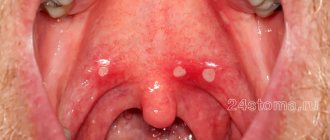
You can see what exostosis looks like on the gum or bottom of the lower jaw in the photo. If the formation is small, then it is difficult to notice it visually.
Main reasons
Exostosis is the result of overgrowth of jaw tissue. Experts say that the main cause of this disease is a hereditary predisposition. We also list a number of indirect factors:
- Changes in hormonal levels.
- Careless tooth extraction, during which soft tissue was injured.
- Infection during tooth resection.
According to statistics, these formations appear only in the case of complex operations, when it is necessary to remove several units at once or a multi-rooted tooth.
Possible complications after surgical treatment
Complications after surgery often arise due to the fault of the patients themselves. Problems usually appear as a result of non-compliance with doctor's recommendations and hygiene rules. Among the possible undesirable scenarios, the following processes and phenomena are distinguished:
- suture divergence due to too intense chewing load or trauma to the causative area with solid food products,
- the development of an inflammatory process, accompanied by swelling of the mucous membrane and even purulent processes. Most often, the cause is unsatisfactory care of the wound and the oral cavity in general.
If such complications occur, it is important to see a doctor as soon as possible. Otherwise, ignoring problems will lead to even more serious consequences.
In what cases is removal prescribed, what are the contraindications?
The presence of pronounced bone formation in the mouth requires radical measures, namely surgical removal of the exostosis. Such a measure is resorted to in the presence of the following indications:
- rapid increase in the size of the bone nodule,
- severe discomfort during eating and talking,
- violation of aesthetics,
- as a prophylaxis before implantation and prosthetics.
For large sizes, removal of the growth is indicated
. As for contraindications, such an operation cannot be performed if the patient has problems with the adrenal glands and the functioning of the endocrine system, blood clotting disorders, or diabetes.
Treatment of cones
The treatment methodology depends on the specific case. The approach is determined by:
- the presence of pain;
- part of the cavity that has been infected (upper, lower);
- time elapsed since the onset of the disease;
- reaction of the cone to pressure.
Treatment of angioma
Removing a lump of this type occurs in three stages:
- Drug treatment and treatment;
- Surgery;
- Radiation therapy.
At the first stage, alcohol is used: it constricts blood vessels and helps eliminate high blood loss during surgery.
Then, in the case of the capillary form, radium therapy is used, it consolidates the effect of treatment, and sometimes can act as an independent drug and cure the disease completely.
It is prohibited to treat the cavernous form of angioma with radium. It can help transform the lump into a malignant one, which can greatly worsen the patient’s condition.
Treatment of pemphigus
A lump of this type that appears on the palate is treated mainly with antibiotics and a diet that includes a high content of proteins and vitamins, but without salt.
If such a lump appears, it is necessary to use disinfectant solutions. In severe forms, blood transfusions are used.
Treatment of myxoma and cysts
In these forms of the disease, the attending physician prescribes antiseptic drugs and prepares the cavity for surgery (if there are signs of accelerated growth of the lump into the tissue).
Additional methods of electrical treatment are also used, which make it possible to artificially kill tissue without the use of a scalpel, but all methods of this type are dangerous, thanks to them, a malignant formation may appear in place of a benign one.
Treatment of cancer
Cancer requires immediate intervention by a qualified specialist. The earlier the stage of the disease, the less harm the treatment and the disease itself will cause to the body. If a cancer lump appears, the following treatment approaches are used:
- Radiation therapy – the cancerous growth is irradiated with X-rays. In the early stages, it completely cures the disease.
- Surgical intervention - not only the harmful lump is cut out, but also the tissue around it, in order to exclude relapses. Such an intervention leaves defects on the face, which can later be corrected with plastic surgery.
- Chemotherapy - taking cytostatics.
For the cancer in question, chemotherapy is effective only in combination with radiation and surgery.
It is easier to defeat any disease at its inception stage. But in the case of bumps on the palate, it is better not to have the disease than to treat it later.
The root causes of the disease have not been fully established. Therefore, following the hypothetically formed rules will not completely protect you from the disease, but it will definitely reduce the chance of developing a similar problem. A preventative visit to the dentist will ensure timely treatment, even if the lump pops up unnoticed.
Category Miscellaneous Published by Mister dentist
Exostosis in dentistry - bone tumor in the oral cavity and technology for its removal
The appearance of atypical neoplasms in the oral cavity is a phenomenon that never happens without a reason. Most often, the cause of such troubles is injury or dental disease. One of the possible scenarios is the appearance of a bone growth - exostosis. Despite the fact that in most cases the defect can be easily detected by touch, most patients seek help at an advanced stage. Further in this article we will talk about what it is - tooth exostosis, in what cases it usually appears and how the procedure for its removal is carried out.
Why remove exostosis?
Exostoses, as a rule, do not hurt or cause discomfort. Most often they form in children under the age of 20 during the formation of the skeleton and bone tissue, however, pathology also occurs in adults.
It is necessary to remove bone growths for several reasons:
- violation of aesthetics: exostoses look like white balls on the gums, which are noticeable when smiling,
- growths often increase in size,
- possible transition to a malignant form,
- if it is necessary to install implants or prostheses, exostoses will become an obstacle to the procedure, since the prostheses will injure the growths, and the implants simply cannot be fixed due to a violation of the quality of the bone tissue.
Diagnosis of the disease
Expert opinion
Vasiliev Alexander Alexandrovich Implant surgeon Work experience 9+
“Exostoses do not always have external manifestations - often they can only be detected on photographs when diagnosing other diseases. However, the examination uses an x-ray - a targeted image of one tooth. Or, more effectively, an orthopantomogram. It allows us to examine the condition of the jaw bone and tooth roots along the entire row, because growths are often formed in multiple quantities.”
Indications and contraindications
If the formation is large, leads to inflammation of the mucous membranes of the oral cavity, jaw deformation, or interferes with talking or chewing food, then it is removed. The operation is also prescribed before prosthetics, since even small bumps become an obstacle to the installation of implants.
The patient himself can contact the surgeon if the bone spike spoils the aesthetics of the face. In this case, it is located on the buccal side and the protruding part is visible externally. An aesthetic defect gives a person more psychological discomfort than physical discomfort.
Before the procedure, the surgeon excludes contraindications. The list of restrictions includes infections in the mouth, acute respiratory infections, diabetes mellitus in the stage of decompensation, poor blood clotting, exacerbation of chronic diseases, mental disorders, childhood, pregnancy and a number of other contraindications.
For children, surgery is delayed until they are 18 years old. This is due to the fact that the bones are still growing and the pathology can resolve on its own.