Stomatitis is a disease characterized by the appearance of ulcers on the gums. The reasons for its occurrence are different. Aphtha, or as it is called in simple terms - a sore on the gum - can appear due to allergies, herpes virus, bacteria, etc.
Mouth ulcers or gum stomatitis most often occur in children, since the child most often comes into contact with dirty toys, household items and, at every opportunity, puts everything into his mouth. It occurs less frequently in older people.
Varieties of stomatitis have different causes, symptoms and require different treatments. We will provide more detailed information about them below, this will help you navigate.
Stomatitis is a disease characterized by the appearance of ulcers on the gums.
What is stomatitis
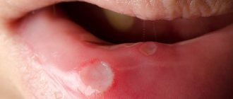
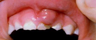
Stomatitis is an inflammatory disease of the gums that appears externally as white spots or sores on the gums.
You should not ignore stomatitis or try to treat it yourself. Mouth ulcers occur for various reasons, sometimes quite serious. If a white sore appears on the gum, it is important not only to get rid of the symptoms, but to understand the cause of its appearance, because stomatitis in adults often manifests itself as a secondary process of some disease, which is best diagnosed with the help of doctors in a timely manner.
What does stomatitis look like?
Stomatitis is an inflammation of the lining of the mouth that causes ulcers, blisters and spots.
White sores can appear throughout the mouth: a white spot can be on the gum, tongue, lips, cheeks, and so on. Ulcerative damage to the integrity of the oral mucosa can cause severe discomfort, pain, provoke an increase in the sensitivity of nerve endings, and an increase in body temperature. It would seem that a minor white sore on the gum not only prevents you from eating normally, but also from sleeping and drinking, which can lead to problems with the gastrointestinal tract. If you have developed stomatitis, its signs are quite obvious - a white sore appears on the gum, and your general condition may worsen significantly. In this case, you should not delay visiting a doctor.
Stomatitis causes inflammation of the oral mucosa, which causes ulcers, blisters and spots to appear.
Symptoms of the disease
Stomatitis has its own characteristic signs, by which a specialist quickly identifies the disease:
- sores, vesicles, blisters on the gums;
- inflammation of the gums;
- swelling, pain, redness of the oral mucosa;
- hyperemia of the gums and mucous membranes;
- white or yellow coating;
- increased salivation (hypersalivation);
- bleeding gums;
- bad breath;
- unpleasant putrid taste in the mouth;
- enlargement of the submandibular lymph nodes;
- increase in temperature;
- general deterioration of health.
With stomatitis, the patient's well-being can seriously deteriorate.
Where can stomatitis occur:
- most often stomatitis occurs on the gums, both the upper and lower jaws;
- buccal mucosa;
- lips;
- sky.
The mechanism of development of stomatitis and causes of occurrence
The mechanism of development of this disease has not been precisely established, and today it is believed that it is based on an immune reaction to various irritants - presumably, their role is played by molecules that the immune system is not able to recognize. As a result, lymphocytes attack unidentified molecules, which leads to stomatitis on the gums in adults and children.
Stomatitis is often a manifestation of a number of systemic diseases, such as vitamin deficiency, endocrine diseases, blood diseases, gastrointestinal tract and others.
Among the local factors for the formation of ulcers on the gums, the following causes of stomatitis are distinguished:
- microorganisms (viruses, mycoplasmas, bacteria);
- diseases of the oral cavity and gastrointestinal tract (for example, colitis, gastroduodenitis, gastritis) can provoke catarrhal stomatitis;
- poor oral hygiene;
- herpes on the lips;
- dehydration;
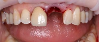
- dentures of low quality, as well as incorrectly installed ones;
- use of certain medications;
- mechanical injury to the gums and mucous membranes of the mouth;
- vitamin deficiency and hypovitaminosis;
- insufficient intake of vitamins;
- smoking;
- chronic systemic diseases;
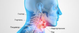
- oncological diseases of the neck and nasopharynx;
- side effects of chemotherapy;
- hormonal imbalances.
Sometimes stomatitis appears after dental treatment. If the cause of the disease is identified and eliminated in time, it is possible to cure it quickly and effectively.
Is stomatitis contagious and how is it transmitted?
Stomatitis itself is not a contagious disease, but the underlying cause is contagious, for example, the herpes simplex virus in herpetic stomatitis or an RNA virus in vesicular stomatitis. In these cases, stomatitis can develop as a consequence, as a variant of the body’s response to the causative virus.
If stomatitis occurs due to other reasons: mechanical damage, vitamin deficiency, hormonal imbalance, it does not pose any danger in terms of infection.
Forms of lichen planus
Six main clinical forms of lichen planus should be distinguished:
- Typical form of lichen planus;
- Keratotic form of lichen planus;
- Exudative - hyperemic form of lichen planus;
- Erosive - ulcerative form of lichen planus;
- Bullous form of lichen planus;
- Atypical lichen planus.
Although lichen planus is a chronic disease, there are moments of exacerbation and then the clinical picture may worsen, depending on the form of lichen planus, immunity and the presence of other somatic diseases in the patient.
Typical form of lichen planus
The typical form of lichen planus, also known as the classic form, is recognized by clinicians. The patient does not report any complaints in the typical form of lichen planus. There may be complaints of pain when eating spicy or hot food. More often, such complaints arise a couple of days before the appearance of visible papules on the oral mucosa.
The typical form of lichen planus is characterized by the appearance of papules on the unchanged oral mucosa, and, as you remember, these papules are grouped, resembling lace or fern leaves. Papules in the typical form of lichen planus are small, up to 2 mm in diameter. In the center there is an area of keratosis, so the papules are dense on palpation.
Hyperkeratotic form of lichen planus
The hyperkeratotic form of lichen planus consists of huge foci of keratinization at typical sites of localization for lichen planus. Patients with the hyperkeratotic form do not make any complaints. They may complain about aesthetic defects, dryness and flaking.
Exudative-hyperemic form of lichen planus
The exudative-hyperemic form of lichen planus occurs in 25% of all patients diagnosed with lichen planus. Complaints, as with other forms of lichen planus, and with the exudative-hyperemic form, are vague: burning and pain when eating spicy food, dry mucous membranes. Papules have a typical structure, shape and location. Only in the exudative-hyperemic form is the mucous membrane swollen and hyperemic.
Erosive-ulcerative form of lichen planus
The erosive-ulcerative form of lichen planus is the most severe clinically and difficult to treat form of lichen planus. More often it occurs as complications of typical or exudative-hyperemic forms of lichen planus. With this form of lichen planus, patients report complaints of pain when eating, talking, and a burning sensation.
On the hyperemic and edematous mucosa, in typical places of localization of lichen planus papules - erosions of irregular and unclear shape, often covered with fibrinous plaque, there may be granulations. If these are single ulcers, then the pain will not be so intense. But in most cases, the ulcers merge with each other, affecting most of the oral mucosa. If erosions are left untreated for a long time, they turn into ulcers with keratinization lines.
Bullous form of lichen planus
The bullous form of lichen planus is the rarest form of lichen planus, recorded in 3% of patients with lichen planus.
The bullous form of lichen planus is described not only by the presence of papules, but also by the presence of blisters, the size of which can range from the head of a pin to a kidney bean. The blisters can remain on the mucous membrane for up to several days, then they open, and erosion occurs in the place of the opened blisters. Erosion in bullous form of lichen planus is irregularly shaped, with a bleeding bottom. Often covered with fibrinous plaque.
Atypical form of lichen planus
An atypical form of lichen planus is most often observed on the mucous membrane of the lips and gums. On the lip, two symmetrical areas with congestive hyperemia are often identified; these areas are slightly higher than the normal mucous membrane of the lips and gums. Often areas of the epithelium are covered with a whitish coating, which is difficult to remove during diagnosis.
You need to understand that the described forms of lichen planus can transform one into another depending on the influence of both local and general factors. It can occur simultaneously on the mucous membranes of the mouth, lips, and skin. Or vice versa. Forms of lichen planus can become malignant, especially when localized on the mucous membrane of the cheeks and the red border of the lips (erosive-ulcerative form of lichen planus).
Types of stomatitis
In modern periodontology, several types of stomatitis are distinguished:
- Aphthous stomatitis. It may be accompanied by the appearance of single ulcers or multiple rashes. At the initial stage, these ulcers have a pinkish tint with pronounced red edges, but after an exacerbation, yellowish aphthae form with general swelling of the oral mucosa. Aphthous stomatitis occurs with high fever, enlarged lymph nodes, sharp pain and burning sensation while eating. There is also a chronic form that can occur in a person with a weakened immune system. In this case, this disease can manifest itself from period to period.
- Ulcerative stomatitis. It is an inflammatory disease characterized by focal destruction of the mucous membrane.
- Catarrhal - caused by poor oral hygiene, insufficient hydration of the oral cavity, the introduction of bacteria and fungi of the genus Candida. In the latter case, the disease is called candidal or fungal stomatitis, popularly known as thrush. The immediate cause of inflammation is the activity of fungi and their penetration into the mucous membrane. Candida is normally always present on the body and mucous membranes in small quantities. The disease appears when their active reproduction begins. Candidiasis is a contagious disease. It can be infected by contact, by using the same utensils and hygiene products. A characteristic manifestation of the disease is a cheesy white coating. It appears in different areas of the mucosa in the form of spots of different sizes. Associated symptoms are dryness, burning, itching. The latent period of the disease can last up to 20 days. With proper treatment, the disease resolves without complications.
- Traumatic. Injuries that cause stomatitis can be mechanical, chemical or physical and may be acute or long-term, such as improperly manufactured or installed dentures. The sharp edges of the prosthesis can irritate the mucous membranes and tongue. This provokes the appearance of stomatitis.
- Vesicular. This is one of the types of contagious stomatitis. It is caused by an RNA virus of the genus Vesiculorus of the Rhabdoviridae family. Patients should use separate hygiene products and utensils so as not to infect their household.
- Enteroviral vesicular stomatitis is caused by enteroviruses.
- Herpetic or cold sore. The disease lasts 8 days, is accompanied by weakness, drowsiness, a sharp increase in body temperature, and possible inflammation of the lymph nodes. In addition, with this type of stomatitis, gingivitis occurs and the saliva becomes viscous. The mucous membranes of the oral cavity become covered with a rash consisting of blisters that burst after three days. Accompanied by gingivitis, viscous saliva.
- Allergic. Autoimmune reactions and allergies can manifest themselves in the oral cavity. The frequency of this disease is increasing, which is associated with the emergence of new allergens and environmental deterioration. The drugs and materials used in dentistry also play a role—some people may also be allergic to them. The symptoms of allergic stomatitis in general do not differ from the manifestations of other types of inflammation of the oral cavity. Swelling, redness, pain, and the formation of various elements of a rash are observed. Ulcers and erosions may also appear. Allergic stomatitis can develop against the background of autoimmune disorders. At the same time, many variants of mucosal lesions are associated with them, when it is not possible to establish connections with other possible causes.
In addition, stomatitis can take acute and chronic forms, depending on the characteristics of its course:
- acute stomatitis is accompanied by a significant deterioration of the condition,
- Chronic is a disease that occurs periodically.
Etiology of lichen planus
As I said earlier, the etiology of lichen planus is based on several theories.
Of these, the following etiological theories are important:
- Neurogenic theory of the occurrence of lichen planus;
- Toxic-allergic theory of the occurrence of lichen planus;
- Viral theory of the occurrence of lichen planus.
Despite a number of theories put forward about the occurrence of lichen planus, this type of allergy is considered semi-etiological, that is, one specific factor that provokes the occurrence of pathology is not identified.
Attempts to isolate a specific type of virus that is present in patients diagnosed with lichen planus have also been unsuccessful.
Doctors consider the presence of a history of previous neurological diseases or conditions to be important in the occurrence of lichen planus. That is, the possibility of a person developing lichen planus is influenced by stress, lack of sleep, depression, neuroses, panic conditions, epilepsy, etc.
Interesting fact: it was noticed that in people with diseases of the gastrointestinal tract (erosions, gastritis and stomach ulcers), that is, those diseases where there are changes in the mucous membrane, the risk of developing an erosive form of lichen planus is higher than in those who suffer other gastrointestinal disorders. However, the mechanism of dependence has not been established.
In the etiology of lichen planus, it should also be noted that the patient has:
- Diabetes mellitus;
- Cardiovascular failure;
- Hypertension;
- Pathologies of the liver and kidneys;
- Genetic predisposition;
- Immune suppression;
- Trauma to the oral mucosa (poor quality fillings, dentures, chronic trauma, chemical, mechanical trauma);
- Long-term use of medications;
Lichen planus most often occurs in middle-aged women, or in young people, children and the elderly.
Elements of defeat
Elements of the lesion in lichen planus may appear first on the oral mucosa, then on the red border of the lips, and only then on the skin. But it should be remembered that there is NO specific order and dynamics of occurrence. I have given only the more common options.
In general, lichen planus is a disease related to papular diseases in which metabolic processes in the oral mucosa and skin are disrupted. That is, it is a complex of dystrophic and inflammatory processes at the same time.
Skin papules in lichen planus
Skin papules with lichen planus are violet-bluish in color, up to 2 mm in diameter, dense in the center, with the phenomenon of keratinization.
Papules of the red border of the lips in lichen planus
Papules of the red border in lichen planus first appear in separate forms, which over time are connected to each other by keratinization bridges. After which they imagine a single huge papule rising above the intact surface of the red border of the lips.
Papules on the oral mucosa with lichen planus
Papules on the oral mucosa with lichen planus are most often grouped, forming circles, lines, and lace. Individual papules are connected to each other by keratotic bridges. The tops of the papules become keratinized and acquire a milky bluish color (this is the most accurate sign for the differentiated diagnosis of lichen planus and leukoplakia, since in leukoplakia the plaques have a yellow tint).
In some cases, the process of keratinization does not occur, but rather necrosis. Then there will be erosion in place of the papule, followed by an ulcer.
If we talk about the more frequent localization of papules in the oral cavity, the order is as follows:
- Buccal mucosa in the retromolar region;
- Tongue (lateral and dorsal surfaces);
- Lips;
- Gum;
- Mucosa of the alveolar processes;
- Solid sky.
How to distinguish stomatitis from other diseases
Since stomatitis can appear for a variety of reasons, and the forms of its manifestation can be very different, it is highly not recommended to self-medicate, since in this case you can only aggravate the course of the disease and cause the appearance of new symptoms. Due to such diversity, methods of treatment and prevention of this disease can also be very different.
Manifestations of stomatitis may be similar to a number of diseases, from each of which a doctor, when examining a patient, will definitely be able to distinguish this ailment:
- Sore throat or stomatitis: you can determine a sore throat, which may be similar to stomatitis, by damage not only to the mucous membranes of the mouth, but also to the pharynx. It is very painful for the patient to swallow.
- Herpes or stomatitis: In the case of herpes, the rashes first look like blisters and appear later.
- Cancer or stomatitis: the first disease is distinguished by the fact that ulcers appear, which grow together over time. As a result, one ulcer forms and begins to grow.
- Thrush or stomatitis: when examining the mouth, if there is a disease such as candidiasis, a yellow or white coating is detected, redness, the wounds are round in shape, and the ulcers are small.
- Syphilis or stomatitis: the first disease is distinguished by the nature of the ulcers. They appear in the form of eosis and do not hurt. Sometimes there is a depression in the center of the wound.
The doctor selects the treatment for stomatitis based on the cause of the disease, the complexity of its course, the form of manifestation, the individual characteristics of the body, and so on - in general, you will have to take into account a lot of things in order to choose the correct and most effective treatment. A specialist usually easily determines that it is stomatitis and not another disease.
A rash in the mouth may indicate the presence of coronavirus in a patient
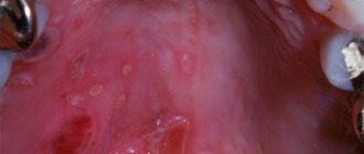
Mucocutaneous lesions occur on average 12 days after the onset of other symptoms of the disease. Previously it was reported that infection with coronavirus can manifest itself in the form of a rash on the skin and even lead to the appearance of ulcers inside the mouth, the authors of this study for the first time stated that one of the symptoms of the virus may be enanthema - a rash on the mucous membrane.
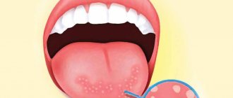
Enanthema manifests itself as multiple small spots on the mucous membrane. The authors suggested that dentists can regard this sign as one of the symptoms of coronavirus and protect themselves if they inspect the mucous membranes for the presence of a rash before starting treatment.
“The presence of enanthema on the mucous membrane most likely indicates the viral nature of the rash,” says the author of the study, MD. Juan Jiménez-Coch, representing the Department of Dermatology at the Ramon y Cajal University Hospital in Madrid.
At the moment, researchers continue to work on finding drugs to fight the virus and developing a vaccine. At the same time, the list of symptoms of the disease is expanding, among which the most common are: headache, fever, sore throat. Less common symptoms include diarrhea, loss of smell and taste, vasculitis, and a rash similar to chickenpox. Enanthema will probably soon be added to this list.
At the university hospital, among patients with confirmed coronavirus, 21 patients were identified who also had manifestations of a rash. Among this group, enanthema variants were found in 6 patients. The age of patients from the study group was from 40 to 69 years, among them there were 4 women and 2 men.
Different types of enanthems
One patient had a macular type of enanthema, two had a petechial type, and three had both types of enanthema. The rash was localized to the palate.
In patients with petechial enanthema, the rash appeared faster than in patients with the macular type of rash. Laboratory tests of the patients did not reveal any abnormalities that could explain the appearance of the rash by another cause. The use of medications by patients also could not be associated with the appearance of enanthema. It is known that such rashes often occur under the influence of a viral load, although sometimes they can act as an adverse reaction to taking medications.
A reliable characteristic of the viral origin of the rash is its localization in the oral cavity. Vesicular and petechial types of rash are associated with the entry of the virus into the body, and the petechial type of enanthema is more common in adult patients.
In this study, 83% of patients had a petechial type of enanthema, or a predominantly petechial type. It is noteworthy that in two patients the rash appeared either 2 days before the onset of the main symptoms of coronavirus, or 2 days after the onset of these symptoms. In this regard, the authors made a convincing assumption that the rash in these patients could not appear as a reaction to taking medications.
The study authors report that the validity of the results may have been affected by the small number of participants and the lack of a control group of patients. “Although doctors are increasingly reporting cases of rashes in patients with confirmed coronavirus, it remains difficult to determine what the true causes of skin rashes are,” the authors say.
Source: https://stomatologclub.ru/stati/stomatologiya-8/syp-vo-rtu-mozhet-ukazyvat-na-nalichie-koronavirusa-u-pacienta-3368/
Can stomatitis go away on its own?
Patients often delay visiting a doctor, hoping that stomatitis will go away on its own. Stomatitis in non-advanced forms can actually go away on its own within a week. But given the risk of complications and, as a rule, a fairly high degree of discomfort from which the patient suffers with this disease, it is much wiser to seek help from a doctor and receive competent medical care.
Correctly prescribed treatment, following the recommendations of the attending physician, will bring rapid relief and improved quality of life. In addition, this will speed up the process of curing stomatitis, avoid the unpleasant consequences of untimely treatment, and also, thanks to diagnostics, learn about the causes of stomatitis and, if necessary, take care of your health. It is difficult for a patient who does not have special knowledge to assess the severity of his disease and the causes that caused it, so seeking medical help is necessary.
Possible consequences and complications of stomatitis
Recurrent stomatitis can be cured quickly if you follow your doctor’s recommendations. But sometimes patients may experience some complications:
- lung diseases;
- damage to the entire mucosa;
- digestive problems;
- loss of some teeth.
Typically, such complications are the result of self-medication or lack of treatment, which is why you should consult a doctor in a timely manner.
Diagnosis of stomatitis
Only a doctor can make a diagnosis. Typically, the doctor reviews the medical record and then examines the patient's mouth. Today, no special medical diagnostic methods, such as laboratory culture analysis or biopsy, are used to detect stomatitis, since they do not exist for this disease. In diagnosis, the doctor is guided by the main and main signs of stomatitis:
- This is the appearance of the ulcers and their location
- The condition of the oral tissues around the ulcers - with stomatitis they look normal and healthy
- The fact of recurrence of episodes of the disease
- The patient has no deterioration in general health, high temperature and other significant general symptoms. This symptom does not apply to advanced forms of stomatitis, mainly in children, when there are a large number of ulcers and just a high temperature and poor health. The diagnosis of stomatitis can only be made by a doctor.
Treatment of stomatitis
Only a doctor can prescribe correct treatment for stomatitis in adults or children. If you want to quickly cure stomatitis, it is best to consult a doctor in the early stages of the disease. Remember that improper self-medication can lead to generalization of the infection and serious consequences for your health.
Remember that improper self-medication of stomatitis can have serious consequences for your health.
Depending on the type of damage to the oral mucosa, the doctor will prescribe the correct and effective treatment for your stomatitis, most often with medication - these can be antifungal, antiviral, anti-inflammatory, antiseptic, antihistamine and other medications. In addition, the doctor will recommend a gentle diet and prescribe mouth rinses using decoctions of various plants with antiseptic properties or tinctures of them, ready-made rinses. Sometimes the use of medicinal lozenges may be prescribed - in addition to the antiseptic effect, they improve salivation and provide a distracting effect.
In case of severe pain symptoms and the need for rapid treatment of stomatitis, the doctor may prescribe anesthetics for local use. When treating recurrent stomatitis, it is especially important not to miss repeated appointments with the doctor, since specialist supervision is necessary for complete cure and prevention of relapses of the disease.
In some cases, as prescribed by a doctor, a modern and effective method of treating stomatitis can be used - laser therapy. Its essence is the impact of a laser device on areas of the oral mucosa affected by stomatitis, during which the inflamed areas are disinfected and pain is reduced. As a result, recovery occurs very quickly.
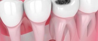
In cases where, in addition to stomatitis, the patient is also concerned about caries, the primary task is to cure stomatitis. Planned dental treatment for stomatitis is not carried out; first you need to cure stomatitis, and then start dental treatment. Of course, this does not apply to acute cases, and the treatment procedure is determined by the attending physician.
Recommendations during treatment of stomatitis
- follow the treatment method for stomatitis prescribed by your doctor,
- It is especially important when treating children: eat food several times a day in small portions, crushed and not hot, so as not to injure the mucous membranes,
- exclude salty, spicy and sour foods during treatment - pickles, marinades, pepper and mustard, tea with lemon, sour juices and fruit drinks, let them wait until better times.
How many days does stomatitis treatment last?
You should not delay visiting a doctor to treat stomatitis in the mouth, because the sooner you contact a specialist, the sooner you will begin proper treatment and the faster you will get rid of the disease. It is worth considering that treatment of chronic stomatitis takes a little longer, since the disease has already developed into a more complex form.
Most often, if the doctor’s recommendations are fully followed, stomatitis is completely cured in 3-10 days, sometimes it may take about 2 weeks.