Effective treatment of stomatitis in adults
Treatment of stomatitis in adults largely depends on the type of this disease, as well as concomitant diseases that provoke the appearance of ulcers and inflammation in the mouth. The most interesting thing is that the diagnosis of stomatitis is still carried out visually: the patient undergoes tests only if other, more serious diseases are suspected. Effective therapy for stomatitis is possible, but only if the type of disease is correctly determined. Recovery usually occurs within one or several weeks, but there are also more severe forms when long-term rehabilitation is required for complete recovery. Let's look at the main types of disease and drugs for the treatment of stomatitis in adults.
Treatment of herpetic stomatitis
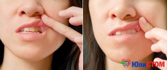
Treatment of stomatitis after detection can be carried out many times (once a year or more often), since once settled in our body, the herpes virus remains there for life. Herpes stomatitis is characterized by the accumulation of reddish transparent blisters on the lips and inside the mouth, so it is quite easy to detect. Especially often, herpetic stomatitis manifests itself against the background of decreased immunity, vitamin deficiency, hypothermia and stress. Remember that herpetic stomatitis in adults is contagious, so you should avoid tactile contact.
Types of drugs
- Antiviral agents.
- Immunostimulants (as a preventative measure).
- Ointments (including to eliminate visual manifestations of the disease).
3.Herpes
Herpes on the lips
appears due to a virus called herpes simplex virus type 1. Unlike ulcers, herpes is contagious from the moment the herpes blisters rupture until complete recovery. Primary infection often occurs during childhood and can be confused with cold or flu symptoms. Once a person becomes infected with the herpes virus, the virus remains in the body and lies dormant most of the time. But during stress, colds, injuries, hormonal changes, under the influence of sunlight, the virus is activated and herpes appears on the lips.
Herpes tends to appear in the same place. And in addition to infecting other people, the herpes virus can spread to other parts of the infected person's body - the eyes and genitals.
About our clinic Chistye Prudy metro station Medintercom page!
Treatment of candidal stomatitis
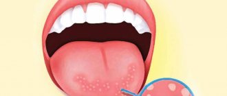
A type of stomatitis that is caused by a special type of fungus – Candida albicans. Very often, candidal stomatitis begins with glossitis, which is why its second name is stomatitis on the tongue. Treatment in this case requires fairly prompt treatment, since in adults it is quite painful and is accompanied by a burning sensation in the mucous membrane and sore throat. On the inner surface of the lips and cheeks, as well as on the tongue, characteristic foci of inflammation appear, covered with a white cheesy coating.
Treatment methods
- Treatment of common diseases (if detected).
- Antimicrobial therapy.
- Preventive rinses.
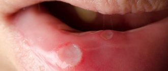
Treatment of aphthous stomatitis
A distinctive feature of this type of stomatitis is the appearance of so-called aphthae on the oral mucosa. These are small round ulcers with redness at the edges, the touch of which causes unpleasant painful sensations. Often occurs as a result of the activity of microorganisms (in particular, staphylococcus). Treating aphthous stomatitis at home is only recommended if it is not caused by a more severe illness. Often aphthous stomatitis occurs with problems with the immune system, liver and gastrointestinal tract.
Treatment methods
- Allergy therapy.
- Antiseptic therapy (treatment of aphthae with anti-inflammatory solutions, gels and ointments).
- Dental treatment and sanitation of the oral cavity (after dental treatment, stomatitis occurs less frequently than in the presence of carious lesions).
- Immunotherapy.
- Diet.
Modern drugs for the treatment of patients with inflammatory diseases of the pharynx
Acute pharyngitis can be an independent disease, and also develop with acute respiratory diseases of the airways. The development of acute pharyngitis can be promoted by ingesting hot and cold food (drinking), inhaling cold air, as well as air containing harmful impurities or industrial dust. With acute inflammation of the pharyngeal mucosa, infiltration with small cell elements may occur, swelling and hyperemia may appear, mucous glands increase secretion production, lymphoid granules may swell and increase in size [2,3]. With acute pharyngitis, the patient first complains of a feeling of severe dryness, heat in the throat, pain when swallowing food and saliva. Subsequently, due to increased mucus secretion, increased frequency and sharp pain in swallowing movements are noted. Chronic pharyngitis can be either an independent disease or one of the manifestations of chronic diseases of the gastrointestinal tract, metabolic disorders, exposure to occupational hazards, alcohol abuse and smoking. The clinical manifestations of chronic pharyngitis are very diverse. In all its forms, patients experience a feeling of dryness, the presence of a foreign body in the pharynx, and persistent moderate pain in this area. Treatment of acute pharyngitis consists of prescribing a gentle diet, rinsing with warm alkaline solutions, which help cleanse the pharyngeal mucosa and warm it. Local antiseptic and antibacterial drugs are prescribed. Treatment is aimed primarily at reducing inflammation and, as a result, reducing pain in the throat. Therapy for chronic pharyngitis requires an individual approach and a preliminary general examination of the patient to determine the reasons that resulted in this disease. The complex treatment of acute and chronic pharyngitis includes local antibacterial drugs. The most famous and frequently prescribed are Bioparox and Grammidin. The active substance of Bioparox is fusafungin, a local antibiotic. Grammidin contains gramicidin C hydrochloride and cetylpyridinium chloride monohydrate, which have an antimicrobial effect against pathogens of infectious diseases of the oral cavity and pharynx. Considering that pharyngitis often develops against the background of a viral infection, antibacterial drugs may simply be ineffective [6-9]. Also, the above drugs do not have anti-inflammatory and antipyretic effects. Even more significant is that, by destroying both pathogenic and non-pathogenic microflora of the oropharynx, they reduce the immunological status of the patient, increase the number of relapses of inflammatory diseases of the pharynx, contribute to the development of allergic reactions, the emergence of resistant strains, dysbiosis of the oral cavity, oropharynx and intestines. Moreover, the severity of the side effects of irrational antibiotic therapy sometimes exceeds the severity of the diseases of the pharynx for which it is prescribed. When treating patients with pharyngitis, local antiseptic drugs are widely used. The forms of release of these drugs are very diverse: rinses, sprays and lozenges. When using dosage forms in the form of a spray, part of the medicine is swallowed, there is minimal contact with the mucous membrane of the pharynx, it is quickly washed away with saliva, its active components, due to the shortcomings of the release form, cannot act on hard-to-reach areas of the mucous membrane of the oral cavity and pharynx. Medicines in the form of a rinse solution have the least contact with the mucous membrane of the pharynx and the shortest duration of action of the active components among all forms of release. Lozenges are effective immediately after use. Such dosage forms, unlike other forms of release, allow the active substance to effectively act on the entire surface of the pharyngeal mucosa, including even hard-to-reach areas. Lozenges have the longest duration of action of active components among all release forms [11]. Some of the most commonly used antiseptic drugs are Lizobact and Hexoral. Lizobact is a complex drug, the effect of which is determined by its constituent components. Lysozyme is a protein enzyme used as an antiseptic (direct effect on gram-positive and gram-negative bacteria, as well as fungi and viruses). Pyridoxine has a protective effect on the mucous membrane (anti-aphthous effect). Hexoral in the form of an aerosol for topical use is based on the antiseptic hexetidine. Active against gram-positive and gram-negative microorganisms, as well as fungi, has a hemostatic effect. Any inflammation is associated with hyperfunction of prostaglandins, caused by activation of the enzyme cyclooxygenase, especially its isoform COX-2. Its activity is best suppressed by the action of nonsteroidal anti-inflammatory drugs (NSAIDs) [3-5]. Therefore, their use for the treatment of acute and exacerbations of chronic diseases of the pharynx is pathogenetically justified. They have anti-inflammatory and analgesic effects. Our clinic conducted studies to compare the effectiveness of the most popular drugs for topical use in patients with pharyngeal diseases. The effect of the drugs Grammidin, Tantum Verde, Hexoral, Lizobakt and Strepsils® Intensive was studied. Inclusion criteria: patients with diseases of the pharyngeal mucosa (acute and chronic pharyngitis, tonsillitis; in the literature such conditions are often referred to as tonsillopharyngitis) with pain in the throat due to a cold or exacerbation of chronic pharyngitis, with complaints of a feeling of rawness, dryness and soreness in the throat ; increased pain when swallowing saliva; with local signs of the disease, such as hyperemia and swelling of the mucous membrane of the posterior pharyngeal wall, the presence of hypertrophied granules on the posterior pharyngeal wall and an increase in the lateral pharyngeal ridges. The duration of the disease was no more than a day (from the onset of the disease or exacerbation of the chronic process) at the age of 18 to 60 years, regardless of gender. Exclusion criteria: any other reason for the development of similar clinical symptoms (trauma, burn); treatment with any systemic or local antibacterial drug, systemic and local NSAIDs; use of local anesthetics; history of allergic reactions to any component of the drug being studied; pregnancy and lactation; presence of diabetes mellitus; a history of alcohol or drug addiction, psychological or other emotional problems that may limit the patient's ability to comply with protocol requirements; the presence of other infectious diseases requiring combination or systemic antibacterial therapy (sinusitis, pyelonephritis, endocarditis, etc.); participation in another clinical trial, non-compliance. The study involved 100 patients aged 18 to 60 years (regardless of gender) with symptoms of sore throat due to colds, sore throats, acute pharyngitis or exacerbation of chronic pharyngitis. The total duration of the study was 10 days, the duration of the treatment phase (the period of taking study drugs) was 5 days, the follow-up period was 3 days. The study design included 4 visits: 1st – randomization, 2nd – after 2±1 days, 3rd – after 3±1 days (end of taking the drug), 4th – after 3±1 days (end of the observation period for the patient). The effectiveness of therapy was assessed using subjective and objective criteria (Tables 1, 2, Fig. 1, 2). During each visit, the following were assessed: clinical local symptoms (sensation of rawness, dryness, sore throat, sore throat at rest, when swallowing saliva, when eating); symptoms of intoxication (general weakness, weakness, adynamia, increased sweating, muscle pain, headache, discomfort in the eyeballs, aggravated by eye movement - photophobia, lacrimation), body temperature. Local signs of acute inflammation during pharyngoscopy were also assessed (hyperemia of the pharyngeal mucosa, including the tonsils, mucous or mucopurulent deposits on the pharyngeal mucosa, individual follicles on the posterior wall of the pharynx protrude in the form of grains, swelling of the uvula). All symptoms were assessed on a 4-point scale, where 1 point is the absence of symptoms; 2 points – mild symptoms; 3 points – moderate severity of symptoms; 4 points – severe severity of symptoms. The final assessment of the effectiveness of therapy was carried out based on the results of the 4th visit. For each patient, 1 treatment outcome was selected: – recovery – disappearance of all initial symptoms and signs of the disease (absence of clinical symptoms of intoxication, body temperature <37.0 °C); – improvement – improvement of the condition, but without complete disappearance of all signs and symptoms of the disease noted before the start of the study; – lack of effect – lack of dynamics of disease symptoms or worsening of the condition; – relapse – improvement or disappearance of initial symptoms during treatment, followed by their worsening or reappearance within 3 days of follow-up after the end of therapy; – cannot be assessed – treatment with the drug was stopped due to the development of an allergic reaction or other side effects, as well as the progression of another inflammatory process that is not treatable with the study drug. From October to December 2012, a total of 100 patients were enrolled in the study; most of them required medical attention for inflammation in the throat. 2 patients dropped out of the study: 1 due to complete recovery, 1 for unknown reasons. The study population was predominantly female (59% female and 41% male); the average age of the patients was 39.1 years. Among the patients who completed the study, there were 58% women and 42% men, with an average age of 38.9 years. At the beginning of the study, approximately 2/3 of patients (65%) noted severe inflammation in the pharynx, 22% of patients noted moderate inflammation. The patients were divided into 5 groups of 20 people. Group 1 received Strepsils® Intensive. Patients were advised to dissolve the tablets in the mouth until completely dissolved. Patients were also drawn to the fact that during resorption it is necessary to move the tablet throughout the oral cavity to avoid damage to the mucous membrane at the site of resorption. The frequency of administration did not exceed 5 tablets within 24 hours. In the 2nd group they received Lyzobact. Prescribed 2 tablets 3–4 times/day. It was recommended to dissolve the tablets slowly and keep the dissolved substance in the mouth for as long as possible. Group 3 received Hexoral. Prescribed 1 injection for 1–2 days. 3 rubles/day In the 4th group they received Grammidin. Prescribed 2 tablets (one after the other) with an interval of 20–30 minutes. It was recommended to slowly dissolve in the mouth 4 times a day. In the 5th group they received Tantum Verde. Prescribed in the form of an aerosol, 4 doses every 3 hours. Patients of the 1st group (receiving Strepsils® Intensive) 1st visit. During pharyngoscopy, 18 patients (90%) had severe hyperemia and swelling of the pharyngeal mucosa (3–4 points on a 4-point scale), 2 (10%) had an increase in granules on the posterior wall of the pharynx and the lateral pharyngeal ridges. Patients rated pain in the throat on a 10-point scale: 14 (70%) as severe (from 7 to 9 points), 4 (20%) as mild (from 4 to 6 points). 2 patients (10%) complained of soreness and slight pain in the throat and rated the pain as 1–2 points. After 15 min. after taking the 1st Strepsils® Intensive lozenge, a marked improvement was noted, as indicated by a decrease in the overall level of discomfort in the throat. 2nd visit. During the survey, all patients noted that after taking the 1st Strepsils® Intensive lozenge, the average level of discomfort in the throat decreased for a long time and reached a stable level after 45 minutes. After 60 min. The average level of pharyngeal discomfort decreased from moderate to mild or moderate. All mean changes from baseline were significantly different from zero. After 45 min. the proportion of patients reporting improvement after taking Strepsils® Intensive lozenges increased to 60%, with maximum improvement achieved after 60 minutes. The proportion of patients whose pain relief was rated as good or excellent increased over time to 30%, but after 75 minutes. decreased. 3rd visit. During pharyngoscopy, in 16 patients (80%), the mucous membrane of the posterior pharyngeal wall was pink and not swollen. In 2 patients (10%), moderate hyperemia of the mucous membrane of the posterior pharyngeal wall remained. In 2 patients (10%) (with exacerbation of chronic hypertrophic pharyngitis), the granules on the posterior wall of the pharynx and the lateral pharyngeal ridges decreased significantly. Patients rated pain in the pharynx on a 10-point scale: in 16 (80%) - as minor (from 1 to 3 points), 3 (15%) patients did not experience any complaints from the pharynx. 1 patient dropped out of the study. 4th visit (assessment of the overall result of treatment effectiveness): absence of symptoms of inflammation in 100% of patients. Patients of the 2nd group (receiving Lysobact) 1st visit. During pharyngoscopy, 16 patients (80%) had severe hyperemia and swelling of the pharyngeal mucosa (3–4 points on a 4-point scale), and 4 (20%) had an increase in granules on the posterior wall of the pharynx and lateral pharyngeal ridges. Patients rated pain in the throat on a 10-point scale: in 15 (75%) it was severe (from 7 to 9 points), in 4 (20%) it was not intense (from 4 to 6 points). 1 patient (5%) complained of soreness and slight pain in the throat and rated the pain at 1–2 points. 2nd visit. During the survey, patients noted the following: in 7 (35%) the pain in the throat decreased significantly; in 10 (50%) – without significant changes in the pharynx (reduction of pain); in 3 (15%) – increased pain. 3rd visit. During pharyngoscopy, in 10 patients (50%), the mucous membrane of the posterior pharyngeal wall was pink and not swollen. In 6 patients (30%), severe hyperemia of the mucous membrane of the posterior pharyngeal wall remained. In 2 patients (10%) (with exacerbation of chronic hypertrophic pharyngitis), the granules on the posterior wall of the pharynx and the lateral pharyngeal ridges significantly decreased; in another 2 (10%) the granules and lateral pharyngeal ridges remained at the same level. Patients rated pain in the throat on a 10-point scale: in 10 (50%) - as insignificant (from 1 to 3 points), in 10 (50%) pain in the throat remained at the same level. 4th visit. Recovery was noted in 10 patients (50%). In 2 patients (10%) with exacerbation of chronic granulosa pharyngitis, significant improvement was noted. 8 patients (40%) were offered a different treatment regimen due to the lack of effect from the therapy. Patients of the 3rd group (received Hexoral) 1st visit. During pharyngoscopy, 15 patients (75%) had severe hyperemia and swelling of the mucous membrane of the pharynx (3–4 points on a 4-point scale), 1 (5%) had moderate hyperemia and swelling of the mucous membrane of the posterior pharyngeal wall, 4 (20 %) - an increase in granules on the posterior wall of the pharynx and the lateral ridges of the pharynx. Patients rated pain in the throat on a 10-point scale: 16 (80%) as severe (from 7 to 9 points), 4 (20%) as mild (from 4 to 6 points). 2nd visit. During the survey, 8 patients (40%) noted a significant decrease in pain in the throat; 10 (50%) – no significant changes in the pharynx (reduction of pain), 2 (10%) – increased pain in the pharynx. 3rd visit. During pharyngoscopy, in 8 patients (40%), the mucous membrane of the posterior pharyngeal wall was pink and not swollen. In 8 patients (40%), severe hyperemia of the mucous membrane of the posterior pharyngeal wall remained. In 3 patients (15%) (with exacerbation of chronic hypertrophic pharyngitis), the granules on the posterior wall of the pharynx and the lateral pharyngeal ridges significantly decreased; in another 1 patient (5%), the granules and lateral pharyngeal ridges remained at the same level. Patients rated pain in the throat on a 10-point scale: 8 (40%) – as insignificant (from 1 to 3 points), 10 (50%) – at the same level, another 2 (10%) had pain in the throat intensified. 4th visit. Recovery was noted in 11 patients (55%), in 3 (15%) with exacerbation of chronic granulosa pharyngitis - a significant improvement; 6 patients (30%) were offered a different treatment regimen due to the lack of effect from the therapy. Patients of the 4th group (receiving Grammidin) 1st visit. During pharyngoscopy, 12 patients (60%) had severe hyperemia and swelling of the mucous membrane of the pharynx (3–4 points on a 4-point scale), 5 (25%) had moderate hyperemia and swelling of the mucous membrane of the posterior pharyngeal wall, and 3 (15) %) - an increase in granules on the posterior wall of the pharynx and the lateral ridges of the pharynx. Patients rated pain in the throat on a 10-point scale: 14 (60%) as severe (from 7 to 9 points), 6 (30%) as mild (from 4 to 6 points). 2nd visit. During the survey, patients noted the following: in 7 (37%) - a significant reduction in pain in the throat; 11 (53%) had no significant changes in the pharynx (reduction of pain); in 1 (5%) – increased pain in the throat; 1 patient (5%) has dropped out of the study. 3rd visit. With pharyngoscopy in 7 patients (37%), the mucous membrane of the posterior wall of the pharynx was pink, not swollen. In 9 patients (47%), pronounced hyperemia of the mucous membrane of the posterior wall of the pharynx was preserved. In 2 patients (11%) (with exacerbation of chronic hypertrophic pharyngitis), granules on the back wall of the pharynx and side rollers of the pharynx were reduced, in another 1 patient, granules and side rollers remained at the same level. Patients evaluated the pain in the throat on a 10-point scale: 7 (37%)-as insignificant (from 1 to 3 points), in 11 (58%) the pain in the throat remained at the same level, in the 1st patient, the pain in the throat intensified . 4th visit. Recovery was noted in 10 patients (53%). In 2 patients with exacerbation of chronic granule pharyngitis, a significant improvement was observed. 7 patients (37%) were proposed by another treatment regimen due to the lack of the effect of the therapy. Patients of the 5th group (receiving Tantum Verde) 1st visit. With pharyngoscopy in 16 patients (80%), pronounced hyperemia and swelling of the mucous membrane of the pharynx (3-4 points on a 4-point scale) were observed, in 1 (5%)-moderate hyperemia and swelling of the mucous membrane of the posterior wall of the pharynx and 3 (15 (15 %) - an increase in granules on the back wall of the pharynx and side rollers of the pharynx. Patients evaluated the pain in the throat on a 10-point scale: 15 (75%)-as strong (from 7 to 9 points), 5 (25%)-as non-intensive (from 4 to 6 points). 2nd visit. During the survey, patients noted the following: in 9 (45%) the pain in the throat decreased significantly; in 10 (50%) - without significant changes in the throat (pain reduction); 1 (5%) has an increase in pain. 3rd visit. With pharyngoscopy in 8 patients (40%), the mucous membrane of the posterior wall of the pharynx was pink, not swollen. In 8 patients (40%), pronounced hyperemia of the mucous membrane of the posterior wall of the pharynx was preserved. In 3 patients (15%) (with exacerbation of chronic hypertrophic pharyngitis), granules on the back wall of the pharynx and side rollers of the pharynx were significantly reduced, in 1 patient (5%) granules and side rollers remained at the same level. Patients evaluated the pain in the throat on a 10-point scale: 8 (40%)-as insignificant (from 1 to 3 points), 10 (50%)-at the same level, in another 2 (10%) pain in the throat intensified. 4th visit. Recovery was noted in 12 patients (60%). In 2 patients (10%) with exacerbation of chronic granule pharyngitis, a significant improvement was observed; 6 patients (30%) were proposed by another treatment regimen due to the lack of the effect of the therapy. Positive results of the treatment were obtained when using all of the above drugs, but our study showed that Strepsils® tablets for resorption intensively have a quick onset (within 15 minutes), eliminate the pain after the completely dissolved of the resorption for at least 90 minutes. Providing the patient with a decrease in pain. In the study, it was revealed that the efficiency of pills for resorption of Strepils® was significantly higher compared to other mentioned drugs (Lizobakt, Haecrasoreal, Grammidin, Tantum Verde).
References 1. Little P Williamson I., Warner G. et al. Open randomized trial of prescribing strategies in managing sore throat // BMJ. 1997. Vol. 314. R. 722-727. 2. Benrimoj SI, Langford JH, Homan HD et al. // Fund Clin Pharmacol. 1999. Vol. 13. R. 189. 3. Burke P. // Practitioner. 1993. Vol. 237. R. 854-856. 4. Butler CC, Rollnick S, Pill R et al. // Br Med J. 1998. Vol. 317. R. 637-642. 5. Butler CC, Rollnick S, Kinnersley P et al. // Br J General Pract. 1998. Vol. 48. R. 1865-1870. 6. Christian J., Largey PM, Shaw H. et al. // Pharmacol Res. 1999. Vol. 39. R. 104. 7. Battist N. The evaluation of the analgesic and anti-inflammatory effects of flurbiprofen mouth wash and 100 mg tablets in oral medicine // Minerva Stomatol. 1994. Vol. 43. N 4. R. 141–144. 8. Benzimoj SI, Langford JH, Homan HD et al. Efficacy and safety of the anti-inflammatory throat lozenge flurbiprofen 8.75 mg in the treatment of sore throat // Fundament. Clin. Pharmacol. 1999. Vol. 13. R. 189. 9. Benzimoj SI, Langford JH, Christian J. et al. Efficacy and tolerability of the anti–inflammatory throat lozenge Flurbiprofen 8.75 mg in the treatment of sore throat // Clin. Drug Invest. 2001. Vol. 21. N 3. R. 183–193. 10. Bulter CC, Rollincr S, Pill R et al. Understanding the culture of prescribing; qualitative study of general practitioners' and patient prescriptions of antibiotics for sore throats // BMJ. 1998, Vol. 3. N 17. R. 637–642. 11. Critchley IA, Sahm DF, Thornsberry C. et al. Antimicrobial susceptibilities of Streptococcus pyogenes isolated from respiratory and skin tissue infectious: United States LIBRA surveillance date from 1999 // Diagnostic microbiology and infectious disease. 2002. Vol. 42. R. 129–135. 12. Dagnelie CF, Toun–Otten F., Kuyvenhoven MM et al.. Bacterial flora in patients presenting with sore throat in Dutch general practice // Fam.Pract.1993. Vol. 10 (3). R. 371–377. 13. Del Mar CB, Glaszion PP Do antibiotics shorten the illness of sore throat. Oxford: Cochrane Collaboration, 1997. 14. Gossel TA Sore throat // USParmacist.1985. Vol. 10. R. 24–29.
Treatment of ulcerative stomatitis
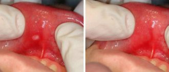
Ulcerative stomatitis very often occurs due to poor oral hygiene and bad habits (especially smoking). Often this type of stomatitis at its onset is confused with periodontitis and gingivitis, since it is characterized by inflammation of the gums, a grayish coating around the teeth and death of soft tissues. Often accompanied by fever and fever. One of the few forms of stomatitis, which often requires treatment by a doctor, and in the most advanced cases, surgical intervention.
Treatment methods
- Antibiotic therapy.
- Antibacterial treatment of affected areas.
- Surgical intervention (in severe cases, removal of areas of dead tissue and gum grafting).
Reasons for development
Stomatitis often occurs in people with reduced immunity and impaired microflora. If these 2 conditions are observed simultaneously, then even opportunistic microorganisms can cause severe inflammatory processes. Many other factors also influence the development of the disease.
Inflammation of the oral mucosa can occur in the following cases:
- mechanical, chemical, thermal injuries - hot burn, injury from solid food, cheek bite, etc. leads to tissue damage, penetration and spread of infection;
- poor nutrition, lack of vitamins, especially C, group B, minerals - zinc, iron and others;
- failure to comply with hygiene rules, eating unwashed fresh berries, vegetables, fruits, dirty hands while eating;
- treatment with medications that reduce salivation, antibiotics that destroy beneficial microflora, and a number of other medications;
- Frequent brushing of teeth with a paste containing sodium lauryl sulfate leads to a decrease in saliva secretion and dry mucous membranes, which become more susceptible to aggressive substances;
- poor-quality prosthetics and dental treatment due to which the gums are constantly injured;
- smoking, drinking alcohol;
- dehydration with prolonged diarrhea, vomiting.
Stomatitis often occurs during pregnancy due to hormonal imbalance. At risk are patients with diseases of the heart, blood vessels, gastrointestinal tract, pathologies of the immune and endocrine systems, and those with helminthic infestation. In diabetes mellitus, the aphthous form is diagnosed. People who use hormone inhalers to relieve asthma attacks suffer from candidiasis.
To answer the question whether stomatitis is contagious or not, differential diagnosis is necessary. Bacteria, viruses, and Candida fungi can be transmitted from person to person. The most contagious is the viral form of the pathology. However, with good immunity, the infection will not necessarily develop. It is also important how stomatitis is transmitted. If this is direct contact, for example a kiss, then the risk of getting sick is higher.
Treatment of bacterial stomatitis
This type of stomatitis is also called prosthetic stomatitis, since it occurs due to unsatisfactory care of the orthopedic structure, when many pathogenic microorganisms accumulate in the area of contact between the prosthesis and soft tissues. After treating foci of inflammation, it is necessary to carry out complete cleaning and antibacterial treatment of the prosthesis or replace it. This type of stomatitis should not be confused with allergic stomatitis, which develops against the background of an allergy to the prosthetic material. In this case, the prosthesis must be changed to a hypoallergenic one.
Treatment methods
- Anti-infective therapy.
- Mouth rinse.
Treatment of allergic stomatitis
This type of stomatitis is caused by the body's immunological response to contact with an allergen. The role of the latter can be played by anything: food products, new oral hygiene products, dentures, animal hair and much more. At risk are patients who are predisposed to allergies or suffer from various autoimmune diseases. A distinctive feature of allergic stomatitis is the acute onset of the disease, accompanied by an increase in temperature, severe pain and putrid odor from the mouth, which does not disappear even after thorough brushing of the teeth.
Treatment methods
- Identifying the allergen and eliminating it (or minimizing contact).
- Antihistamine therapy.
- Relief of inflammation with corticosteroids.
- Taking analgesics to relieve pain.
- Local antiseptic drugs
- Diet.
Prevention
To ensure that candidal stomatitis never bothers you, you should follow a few simple but very useful rules:
- Follow basic hygiene rules. Namely: wash your hands well with soap, especially when returning from the street, brush your teeth, use dental floss at least once a day, and also rinse your mouth with special means.
- You should definitely buy a new toothbrush. For those who have dentures, it is recommended to place them in a disinfectant solution overnight.
- After consuming liquid antibiotics, you should immediately rinse your mouth with water.
- When using spray corticosteroid drugs, you should wear a special nozzle.
Treatment of stomatitis in children
Treatment of childhood and adult stomatitis follows the same methods. At the same time, some drugs used for adults are replaced with those that are better suited for the child’s body. First of all, we are talking about antibiotics, antiseptics and drugs to enhance immunity. Children are often prescribed more gentle methods, which include the use of herbs and natural tinctures for rinsing. At an early age, dentists recommend preventing stomatitis using traditional medicine.
| Age | Type of disease |
| From 0 to 3 years | Candidal stomatitis, or childhood oral thrush. This type of stomatitis is especially common in infants. |
| 1 – 4 years | Aphthous and herpetic stomatitis caused by external infections and mechanical trauma to the oral cavity. |
| Children of primary and secondary school age | Allergic and aphthous stomatitis. |
Treatment of stomatitis at home
I would like to immediately note that treating stomatitis with folk remedies at home is possible, but you need to understand that in many cases it is impossible to do without a visit to a specialist and the use of antibiotics. Ignoring these points can lead to worsening of the disease and longer rehabilitation. In addition, on the Internet you can find a whole lot of “revolutionary” recipes for stomatitis, so you need to be able to separate the wheat from the chaff and understand which remedies can really help, and which are the fruit of the inflamed imagination of “couch healers”. Below we publish the most common methods of preventing and treating stomatitis with folk remedies, but please note that only an experienced specialist can draw up an optimal treatment plan.
In the case of children, treatment of stomatitis with folk remedies at home is used quite often. This is especially justified as a preventive measure, since the child’s body is more susceptible to the influence of the external environment. The most popular remedies are considered to be all kinds of decoctions, for the production of which chamomile, calendula, burdock, propolis and blackberry leaves are used. A homemade ointment is also used to treat stomatitis, which is made from novocaine, egg white and honey. Be that as it may, if your child shows signs of stomatitis, it is best to immediately take him to a specialist who can make the correct diagnosis and plan treatment.
Treating stomatitis with home remedies
| Facilities | Description |
| Hydrogen peroxide | Rinsing with a solution of hydrogen peroxide helps reduce discomfort and has an antiseptic effect. |
| Baking soda | Treatment of stomatitis in adults with soda is recommended when the disease is detected in the initial stages. A solution of the substance (a spoon in a glass of water) is recommended for rinsing. |
| Decoctions of chamomile, rose hips and calendula | They have disinfecting properties and contain vitamins and nutrients to maintain oral health. |
Diet for stomatitis
An adult with stomatitis is recommended to consume fermented milk products with a soft consistency without preservatives or additives, neutral vegetables and fruits rich in vitamins, and lean boiled meat. Low-fat broth is suitable as a first course. If stomatitis is caused by gastrointestinal diseases, it is useful to add cereal porridge to the diet. It is better to replace tea and coffee with plain water, compotes and decoctions. To avoid painful sensations, you should give preference to soft and warm foods. After a meal, it is recommended to rinse your mouth with clean water or a special solution.
Up to contents