A convex formation on the eyelid in the form of a lump is not uncommon. The causes of this pathology may be different, but in any case, a lump on the eyelid should not be ignored. The formation can develop on both the lower and upper eyelids. It may not cause any inconvenience other than aesthetic discomfort, but it may hurt and fester. The bulge usually has a round or elongated shape. It may not change in size for a long time, and sometimes, on the contrary, it increases rapidly. Let's look at the reasons for the formation of lumps on the eyelid and how to treat them.
Chalazion
Cones of this type are quite common. They develop from a sebaceous gland whose duct is blocked. This formation is also called a “grading lump” or “cold barley.” The continued production of sebaceous gland secretion leads to the accumulation of a viscous mass in the capsule, which stretches and thickens, taking the form of a dense lump. On palpation, the contents under the skin feel like a moving ball.
Cold barley develops at a slow pace, so it does not cause pain. Only a formed hard capsule can cause pain when squeezed. If a chalazion is not treated, it can develop into a cyst. As the lump develops, the risk of complications increases: inflammation, formation of a purulent fistula, granulation.
Diagnosis of cheek cancer
If a tumor of the mucous membrane of the cheek is suspected, oncologists at the Yusupov Hospital conduct an examination using a mirror, palpation of the tumor and lymph nodes. If a long-term non-healing ulcer is detected, a biopsy is performed. If a negative result of histological examination of the material obtained during the biopsy is obtained, the suspicion of the malignant nature of the tumor remains, the biopsy is performed again.
Oncologists clarify the stage of the tumor process, assess the extent of spread of the malignant tumor from the buccal mucosa to adjacent tissues, and find out whether there are metastases to regional lymph nodes and distant organs. If the presence of metastases is suspected, computer and magnetic resonance imaging, ultrasound, and scintigraphy are used. Distant metastases are found in 20% of patients at the time of diagnosis.
Computed tomography and magnetic resonance imaging make it possible to assess the condition of the deeper anatomical structures of the oropharynx and surrounding tissues. If there is a suspicion of metastases to the lymph nodes or tumor infiltration of the floor of the mouth, a cytological examination of the aspirate obtained under ultrasound guidance is performed. To exclude distant metastases, a chest x-ray in two projections and an ultrasound examination of the abdominal organs are done.
Considering that the prognosis for cheek cancer is serious, a tomography of the neck, chest and upper abdominal cavity is performed. Using bone scintigraphy, I exclude bone metastases. Positron emission tomography allows one to identify the source of metastases in cases of undetected primary tumors.
Video from our specialist about the disease and its treatment
There are cases where chalazion spontaneously resolved without medical intervention. However, most often this formation does not develop back and requires prompt and conservative help. Treatment for such a lump on the eyelid is prescribed by an ophthalmologist. If the chalazion is small and not old, you can limit yourself to UHF therapy, ointments and eye drops. More severe cases are treated by injecting corticosteroids into the capsule cavity. Local drugs (ofloxacin, dexamethasone, sodium sulfacyl, hadrocortisone, levofloxacin, tetracycline ointment) can also be used as an addition to the injection.
If drug therapy is ineffective, the doctor decides on surgical treatment. The operation to remove a chalazion is performed under local anesthesia and lasts no longer than 15 minutes.
Barley
Styes are more common than chalazions. This type of lump on the lower or upper eyelid is caused by inflammation of the follicle (bulb) of the eyelash. This also clogs the sebaceous gland duct. Styes develop over several days or even hours and can occur in both adults and children. More often, the systematic appearance of barley is observed in people with weakened immune systems or who have changed their place of residence to an area with a more severe climate, as well as in people exposed to constant stress factors.
Based on their origin, there are two types of barley. Inflammation can be external (when the sebaceous gland suppurates) and internal (when the source of inflammation is located in the membolic gland).
The development of external styes is characterized by subjective sensations similar to a foreign body entering the eye. The initial stage may also be accompanied by stabbing pain. External stye visually manifests itself as redness and swelling of the eyelid. The internal one is usually not so noticeable, but it causes even more discomfort and pain.
Without treatment, barley develops within a few days into an abscess, which opens with the release of purulent contents. This brings relief, but an open wound is dangerous due to the possibility of re-infection.
It is better to start treating barley without waiting for the abscess to spontaneously break through. This allows you to get rid of the painful lump faster and with less risk of complications. If you still don’t have the courage or time to visit an ophthalmologist, you should remember that prolonged suppuration of the eyelid is very dangerous. If the stye does not open for more than two weeks, surgical treatment is necessary. An ophthalmic surgeon will remove the abscess under local anesthesia and give recommendations for further treatment of the eyelid. Most often, therapy for developing or already opened barley includes drops and ointments that contain antibiotics (albucid, gentamicin, erythromycin, tetracycline ointment).
Furuncle
A boil on the eyelid is one of the most dangerous lumps. The disease causes purulent necrotic inflammation of the ciliary follicle, sebaceous glands, and surrounding connective tissue. The main cause of boils is staphylococcus.
Typically, the localization of the boil is the part of the upper eyelid close to the eyebrow. Much less commonly, a lump appears on the edge of the eyelid, very close to the eye.
In its development, the disease goes through the following stages: first, a small painful nodule appears and the surrounding tissues swell (sometimes both the eyelid and the entire side of the face swell); after a few days, a yellowish abscess appears at the top of the nodule. Then the node fluctuates, the abscess opens with emptying of the contents. On the eyelid, after opening the boil, a plug (rod) remains, consisting of purulent masses and necrosis products, which after some time spontaneously comes out. As a rule, a small scar remains at the site of the boil.
A painful lump and swelling are not the only signs of a boil; the patient often feels weakness, headache and fever.
Treatment of a boil must be carried out by a doctor who will prescribe the necessary medications (antibiotics, sulfonamides, painkillers) and, if necessary, open the abscess. It is prohibited to squeeze out boils yourself.
Millums (millet)
This type of bump occurs with equal frequency on the upper and lower eyelids. Millet grains can range in size from a poppy seed to a grain of rice and usually form in groups. Millums are the most harmless of all formations and cause only aesthetic discomfort. At its core, these are whiteheads localized in the eyelid area.
Millet removal should only be done by a cosmetologist. Since they do not carry the risk of complications, they are not considered an ophthalmological disease, but fall within the competence of a dermatologist-cosmetologist.
Prevention of millums includes caring for the eyelids, timely removal of dead epidermal cells, ensuring the cleanliness of the ducts of the sebaceous glands and pores of the skin around the eyes, as well as a balanced diet that excludes excessive consumption of fatty foods.
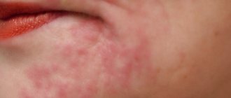
Xanthelasma
This type of eyelid bump looks more like a flat plaque. A connection between the development of such formations and chronic metabolic disorders has been revealed. Most often, xanthelasmas appear in women suffering from diabetes, hypercholesterolemia, and pathologies of the endocrine system.
These flat formations only partially rise above the skin and have a yellowish tint. They can be located not only on the eyelid, but also in the area around the eyes. In most cases, xanthelasmas appear on the skin in groups. They do not resolve on their own. Cosmetic removal is possible, but it should be understood that if the cause of their formation is not eliminated, it can lead to the appearance of new plaques. You should focus on treating the underlying disease, and only after that seek cosmetic help.
Bumps on the face: causes and provoking factors
Subcutaneous formations are due to various reasons. Sometimes blockage of the sebaceous glands is caused by pathogenic microorganisms - bacteria. Their accumulation leads to the development of inflammatory processes; accordingly, the clinical picture worsens and pain occurs.
Many people believe that the formations appeared as a result of improper skin care, but this is not always true. The human body is the most complex mechanism known to mankind.
All processes in it are interconnected; a failure in one area can lead to disruption in another. Why do bumps appear under the skin?
1xBet application for Android phones
The etiology of occurrence is based on the following factors:
- Hormonal imbalance (excess or deficiency of hormones).
- Poor nutrition, bad eating habits.
- Alcohol abuse, smoking, drug use.
- Allergic reaction to food, medications, etc.
- Disruption of the central nervous and endocrine systems.
- Overheating or hypothermia.
- Infectious processes in the body.
Another reason is the excessive use of various cosmetics of synthetic origin. Unfortunately, many care products do not meet standards and contain harmful substances.
For your information, a squeezed pimple or blackhead can lead to unforeseen consequences in the form of sepsis, suppuration with a characteristic thickening. As a result, unsightly scars remain on the face, which are quite difficult to get rid of.
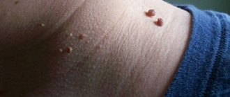
Papilloma
The causative agent of benign formations called “papillomas” is human papillomavirus. Most often, infection with this virus occurs during birth from mother to child, but the disease can also be acquired during life through contact with infected people. The virus can appear on almost any area of the skin and mucous membranes in the form of round growths. Papilloma is usually painless. However, it can hardly be called “aesthetically attractive”, so it causes significant discomfort.
You can distinguish papilloma from other types of formations by the following characteristics:
- the cone is more like a ball on a stalk or has a mushroom-like shape;
- the wart-like growth has a rough surface that resembles the surface of cauliflower to the touch.
It is worth noting that a growth with a smooth surface is not a papilloma, and it must be examined by an oncologist.
Treatment of papillomas should be comprehensive. Surgical removal is performed by a dermatologist. He also examines the type of virus and prescribes drug treatment. The fact is that the manifestation of a disease at one point does not mean that the entire body is not infected. The virus is suppressed by the immune system, but travels through the bloodstream. Some types of human papillomavirus are very dangerous with a high probability of degeneration of skin formations. Only an experienced dermatologist can prescribe adequate treatment based on diagnostic results. Measures to improve overall immune status are essential in the treatment of papillomas.
Causes of cheek cancer
Cancer of the buccal mucosa develops under the influence of the following provoking factors:
- Use of tobacco in any form (cigarettes, cigars, pipes, chewing tobacco);
- Alcohol abuse (the risk of developing cancer increases when the use of alcohol and tobacco is combined);
- Infection with carcinogenic forms of human papillomavirus.
One risk factor is exposure to sunlight. Both family history and genetic predisposition, as well as exposure to mutagenic environmental factors, play a role in the development of cheek cancer. The formation of a malignant tumor occurs in several stages. The most important is the disruption in the functioning of oncogenes and genes that inhibit tumor growth. The development of malignant neoplasms of the cheek is associated with inactivation of the p16 gene, mutations in the p53 gene, and the introduction of the human papillomavirus.