Causes of darkening of tooth enamel
The cause of tooth discoloration can be not only staining of the enamel, but also darkening of the hard tissue underneath. While the first may simply be the result of eating habits, the second indicates the presence of an internal problem.
Dark enamel in all or most of the teeth may appear due to the following reasons:
- Excessive consumption of coffee and strong tea,
- Smoking
- Metabolic disease
- Long-term use of certain medications, such as tetracycline-based antibiotics
- Reaction to filling material
- thinning of the enamel after nerve removal.
From smoking, tobacco tars accumulate on the teeth, which are not completely removed during normal hygiene procedures. Food dyes from some foods, as well as tea, coffee and other drinks, also tend to “eat” into the enamel over time.
Improper metabolism leads to an imbalance in the acid-base balance in the mouth, which causes bacteria to multiply more actively and plaque to form more actively. That is, a patient whose enamel of all teeth has darkened should pay attention to the condition of the gastrointestinal tract.
Some medications have the ability to change the pigmentation of enamel, for example, antibiotics from the tetracycline group or iron-containing drugs taken in liquid form.
Darkening can also occur when using outdated filling materials containing metals (silver or even tin). But even modern materials, under incorrect storage conditions, change their properties and adversely affect the color of the enamel.
The “wear and tear” factor cannot be ignored. If the tooth has been depulped for a long time, then it is not supplied with useful substances. This means that all mechanical actions lead to abrasion of the enamel and gradual destruction. Careful care slows down this process, but does not completely eliminate it.
Often the patient cannot understand why dark teeth appeared in a row against the background of others that were still light. This may mean that a pathological process has occurred under the still intact tooth crown, which has changed the color of the hard tissue - dentin.
Darkening of dentin can be due to the following reasons:
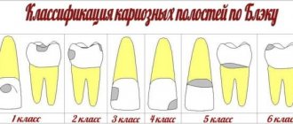
- untreated caries or its relapse
- advanced stage of pulpitis
- poor quality pulp removal
- injury from a strong blow
Caries destroys part of the protective layer of tooth enamel and gets underneath it. As the infection continues to develop, it affects the internal tissues, causing them to turn black. The affected dentin shows through the enamel, so the tooth can stand out noticeably among its white “neighbors.”
Pulpitis is often accompanied by pain, which brings the patient to the dentist even before the pulp dies. But if treatment was not carried out at the acute stage, then the chronic stage begins. At this stage, the pulp (nerve) begins to die, which changes the color of the tooth.
After removal of the nerve, if the operation was performed poorly, the color may also change. Insufficiently thorough cleaning of the canals will cause further decomposition of the remaining pulp. If not all cavities are filled with filling material, then microbes will again penetrate into the remaining voids. All this will again cause darkening of the dentin. Less commonly, the cause of darkening may be the paste used to seal the root after cleaning the canals. Sometimes its composition changes the shade of hard tissues.
Trauma to a tooth causes it to darken due to blood entering the bone tissue.
Sometimes, if a tooth has been restored, the pins may be visible through the filling compound. In this case, repeated restoration may be required.
Medical Internet conferences
The influence of chlorhexidine on the microbial composition of the bottom of a prepared carious cavity
Tverskova V.Yu.
Scientific supervisor: candidate of medical sciences, assistant Kazakova L.N.
GBOU VPO Saratov State Medical University named after. IN AND. Razumovsky Ministry of Health of the Russian Federation
Department of Pediatric Dentistry and Orthodontics
Relevance of the problem. Among oral diseases, caries ranks first in prevalence. Currently, caries is diagnosed in children under 2 years of age in 36% of cases, and in 3-year-old children in 53% of cases[1]. The incidence of deep caries in children is 23% of the total number of caries diseases. According to many authors, caries can be classified as an infectious disease [2], the leading factor in which is Streptococcus mutans. Str. mutans attaches to the smooth surfaces of teeth, participates in the formation of dental plaque, and this action is mediated by the synthesis of glucose polymers from sucrose present in food. Glucan formation causes intercellular aggregation of Str. mutans and other bacteria present in the plaque. The sticky glucan matrix of dental plaque prevents the diffusion of large amounts of acid produced by microorganisms, which prolongs its residence on the surface of the teeth and leads to demineralization of the enamel, causing dental caries. The emerging focus of demineralization of the surface layer of enamel is the “entry gate” for further infection of deeper located hard tissues and dental pulp, this is especially true immediately after eruption, at the first stage of secondary mineralization. The special structure of hard tissues, characterized by less mineralization at this stage of tooth development, creates a number of difficulties in the treatment of acute deep caries with a favorable outcome. Since it is impossible to create conditions close to sterile at the border with the pulp using standard irrigation of the prepared cavity.
Goals and objectives of the study. The purpose of our work was to study the effect of chlorhexidine bigluconate, used in different forms, on the qualitative microbial composition of the prepared carious cavity in acute deep caries.
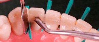
Materials and methods of research. During the study, a group of children (20 people) was identified with a diagnosis of acute deep caries of permanent teeth at the stage of ascending development. To determine the microbiocinosis of the bottom of the carious cavity, a bacteriological study was carried out.
Acute deep caries was treated conservatively only in children with a good level of oral hygiene and with a compensated form of caries in two visits.
On the first visit, rational anesthesia was carried out, the carious cavity was prepared under a bath of the same antiseptic, this was done carefully, but with extreme caution. After the final preparation of the carious cavity with a sterile bur, a section of dentin was taken from the bottom and a bacteriological examination was carried out. Then the children were blindly divided into 2 groups of 10 people. In the first group of patients, a tampon moistened with 0.05% chlorhexidine was placed on the bottom of the carious cavity under a temporary dressing. In the second group, a “patch” of self-adhesive film “Diplen Denta” containing chlorhexidine bigluconate 0.01-0.03 mg/cm² was applied to the bottom of the carious cavity under a temporary dressing. Further research was carried out after 48 hours.
On the second visit, having previously isolated the tooth from saliva, a temporary bandage and a tampon with an antiseptic were removed in the first group; in the second group, the self-adhesive film dissolved on its own, and in both cases, a sterile spherical bur without pressure was taken from the bottom of the cavity for bacteriological examination. In patients, a calcium-containing preparation was applied to the bottom of the carious cavity, and the cavity was filled with permanent filling material “Vitremer”.
Further bacteriological examination was carried out in accordance with generally accepted rules of clinical microbiology.
Results. Microbiological examination of the bottom of the carious cavity in patients after complete preparation of the carious cavity revealed a significant degree of its contamination with pathogenic and conditionally pathogenic microflora. Streptococci, enterococci and yeast of the genus Candida were sown (Table No. 1). In the group of patients, when using 0.05% chlorhexidine during re-seeding from the bottom of the carious cavity, a decrease in the number of streptococci was observed, the effect of 0.05% chlorhexidine on enterococci was insignificant, the growth of yeast of the genus Candida was not detected (Table No. 1).
When using the film "Diplen Dent" a significant decrease in the colonization of cariogenic species Str. mutans, Str. Sanguis, Str. salivarius. The number of representatives of the resident group of bacteria - enterococci - decreased (Table No. 1).
We associate the higher efficiency of the Diplen Denta film containing chlorhexidine bigluconate in a concentration of 0.01-0.03 mg/cm² with its ability to adhere tightly to the bottom of the cavity, which ensures deeper penetration of the active component through weakly mineralized dentin and dentinal tubules.
Conclusions. Thus, the results of the study confirm that the application of chlorhexidine digluconate in the form of a film for 48 hours, as an intermediate stage in the treatment of acute deep caries in children, leads to a significant decrease in the level of streptococci, enterococci and yeast of the genus Candida in the peripulpal area.
What to do with a darkened tooth
If a tooth has darkened, the first thing to do is to determine the cause of the problem. The situation with coffee lovers and smokers is clear - they should have their teeth professionally cleaned by a specialist and adjust their habits. In the future, you can sign up for whitening, but first you will have to prepare yourself mentally, because after it, smoking and drinking coloring drinks are strictly prohibited for some time.
If the enamel of your teeth has darkened due to medications, then you need to either limit their intake or switch to other medications if the attending physician approves. To reduce your exposure to liquid coloring medications, drink them through a straw. If it is impossible to change the therapy, and the pigmentation causes severe discomfort, then installing veneers will help - plates that replace the top layer of enamel.
Only a qualified dentist can make a correct diagnosis. Therefore, the best thing to do if a tooth has darkened is to go to see a doctor. It will determine the presence or absence of a pathological process. There is no need to postpone your visit. If the cause of the color change is inflammation, then an assessment of the situation by a specialist is necessary. Internal inflammatory processes are not always accompanied by acute pain. If you neglect other signs, then you may subsequently encounter not only chronic pulpitis, but also other unpleasant pathologies, such as granuloma, periodontitis, and radicular cyst.
Granuloma and cyst are formations with purulent or serous fluid inside. Their development can proceed practically asymptomatically for some time, making itself known only by a change in color. But then you cannot do without serious interventions: cutting tissue, removing part of the root, or even the entire tooth. Purulent inflammation can even damage the jaw bone. In this case, you cannot do without tooth extraction.
Removal is necessary in the following situations:
- the inflammation spread to the area around the tooth and affected the jawbone;
- the tooth is loosened and the gums are bleeding due to internal injury;
- the crown is too destroyed, less than 30% of hard tissue remains;
- the tooth is split, and the split continues inside the gum.
In the latter case, it is possible to preserve the roots of the tooth, but the cracked upper part will still be removed and replaced with a prosthesis.
It is clear that if a tooth suddenly darkens, an examination should be done immediately. To save a tooth, you need to see a dentist immediately. If there is inflammation, the doctor will remove the infected tissue and fill the voids. After treatment, it may be necessary to bleach not the outer part, but the inner part of the crown, since it was this part that darkened due to the death of the pulp or its remains. Often after this, the teeth return to their former whiteness and natural appearance. In other cases, when lightening and restoration with filling materials are impractical, but removal is not required, partial or complete “artificial enamel” is created for the tooth - veneers or crowns are installed.
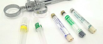
To whiten a pulpless, darkened tooth, the doctor brightens it from the inside. To do this, he opens the canals, cleans them and fills them with a bleaching compound. Until the composition lightens the tissue, the patient will have a temporary filling. This procedure is called endobleaching. In some cases, it may take several sessions.
If the tooth walls are too thin, or there are other contraindications for whitening, then, depending on the situation, the dentist will select alternative methods: direct restoration with composite material, installation of veneers, lumineers or crowns. Positive aspects of restoration with composite material:
- ease of implementation - the tooth is covered with a composite composition, forming one large filling on its surface;
- speed of implementation - can be done in one visit to the doctor;
- affordable price.
Disadvantages of such restoration:
- composite material is easy to paint, you will have to change your eating habits;
- fragility - average service life is about 5 years;
- During repeated restoration, the integrity of the tooth walls suffers.