Content:
- How long does it take to bleed after tooth extraction?
- Why is it important how long it takes to bleed after tooth extraction?
- Types of violation
- Why does it take a long time to bleed after extraction?
- Possible consequences of bleeding
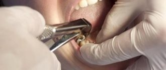
Extraction is one of the most difficult operations performed by dental surgeons. During the recovery period after it, serious complications may develop, one of them is bleeding. Therefore, the gum healing process requires close monitoring by the doctor and vigilance of the patient himself.
If none of the above helps -
If nothing helps, and if you feel weak and dizzy, then you shouldn’t wait any longer. It is necessary to urgently consult a doctor.
- The best option in the conditions of Russian free healthcare is to take a taxi to the nearest 24-hour private dental clinic, where you will get stitches. If you feel weak, then do not drive yourself unless you want to get into an accident. For anesthesia, 2 stitches and a hemostatic sponge you will have to pay approximately 750-1000 rubles.
- Feel free to call an ambulance - when talking to the ambulance dispatcher, complain not only about bleeding, but also about severe dizziness and weakness. With such symptoms, an ambulance will definitely arrive. Usually they will first try to stop the bleeding using tampons with peroxide (which a priori cannot help in this situation). Seeing the failure of such therapy, they will be forced to take you to the hospital, where they will put stitches on your wound and send you home.
Suturing the socket: before and after photos
Why is it important how long it takes to bleed after tooth extraction?
Stopping bleeding indicates the beginning of the formation of a dense clot. It is needed to protect the wound surface from the penetration of bacteria and microbes. It also serves as a mechanical barrier for particles of food and drinks.
If the blood does not clot, the socket will be unprotected. Pathogenic microorganisms will begin to penetrate into the bare bed. It will secrete ichor (this is indicated by the salivary fluid acquiring a characteristic pink hue). In this case, recovery after surgery will take a long time and, most likely, will be accompanied by various complications.
What can you do at home?
If the problem has not resolved itself within three hours, then the following tips will help you:
- Apply a cold compress to your cheek. You can use a cold product or ice previously wrapped in cloth.
- Roll up sterile gauze, apply to the hole and bite for 10-15 minutes.
- Make strong tea. Soak a piece of bandage in liquid and apply to the damaged mucous membrane for 3-5 minutes.
- If you experience high blood pressure, you need to normalize it with medication. The specific drug and dosage are prescribed by the doctor.
Types of violation
Dentists distinguish several types of postoperative bleeding:
- Early or primary. The blood does not stop flowing for a long time immediately after the unit is pulled out. The dentist has to take various measures to stop it.
- Later or secondary. The bleeding quickly stopped at the clinic, but reappeared at home. In such a situation, many people are lost and do not know what to do. There is only one rule - if a negative symptom persists for more than an hour, you should immediately visit a dental surgeon.
Generally speaking, the appearance of blood at the site of the hole should be taken calmly. During extraction, soft tissues and blood vessels are damaged. Therefore, there is nothing strange about bleeding. You need to look at its dynamics. Every day a person’s condition should improve, as should the appearance of the hole in the oral cavity. If this does not happen, then there is some problem. It needs to be identified and eliminated.
The treatment regimen includes:
- systematic professional teeth cleaning – removal of plaque and tartar;
- a course of physiotherapeutic procedures to relieve inflammation;
- sanitation of the oral cavity.
The next stage of treatment is home therapy: rinsing with disinfecting solutions (salt and soda, chlorhexidine).
For a purulent infection, the patient is prescribed antibiotics.
At home, you can independently use traditional medicine methods, including decoctions and infusions of medicinal plants:
- oak bark;
- chamomile flowers;
- sage herbs.
You can use these decoctions and calendula tincture as lotions on problem areas of the mouth.
Use of pharmaceutical products:
- fluoridated toothpastes with the addition of medicinal herbal extracts;
- dental anti-inflammatory gels (Cholisal);
- vitamin complexes.
Why does it take a long time to bleed after extraction?
The reasons may be different, there are many of them. The most common:
- A sharp increase in blood pressure. Occurs in response to stress experienced during dental surgery.
- Vascular injuries. It is impossible to remove a unit without damaging them.
- Hormonal imbalance or increased levels of estrogen in the body. This reason only applies to women.
- Taking medications containing acetylsalicylic acid on the eve of a visit to the dentist. Aspirin helps thin the blood and prevents its rapid clotting. Some other medications work similarly. Therefore, it is important to be cautious about using any medications in the days leading up to the extraction.
- Gum injuries. The patient himself is usually to blame. The desire to touch the blood clot with your hands, tongue, eating too hard foods and chewing them on the inflamed side of the jaw, rinsing the mouth, drinking hot drinks or dishes are all causes of postoperative bleeding.
The wound heals worse if the gum in the area of the extracted tooth has been affected by gumboil or a cyst. In this case, the patient should be constantly monitored. Most likely, he will have to take anti-inflammatory drugs, antibiotics, and applications with antibacterial compounds for some time.
Anemia, hemophilia, alcoholism, diabetes mellitus, and atherosclerosis also increase the likelihood of postoperative complications. You must inform your doctor about the presence of these diseases. It is possible that he will prescribe hemostatic drugs.
Stopping bleeding during dental restoration
November 7, 2013
Introduction
Traxodent paste
It would be great if the gums did not bleed, since blood entering the surgical field greatly complicates the restoration of teeth. The cause of bleeding gums during restoration operations is dental plaque or trauma. Plaque leads to gingivitis, caries or periodontitis. Trauma that results in bleeding may occur during surgery. The wedges exert lateral pressure on the periodontal papilla. The cavity sealing process uses metal or plastic matrices with sharp edges that can injure healthy or inflamed tissue. Dental burs are used to remove caries, excise inflamed tissue and widen the gingival sulcus. Retraction cords are placed to deflect or pull back the gum in an attempt to reach the edges of the cavity. All this leads to blood contamination of the restoration area, which in turn complicates procedures such as taking accurate impressions, preparing the dental cavity, prosthetics and cementing. This problem is typical for orthopedic dentistry.
Dental prosthetics and periodontal condition are interconnected. Poor dentures may have ridges and difficult-to-clean areas that allow plaque to accumulate. In adolescents and older adults, oral hygiene may be poor. Insufficient prevention of plaque formation leads to the need for dentures. In addition, the main contributing factor to dental prosthetics in patients in the following case studies is a diet high in highly refined carbohydrates (carbonated drinks) and lack of nutrients (refined foods).
Rice. 1 Blood got into the first impression, resulting in voids
In order to exclude the development of gingivitis or periodontal pathology, it is necessary to ensure an accurate marginal fit of the prosthesis. A good example is fixed orthopedic restoration. The fit of the prosthesis depends on the completeness of the impression. If the dental laboratory receives an impression that is incorrect due to blood contamination, problems with the prosthesis arise. Based on such impressions, stamps will be inaccurate due to the formation of voids and bubbles. (Picture 1). If a commercial dental laboratory produces a denture using an inaccurate die, the dentist will receive an inaccurate denture that will not fit the patient and will be rejected. Everyone will be disappointed - the patient, the doctor, and the dental laboratory. As a result, everyone loses income. The patient will need to take time off work, the doctor will need to provide additional chair time, and the dental laboratory will need to facilitate the entire process. When a problem occurs, people usually blame it on someone else. For the dentist this may seem like a personal failure, but in reality it is a systemic failure. If this system is not corrected, the dental office's main source of income will suffer. The main task is to understand how blood and gingival fluid enter the surgical field during restoration work, and what can be done to improve the control system.
The defective impression was made according to a standard procedure using a retraction cord soaked in a hemostatic solution and a one-step impression. The thread was placed on the inflamed tissue to treat subgingival caries. Treatment of subgingival caries required subgingival extension of the tooth core, which led to the need for subgingival preparation for a crown. Bleeding of the tissue is typical for such cases. The system used must control bleeding during the impression taking process.
Rice. 2 Traxodent paste is applied to the bleeding groove
To ensure the accuracy of the indirect technique, a new impression must be taken. You should wash the bleeding tissue and try to dry the wet area. A straight cannula is attached to the Traxodent syringe. In this case, the cannula was bent over the handle of the mirror at an angle of 90°. This facilitates positioning in posterior areas of the mouth where direct access is not possible. Traxodent paste should be applied over the bleeding tissue, slowly squeezing over the gingival sulcus along the edge of the preparation.
The paste can remain in the groove for 1-2 minutes (Figure 2), then it must be rinsed with a water/air gun. After washing and drying, no bleeding is observed, the gingival sulcus becomes dry. To obtain a one-stage impression, impression material of low and medium viscosity is used. Other types of impression material are used with no less success.
Fig. 3 The repeated impression, in which no blood has entered thanks to Traxodent paste, has no voids
Traxodent paste is a retraction system based on Hemodent paste. The paste contains aluminum chloride, which causes tissue retraction and contraction. Aluminum chloride promotes protein precipitation, contraction of blood vessels and removal of fluid from tissues. Aluminum chloride-based paste reduces the risk of postoperative inflammation. Aluminum chloride is the least irritating of the retraction medications. This drug, placed in the groove, does not cause noticeable gum recession. Pallor of the gums is the first sign of the paste's action. After this, the oozing blood turns brown and stops flowing. These two signs indicate hemostasis and a positive result of impression taking. (Figure 3).
Thanks to the methods used in this system, a flawless re-impression is obtained without the use of additional retraction cords. A re-impression using Traxodent paste is preferable because a small amount of impression material is left behind the preparation edge. This ensures that the edges of the plaster die are precisely processed in the laboratory, resulting in a precise fitting prosthesis.
Rotational curettage with Traxodent paste
Rice. 4 Blood and fluid in the gingival groove
Let's consider the case when the tooth has an inlay for building amalgam on pins. This tooth requires support from the entire crown. This tooth had a healthy groove and sufficient amount of attached gum. The entire crown requires a clasp to improve prognosis. Creating a clasp requires preparing the tooth to the bottom of the gingival sulcus. Rotary curettage was used to create a groove using a high-speed diamond bur to quickly remove epithelial tissue in the groove adjacent to the margin. Studies have shown that rotary curettage has little effect on gingival margin height if there is adequate gingival keratinization.
Rice. 5 Traxodent paste applied close to bleeding tissue
Rotational curettage is necessary to create 0.2 mm of thickness in the sulcus to maintain adequate thickness of the polyvinylsiloxane impression material. This thickness is required to create tensile strength and prevent breakage when removed from the oral cavity. Removal of the sulcus mucosa resulted in bleeding in the repair area (Figure 4).
Traditional methods would require the application of a retraction cord for 4 to 10 minutes to widen the groove. In this case, Traxodent paste was applied for 2 minutes to control the bleeding caused by rotary curettage (Figure 5). Additional deflection was mediated by the use of the Premier retraction cap (Figure 6). After rinsing with a water/air gun, only amalgam residues are retained. The groove is dry, hemostasis has been achieved (Figure 7).
|
|
Application of Traxodent paste in the aesthetic area
For aesthetic procedures on anterior teeth, creating a groove around the edge with a bur, floss or laser may not be desirable as it may detract from the appearance. Cosmetic restoration procedures are difficult because the preparation line is in close contact with the gum. If the root of the tooth is very dark, a dark line appears in the area of the neck of the tooth. Patients view this as a cosmetic defect.
Applying a thread can lead to ulceration or inflammation of the connective epithelium. The problem is that it is difficult to control the force of thread application. Other trauma, such as mechanical pressure or surgical damage, can cause unwanted migration of the gum line away from the margin. Traxodent paste is the least traumatic method of drying the field and causes slight displacement and retraction of the tissue.
Let's consider a clinical case. The patient is very concerned about the aesthetic outcome and does not like the dark contrasting color along the gum line. She had a cracked porcelain veneer that needed replacing. The tooth has already been prepared at the edge of the gingival sulcus. The tooth has already been prepared at the crest of the gingival sulcus, and the edges will remain in the same position (Figure 8). Due to accidental contact of the bur with the groove, some areas were damaged and began to bleed. Traxodent paste is applied to the groove and remains there for 1-2 minutes (Figure 9). The tooth is washed with an air-water jet and dried. The area is ready to receive an impression (Figure 10). A one-step impression using low to medium viscosity tray material will produce an accurate impression without bubbles or voids. When examined after 3 months, the margin remains stable and within the aesthetic zone (Figure 11).
|
|
|
|
Hemostasis with Traxodent paste for class II fillings
Rice. 12 The gums are accidentally damaged when drilling out caries
The risk of blood entering during the filling of class II cavities is as high as when installing a crown or bridge. This problem may mean longer procedures for the patient and changes in the dentist's chair schedule. Bleeding in the interproximal area is usually associated with caries (Figure 12). Bleeding begins from the inflamed interproximal gingival area if it is touched with a bur during removal of carious dentin.
Bleeding also occurs during isolation of a carious lesion when a ring, wedge or rubber gasket comes into contact with inflamed tissue (Figure 13). Traxodent paste is applied to the bleeding area located in the deepest part of the interproximal pocket (Figure 14). After 1-2 minutes, the paste is washed off with an air-water jet and hemostasis is confirmed. It is obvious that the entire surface of the cavity can be visualized, after which filling is carried out in the usual way.
|
|
Discussion
The value of these case reports is that they demonstrate a new hemostasis technology (Traxodent paste) that can solve common bleeding problems in the area of restorative treatment.
Thanks to its balanced fluidity, the paste can penetrate and remain in hard-to-reach areas of the groove. This balanced flow is important when treating the upper and lower dental arches, as the drug remains in the groove without getting into the laryngeal vestibule or throat. This is important for the patient, as the procedure becomes less unpleasant. Typically, hemostatic agents are strong astringents that create a dry, tight, and rough feeling in the mouth. This new technology is important for the dentist because it allows the drug to remain in direct contact and in its entirety in the gingival sulcus. There is no need to re-apply the drug due to dissolution by saliva or leakage. After application, the drug remains in place, even in contact with the tongue or cheek. Paste differs from liquid hemostatic agents in its ability to absorb liquids. Treatment areas adjacent to the mucous membrane of the alveolar processes of the jaw may become contaminated to deeper spaces. The physician should be aware of the problems that arise in such areas with insufficient adjacent keratinized tissue. As one of the warnings, we can mention the risk of leaching of the hemostatic drug into deep areas between tissues that are not intended for applying the product. The paste is not intended for the treatment of gingivitis, periodontitis and other diseases. The doctor should supervise the application of the paste to the dental treatment area. Traxodent paste is intended to be inserted onto the surface of the tissue, like a bandage, and not to be inserted into the submucosa.
Conclusion
Bleeding is a common problem that occurs during the dental restoration process. Common causes include plaque-induced erythema due to gingivitis, along with the use of instruments in the treatment area or the inadvertent use of instruments that create tears in the gingival tissue during restorative procedures. Any injury to the inflamed tissue leads to bleeding in the restoration area. Bleeding leads to deformation of the impressions, lack of adhesion of the filling and contamination of the cement.
Traxodent paste is a retraction system based on Hemodent paste, which contains aluminum chloride. Traxodent stops gum bleeding and causes temporary soft tissue contraction without further tissue damage.
Dean Eledge DDS, MD Associate Professor, AEGD
see also
September 13, 2013
Traxodent/Traksodent - material for hemostasis and gum retraction, Premier
Retraction system based on Hemodent® paste
March 21, 2012
PREMIER. Unconditional quality of dental materials
The American company Premier was founded in 1913. Soon it will be 100 years since Premier cherishes its traditions and values its reputation, so the company’s specialists carefully monitor the production process and always strive to improve the level of product delivery. UNIDENT presents the Premier line of dental materials, in which you can find materials that are indispensable for a dentist’s work.
October 30, 2011
Traxodent System: Predictive Management of Gingival Tissue
In modern dentistry, control of gingival fluid and gingival tissue is the primary criterion required for a successful clinical procedure. "Traxodent" is a unique system for high-level hemostatic processes. It is available in convenient syringes. Its paste-like consistency improves the retraction of gingival tissue, and 15% aluminum chloride, which is part of the paste, has very high hemostatic properties. Return to section
What could be the consequences?
Complications and consequences can be disappointing. Losing a tooth is not the worst thing that a rotten tooth can lead to. An abundant accumulation of pathogenic microorganisms often provokes problems with the body and disrupts the functioning of internal organs. Let's consider a number of the most common manifestations:
- Blood poisoning.
- Inflammation of the meninges.
- Constant headaches.
- Sleep disturbance.
- Problems with the gastrointestinal tract.
- Lack or significant decrease in appetite.
- Damage to adjacent teeth.
Causes of tooth decay. How to stop him?
To provide adequate treatment, the doctor must first determine the cause of the disease. Otherwise, the measures may turn out to be useless, the process will continue to develop. The causes of tooth decay are external and internal factors. Despite the fact that enamel is considered one of the most durable materials in the body, under unfavorable conditions it quickly deteriorates. As a result, microbes gain unhindered access to the inner, less protected part of the tooth.
To cope with this problem at the initial stage, one visit to the dentist is usually enough. He clears the cavity of affected tissue and closes it with a filling. However, simple neglect of their health and fear of the dental office make people postpone the visit. The result is deterioration of the teeth and their loss.