Why is this happening
The etiology differs from case to case. An infrequent, but one of the most unpleasant options is a genetic predisposition to the disorder. If it is identified, a person will almost inevitably encounter painful dental symptoms, even if he takes very careful care of his oral health. Other possible reasons why the gums recede, move away from the front, molars, and wisdom teeth and become exposed:
- poor hygiene (neglect of brushing, flossing, annual professional maintenance, regular visits to the dentist), as a result of which plaque forms on the neck, and later tartar;
- malocclusion;
- inflammation of the periodontium (tissues that seal the alveoli), provoking the accumulation of pathogenic bacteria, and subsequently purulent contents in the periodontal pocket;
- errors in previous orthodontic or orthopedic treatment, which can lead to injury;
- mechanical, thermal and chemical damage - often pathology is provoked by irritation from the habit of gnawing foreign solid objects, drinking too hot or sour drinks;
- poor-quality prosthetics, in which the dentures do not fit tightly enough to the bases or put undue pressure on soft tissues;
- hormonal imbalances - most often in older women experiencing menopause, carrying a child, breastfeeding or hormonal fluctuations of another etiology;
- poorly balanced diet - lack of solid foods, vegetables and fruits, excess of foods rich in fast carbohydrates (white bread, pastries, sweets, some cereals - for example, semolina);
- The age of the patient, which provokes atrophy, is one of the reasons why the gums near the tooth may begin to recede and recede significantly, become painful and require treatment.
Smokers, especially heavy smokers who smoke a pack or more per day, are also at risk. The exposure of their dental necks is primarily caused by a large amount of plaque formed as a result of nicotine abuse. Often, such patients neglect regular professional (mainly ultrasonic or sandblasting) cleaning, which aggravates the situation and provokes the rapid development of dental pathologies. If a person cannot give up a bad habit, doctors recommend that he be as scrupulous as possible when it comes to hygiene, attend an annual preventive examination and regularly remove harmful deposits.
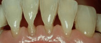
Symptoms of gum recession
In dentistry, there are several types of receding gums, each characterized by its own symptoms, but the general symptoms of gum recession
are as follows:
- tissue volume decreases;
- bleeding during dental hygiene;
- there is swelling, redness of the gums;
- the molar root is exposed;
- there is a high sensitivity to temperature changes;
- caries may develop;
- eating food causes pain and discomfort;
Advanced cases include multiple cervical caries, discoloration of enamel, teeth are located at a far distance from each other. When teeth are exposed, food debris and plaque accumulate in the pockets. Purulent contents are released. The initial stage does not entail unpleasant symptoms, which is why most patients are unaware of the presence of the disease. To prevent gum recession, you need to visit the dentist every six months!
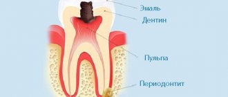
Diseases in which the gums recede from the teeth
There are a number of somatic disorders that are risk factors - in their presence, loss of soft tissue occurs more often and progresses faster than in healthy people. Recession is a common occurrence in patients suffering from:
- diabetes mellitus of any type;
- problems with the kidneys, liver, spinal cord (they can cause circulatory dysfunction in the soft tissues of the oral cavity, as a result of which they do not receive sufficient oxygen and nutrients);
- diseases that are accompanied by immunodeficiency states;
- endocrinological pathologies (disruption of the endocrine glands).
Too frequent and prolonged use of diuretics, prescribed for some diseases, also contributes to the manifestation of the anomaly, since essential nutrients are removed from the body along with the fluid.
Below is a list of dental disorders in which the development of recession, when a piece of gum comes off near the tooth, can be most likely predicted:
- periodontitis - inflammation of the periodontium, which is characterized by destruction of the normal structure of the alveolar process;
- gingivitis - irritation of soft tissues without compromising the integrity of the periodontal junction;
- neoplasms of various etiologies.
Causes
A patient’s complaint that a piece of gum is hanging and causes discomfort when chewing food can be the cause of the following pathologies:
- Gingival hyperplasia. Despite a detailed study of this pathology, there is no definitive answer about the cause of its occurrence. However, provoking factors are identified that become the impetus for the development of this pathology. These include anatomically incorrect bite, lack of treatment for tartar, family history, or long-term therapy with antibacterial or antiepileptic drugs. In women, this condition can cause the onset of menopause or pregnancy (when hormonal changes occur in the body). The disease has characteristic symptoms, which are manifested by swelling and growth of the gingival surface, due to which the tooth can close more than half and the interdental spaces become overgrown. In this case, pain may be absent.
- Gingivitis in hypertrophic (fibrous) form. In the etiology of this disease, the main role is played by hormonal dysfunction, insufficient intake of vitamin C, pathologies of the hematopoietic system, as well as long-term use of certain medications and oral contraceptives. Sometimes this is facilitated by violation of the rules of prosthetics, when the anatomical and physiological parameters of the oral cavity are not observed. In addition to the patient’s complaint that a piece of gum is hanging in the mouth, additional symptoms arise, which are manifested by swelling, the appearance of gum pockets, inside which food particles get stuck, which causes an unpleasant odor between the teeth. The surface of the soft tissues becomes red, and trying to touch them causes increased pain. Hypertrophied interdental papillae cause a cosmetic defect.
- Fibromatosis of the gums. It is characterized by rapid progression, due to which tissue hyperplasia in a relatively short period of time can cover the entire surface of the dental crown. The disease occurs mainly in childhood (from 1 year to 10 years). Sometimes it can be a consequence of hormonal changes in the body in adolescents, women who are expecting the birth of a child, or in menopause.
- Disruption of metabolic processes is another reason for the development of this pathology. The main symptom of gum fibromatosis is the rapid elevation of periodontal tissue, which hides the tooth crown under the soft gum tissue and fills the space between the incisors or molars. Close contact between the tooth and gum tissue causes constant trauma, and as a result, wounds, granulations and bleeding appear.
The appearance of a tissue fold followed by an inflammatory process may resemble acute pulpitis or periodontitis in granulomatous form. Therefore, an adequate choice of therapy can only be made by a dentist after differential diagnosis.
How the pathology manifests itself: symptoms
In the later stages of the disease, the patient can observe its consequences with the naked eye:
- bleeding of the areas surrounding the roots (at first only when eating and cleaning, later constantly);
- formation of noticeable cracks;
- exposure of dental necks;
- swelling and redness of the mucous membranes;
- loosening, suppuration of periodontal pockets (in advanced cases).
Also, the process is often accompanied by a bitter taste of pus and an unpleasant odor from the mouth, which is difficult to mask and does not go away even with the use of paste, chewing gum and rinses.
The primary stages of the disease are sometimes asymptomatic, but even in the first days the patient may feel discomfort. Dentists at the Dentika clinic warn that if you experience the slightest discomfort in your mouth, you should make an appointment with a doctor as soon as possible, since in the early stages of the development of the pathology, its treatment and elimination of the consequences are much easier than when it has already developed sufficiently and causes pain.
How to understand that the gum that has moved away from the tooth has receded, and how to treat it? You can gently pull back the edge of the soft tissue with your finger: even at the beginning of the disease, a slight prolapse under mechanical influence will be noticeable. Therapy in this case can only be prescribed by a professional after a personal examination (there are several options for interventions) - self-medication is unacceptable here.
Associated symptoms
The clinical picture depends on the degree of periodontitis. In mild forms of the disease, the gums do not move, but bleed and are bright red. Signs of progressive pathology are:
- bad breath,
- severe pain,
- tooth mobility,
- swelling and swelling of soft tissues,
- receding and receding gums,
- increased sensitivity of enamel,
- formation of periodontal pockets,
- accumulation of pus between the gum and crown.
If the gum has moved away from the tooth, treatment must be carried out immediately. The problem cannot be ignored, since displacement of the gingival margin increases the risk of infection of the jaw bone tissue, periostitis and other complications. If you don't see a doctor in time, you can lose your teeth.
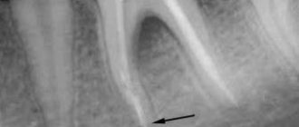
Diagnosis of the disease
Despite the fact that the symptoms are most often noticeable even to the person himself, the dentist, when visiting the clinic, will most likely prescribe additional examinations to clarify the diagnosis and prerequisites. It can be:
- orthopantomogram;
- X-ray examination.
In addition, before choosing a treatment method, it is mandatory to check the depth and condition (for the presence of severe inflammation, suppuration) of the periodontal pocket. Its results determine whether it will be possible to manage with conservative therapy or whether surgical intervention will be required.
Since often the real cause of tissue atrophy is a somatic disease (latent or diagnosed), there is a high probability of prescribing a more in-depth examination (the dentist can refer you to a therapist who will prescribe the necessary tests and issue a conclusion). If you do not start timely treatment and do not achieve remission, the recession will most likely return and you will have to get rid of it again.
Diagnosis and treatment
A dentist will help determine why the gums are moving away from the teeth. The specialist will collect anamnesis, conduct an instrumental examination of the oral cavity, study the results of x-rays and make an accurate diagnosis. Then the doctor will draw up a treatment plan taking into account the stage of periodontitis and the clinical picture. It may include:
- professional teeth cleaning – removal of plaque and tartar;
- curettage of periodontal pockets - cleansing the subgingival area from various deposits;
- anti-inflammatory treatment - rinsing the oral cavity with antiseptic agents;
- Antibacterial therapy – if there is a bacterial infection, the doctor prescribes antibiotics to the patient.
In advanced cases, surgical intervention is indicated:
- removal of the affected part of the mucous membrane followed by suturing;
- flap surgery to restore the required volume of gum tissue.
First aid at home - what to do if gums recede and teeth are exposed
It is unacceptable to refuse to visit the clinic, since in this case the pathological process will continue - it will not be possible to fully recover without a doctor. However, if it is not possible to make an appointment with a doctor immediately when an anomaly is detected, you can slightly delay its progression:
- use only soft toothbrushes and floss;
- Irrigate the oral cavity several times a day with dental (local) antiseptic solutions to prevent the development of severe inflammation;
- use antibacterial gels;
- rinse your mouth with anti-inflammatory herbal teas (chamomile, sage, propolis, etc.) or use them as short-term compresses.
It is better not to disturb the painful area, clean very carefully and do not injure the tissue with hard objects (in particular, avoid hard snacks like seeds, chips or crackers).
How is the treatment carried out if the gum has moved away from the tooth and sank: what to do and why it helps
Therapy in dentistry consists of two stages. The first is cleaning the teeth and getting rid of plaque, which is the root cause of the disease. The second is healing.
Hygienic cleaning can be carried out using one of three methods:
- Instrumental - now it is rarely used. This is a method of removing stone under tissue using a special metal scraper. It is considered quite traumatic and in most clinics doctors no longer undertake it, offering more modern options as an alternative.
- Ultrasonic - using a scaler (also known as a scaler) - an instrument that operates with ultrasound. Used frequently, it is capable of completely removing hard pathogenic deposits.
- Sandblasting. In this case, the hygienist, under high pressure, applies a solution containing abrasive elements to the treated area of the surface, which “knocks” the stone off the crowns and allows the periodontal pockets to be cleaned. As a result (as a “bonus”), you can even get a slight whitening of 1-2 tones.
If the situation is not critical, and the disease has not developed to a serious stage, then within a few months after hygienic treatment, the soft tissues completely heal and return to their normal state. At the same time, pathogenic bacteria are removed, and the oral cavity gradually becomes healthy.
Surgical techniques - how to cure receding gums
If during the pathological process it has become severely separated from the neck, and the periodontal pocket is clearly visible, most likely the structure of the bone has already changed. In this case, surgery cannot be avoided; it is performed under local anesthesia. An open or closed curettage is performed (the so-called “curettage”) - a manipulation in which inflamed or necrotic material is removed using a special instrument - a curette. The opened cavity is cleared of purulent exudate, after which it is sutured or restored in one of the following ways:
- flap restoration - plastic surgery using a donor flap from a neighboring area;
- membrane plasma lifting is a more modern and less traumatic operation that takes longer;
- installation of a collagen implant that promotes regeneration.
In the most difficult situations, it is impossible to do without removing the affected teeth and subsequent implantation or prosthetics.
The method of treatment is chosen by the attending physician together with the patient, depending on the indications and severity of the pathology. The specialists of the Dentika clinic always use the best method for a person, the recovery after which will be as short as possible, and the result will be the most satisfying to the request.
Medicines
Various medications may be prescribed:
- antiseptic;
- antibacterial;
- immunostimulating;
- hormonal.
It depends on the causes of the disease. In some cases, the doctor does not make a final decision, but refers the patient for additional examinations. It must be remembered that self-medication is unacceptable here and before taking medications internally, you must consult a doctor.
Additional treatments
Consultations are usually indicated. If the pathology has developed due to problems with the bite or the position of neighboring teeth, the treating dentist will refer you to an orthodontist for further therapy (for example, installation of braces or aligners). When the cause of the trouble lies in an unbalanced diet, a therapeutic diet is prescribed in addition to the usual manipulations, possibly using dietary supplements (but only a doctor can prescribe them). Some patients are given a prescription for hormonal medications to eliminate the imbalance, and are also advised to use physical therapy to target the painful area.
Alternative reasons
In some cases, the growth of tissue on the gingival surface occurs due to the following two reasons:
Eruption of wisdom teeth (pericoronitis). In this case, a “hood” appears above the dental crown, under which the process of inflammation begins to localize. This is due to the fact that the formed dentition has taken its rightful place, and additional units are not enough. In this case, acute pulsating pain appears, which radiates (gives) to the temple area or to the area of the ear canal. Swelling of the cheek causes changes in the outline of the face (becomes asymmetrical). The tissue fold in the area of the tooth germ becomes hyperemic and is subject to trauma during the act of chewing.
The remainder of the root becomes a consequence of the destruction of the dental crown as a result of mechanical trauma or as a consequence of pulpitis or caries. The lack of adequate therapy (and in this case, it is the removal of the “stump”) becomes the cause of the formation of gumboil. Because of this, the patient experiences an unpleasant putrid odor from the oral cavity. The pathological process may be accompanied by painful sensations, and overhanging tissue may cause bleeding.
Possible complications
Most often, the situation makes patients worry, and they ask what to do if the gums between the teeth droop down, how to restore it? These are true questions to which the doctors at the Dentika clinic are always ready to give competent answers. However, it happens that people do not go to the dentist, preferring to think that the prolapse of soft tissue is just a small cosmetic defect. This is not true: if the disease is started, it, together with the causes of the pathology, can lead to serious consequences:
- lymphadenitis - a condition in which the lymph nodes (usually in the neck) become inflamed;
- periostitis - inflammation of the periosteum;
- periodontitis;
- infectious lesion of the bone of the jaw apparatus.
How is the treatment carried out?
Some people think that they can be cured with regular rinses. Unfortunately, it is not. The first step is to make an appointment with a dental hygienist. The doctor removes soft and hard deposits from the surface of the crowns and subgingival area in one session.
The following methods are used to remove stone:
- Instrumental. Involves the use of special scrapers that remove deposits. It is considered outdated and is extremely rarely used by doctors, as it is traumatic.
- Ultrasonic. Using an ultrasonic scaler, the doctor crushes the stones into tiny particles. This equipment produces waves of the required frequency, which easily destroy dental plaque.
- Sandblasting. It is the most modern and safe. The doctor delivers the cleaning solution under high pressure. Abrasive particles wash away deposits from crowns and effectively flush out existing pockets. The advantage of the technique is to achieve the effect of teeth whitening by one or two shades.
If the hygiene procedure does not help and the problem is serious, the patient is prescribed surgical treatment.
Preventive measures
Preventing a disorder is always easier than curing it, especially when it has already acquired an advanced form. To minimize the risk of recession, when the tooth seems to move slightly away from the gum, and then not treat it, not much is required from the patient:
- eat right, do not abuse foods high in fast carbohydrates, eat enough fruits and solid vegetables, as well as foods rich in calcium;
- maintain the normal state of your immune system;
- carefully monitor compliance with the rules of daily hygiene (use a brush, floss);
- give up bad habits (smoking, frequent consumption of seeds, etc.);
- systematically undergo professional examinations by dentists and undergo annual hygienic cleaning;
- avoid injuries to the oral cavity and dentition (this is especially true for people involved in contact sports).
An important recommendation from experts is to consult a doctor at the first sign of discomfort, without ignoring it: this will help detect pathology at an early stage of development.
Treatment
Therapy directly depends on the type of pathological process, which is why it can be either medicinal or using an operative technique. Depending on the pathology, the following is used:
- therapy during the process of inflammation, when the third molars are erupting, involves the use of methods for excision of the resulting “hood” or complete removal of the figure eight,
- treatment of hyperplasia is carried out using an operable method (gingivectomy) with cutting off the enlarged gingival margin,
- the treatment process for fibrous gingivitis is carried out professionally in a dental office (using a scaler), the lack of positive dynamics allows the use of gingivectomy,
- fibromatous growth of the gingival margins at the initial stage of development is eliminated by prescribing antiseptics and drugs that block the production of histamine. Surgical intervention is considered a last resort and is performed in critical situations or when the process is a congenital pathology,
- the remains of the root system cannot be treated; they are removed in the surgical office of the dental clinic.
The hypertrophied tissue fold above the tooth surface creates favorable conditions for the development of pathogenic microflora. Therefore, the lack of proper therapy or prolonged refusal to visit the dentist’s office can cause the development of periostitis (with subsequent development into osteomyelitis), abscess or phlegmon.
An increase in the gingival margin is a problematic situation that can mainly be resolved through surgical intervention. The use of traditional healing recipes is allowed at an early stage of the emerging pathology, but when the disease becomes chronic, they become useless.