This article uses excerpts from the chapter of the same name in the Face Anatomy atlas. It is of particular importance for specialists involved in the correction of age-related changes and contouring using fillers. The title “Dangerous triangle of the face” was not chosen by chance; it is in this zone that there are prerequisites of an anatomical and functional nature that can interfere with medical manipulation and cause side effects, including severe ones. Let's look at the topography of this triangle and the reasons for its danger.
Introduction
So, a dangerous triangle of the face. We can describe this zone in this way: the apex of the triangle is located in the glabella area, its legs enclose the nasolabial folds and reach the base, which is located under the lower lip [Fig. 1].
Rice. 1. Dangerous triangle of the face.
The area within this zone is often corrected with filler: just think about glabellar lines, nasal hump correction, nasolabial folds and lip remodeling. The anatomical feature, or originality of this zone, which makes it so “insidious”, lies in its blood supply and especially in the topography of the arteries.
Anatomy of speech
The vocal apparatus is a set of anatomical structures that provide the formation of voice and speech. In its structure, it is customary to distinguish two large sections: peripheral and central. The central section is represented by the brain, in particular, the cerebral cortex, a number of subcortical nodes and pathways connecting them together. In addition, it includes the nuclei of the cranial nerves, which are involved in sound production. The anatomy of the peripheral vocal apparatus includes bone, cartilage and muscle formations, ligamentous apparatus and peripheral nerves that perceive or transmit any information to the organs of articulation.
The peripheral department is divided into three functional departments that perform different tasks: respiratory, vocal and articulatory. If any of them malfunctions, speech disorders occur.
Respiratory section
The main sound-producing factor is air passing through the respiratory tract. In this regard, vocalists always practice proper breathing to improve the acoustics of their voice. Breathing movements are carried out reflexively; as a rule, in ordinary life a person does not think about when to inhale or exhale. The regulatory mechanism is associated with the respiratory center in the medulla oblongata.
The respiratory section includes both lungs, the tracheobronchial tree, the diaphragm and the intercostal muscles. The movements of the latter lead to expansion of the chest during inhalation and its narrowing during exhalation. The diaphragm is involved in the abdominal type of breathing, which is carried out mainly due to its stretching.
The formation of sounds and words occurs during exhalation. The air passing through the vocal and articulatory departments causes their structures to vibrate. If a person has lung diseases, the sound of speech is distorted.
Voice department
In phoniatry, there are three characteristics of any person’s voice: timbre, pitch and strength. All of them are created when the vocal cords vibrate as air passes through them. The amplitude of the vibration determines the strength of the voice. The stronger the vibration, the higher the sound. Timbre is determined by voice coloring, specific to each individual person. The tension of the folds and the degree of air pressure on them create a certain pitch of the voice.
One of the most common complaints from patients is changes in voice characteristics. Most often, this condition is associated with functional disorders that occur against the background of infectious and non-infectious diseases.
Sound department
The structure of the articulatory part of the vocal apparatus includes anatomical formations that are located in the oral cavity: the lower jaw, tongue, soft palate and lips. Movements of these structures are carried out with the help of muscles. If their work is disrupted, a person may develop various speech defects. Speech therapy exercises and massage allow you to train muscles, improving speech.
The tongue is the main articulatory organ. Its basis is striated muscles, which provide movement and change in shape. During a conversation, it can become longer, shorter, and also change its width. The structure of the tongue is divided into three parts: the root, fixed to the floor of the oral cavity, the back and the tip.
The lower and upper lips are movable structures. They participate in the pronunciation of almost all sounds, as they determine the speed of air flow from the oral cavity. Thanks to facial muscles, they can change their shape, which also plays an important role in the physiology of speech in humans. The mandible is located at the bottom of the skull and has a limited range of motion. Participates in the pronunciation of stressed vowels: “O”, “U”, “I” and a number of others.
The soft palate forms the upper border of the oral cavity and is tightly connected to the hard palate. Thanks to developed muscle fibers, it can rise and fall down. Anatomically separates the oral cavity from the nasal pharynx. It has a small tongue at the end. When pronouncing the consonants “N” and “M”, the velum is lowered. All other sounds are pronounced with the soft palate lowered. When movement is impaired, the voice becomes nasal, which is associated with the direction of air flow into the nasal cavity.
If a speech defect appears, you should not try to eliminate it yourself. Such treatment should be carried out by a speech therapist or doctor.
The articulatory apparatus also includes a number of passive structures: dentition, nasal cavity, hard palate and pharynx. They participate in sound pronunciation, acting as support points for the tongue and soft palate.
Triangle and arteries
One of the main arteries in this area is the facial artery, a branch of the external carotid artery. The facial artery, immediately after its origin, goes upward and passes to the face in front of the masticatory muscle, where its pulsation can be determined by palpation. Next, it is directed medially and towards the lips, giving off branches - the superior and inferior labial arteries, then passes under the muscle that lifts the upper lip, and reaches the wing of the nose, where its course becomes superficial and ends with two terminal branches - the arteries of the wing of the nose and the angular . Up to this point, everything is clear and clear, but in reality this is not always the case [Fig. 2].
Rice. 2. Facial artery - angular artery.
I will explain more clearly: there are many works with cadavers that emphasize the fact of the existence of frequently encountered anatomical variations and the place of origin of the facial artery, the course of this vessel and its branches. Often such variations are more the norm than the exception. Therefore, it can be argued that even with in-depth knowledge of anatomy, there is a possibility of complications due to the abnormal arrangement of blood vessels. Therefore, it is necessary to use techniques that help to avoid possible side effects as much as possible.
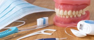
Oral cavity examination methods
Methods for examining the oral cavity come down, first of all, to a thorough examination using directional (preferably shadowless) lighting and special instruments (see Dental Instruments) - a spatula, wide hooks for retracting the cheeks, lips, tongue and stomatol. mirrors for inspecting hard-to-reach areas. Sometimes, to identify luminescent compounds in the oral mucosa (see Luminescence), an inspection of the oral cavity is performed under UV light. During the examination, pay attention to the presence of bad breath. With the help of palpation, the mobility, density, consistency and soreness of various parts of the mucous membrane and patol are determined. formations.
To study the organs surrounding the oral cavity, various X-ray diagnostic methods are used (see). Research methods such as ultrasonic echolocation (see Ultrasound diagnostics) and thermography (see) are also used. According to indications, Citol is produced. examination of swabs and prints from pathologically changed areas of the mucous membrane (see Cytological examination), as well as the study of the microflora of the oral cavity. According to strict indications, a biopsy is performed (see).
In some cases, there is a need to conduct immunological and biochemical studies, as well as to determine various types of sensitivity: tactile, pain, temperature, taste (see Esthesiometry).
See also Patient examination, dental examination.
Possible side effects
Basically, complications arise not so much due to rupture of a venous vessel, but due to interruption of arterial blood flow due to compression or when the drug is administered into the lumen of the vessel with subsequent embolization of the terminal branches with small fragments [Fig. 3].
Rice. 3. Reasons for the development of skin necrosis. Intravasal injection (top), vessel compression (bottom).
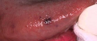
What could happen next? Stopping arterial flow leads to necrosis and tissue death in the blood supply zone of a given vessel. Cases of necrosis of the skin of the wing and tip of the nose, lip tissue and glabella area have been described [Fig. 4–5].
Rice. 4. Necrosis of the tip or wing of the nose.
Rice. 5. Zones of necrosis.
An even more serious risk factor is associated with the fact that the facial artery represents the communication between the external and internal carotid arteries. Its terminal branch, the angular artery, anastomoses with the ophthalmic artery, a branch of the internal carotid artery. It is this connection between the vessels that can lead to embolization of the ophthalmic artery with small fragments of filler and further penetration into the central retinal artery with a possible decrease in vision up to blindness [Fig. 6–7].
Rice. 6. Iatrogenic retinal artery occlusion caused by the injection of fillers.
Rice. 7. Microembolism of the ophthalmic artery: etiopathogenesis.
Microflora of the oral cavity
Over 100 different types of microbes have been found in the oral cavity. The oral microflora includes aerobic and anaerobic bacteria, yeast-like fungi, mycoplasmas, and protozoa. According to S. Neychev (1977), the concentration of aerobic bacteria in 1 ml of saliva is 107, anaerobic - 108.
Among the permanent flora of the oral cavity, streptococci, veillonella, lactic acid bacteria, and actinomycetes predominate. In addition, the permanent flora includes saprophytic Neisseria and diphtheroids. bacteroides, fusobacteria, leptotrichia, spirochetes, etc.
Non-permanent, or random, microflora includes gram-negative bacteria, including Escherichia, Klebsiella, Pseudomonas, Proteus, and Clostridia. The detection of these microorganisms in the oral cavity indicates dysbacteriosis (see).
The microbial flora of the oral cavity, as well as the normal flora of other body cavities (see Human microflora), is a consequence of mutual adaptation of the body and microbes. Despite the known constancy, there are fluctuations in the quantity and composition of the microbial flora associated with hygienic skills, age, dental condition and other factors. It should also be noted that microorganisms are distributed unevenly in the oral cavity. Most bacteria are found on the root of the tongue, on the surface of the gingival margin and in dental plaque (plaque). According to V.G. Petrovskaya and O. Marko (1976), there is a certain specificity in the distribution of flora in the oral cavity, so. for example, Streptococcus salivarius is more often found on the mucous membrane of the tongue, Str. mitis - on the mucous membrane of the cheeks and on the surface of the teeth, Str. sanguis and Str. mutans is isolated primarily from dental plaque (see Teeth, dental biochemistry).
The microflora of the oral cavity performs a number of physiological functions. In a healthy body, due to its antagonistic properties, microflora acts as a “biological barrier”, preventing the proliferation of random microorganisms, including pathogenic ones, entering the oral cavity from the environment. The beneficial value of oral microflora is also associated with its participation in the decomposition of organic substances (food residues), i.e., in the self-cleaning of the oral cavity. In addition, the oral microflora is a constant stimulator of local immunity.
A decrease in the resistance of the oral mucosa and a change in the body's reactivity (see), caused by various factors, can lead to a persistent change in the composition and properties of the oral flora, or dysbacteriosis (see). The altered microflora loses its protective functions and often becomes a source of autoinfection (see). Disruption of microbial balance under the influence of certain therapeutic effects (irradiation, antibiotics, immunosuppressants, dental prosthetics, etc.) can lead to diseases of the mucous membrane such as stomatitis (see), glossitis (see), gingivitis (see) , which are more often of a fungal nature. The generalization of the process is possible - visceral candidiasis (see Candidiasis).
The microflora of the oral cavity is important in the development of dental caries (see Dental caries), periodontal diseases (see). In caries, the most significant role is given to acid-forming microorganisms (streptococci, lactobacilli, actinomycetes) that form dental plaque. In the development of periodontal disease, the most important is given to gram-negative anaerobic bacteria (bacteroides, fusobacteria, spirochetes, veilonella, etc.). It is believed that the endotoxins produced by this flora, which have antigenic activity, stimulate immune responses that support hron. inflammation in periodontal tissues. For example, in the pathogenesis of such pathol. processes such as pulpitis (see), periodontitis (see), often developing as a complication of dental caries, sensitization of the body by microbial metabolic products plays an important role. Chronic inflammatory processes in the oral cavity cause allergic changes in the body and can contribute to the development of foci of infection, sometimes developing into sepsis (see).
The vital activity of microorganisms in the oral cavity is largely determined by the state of local protective factors. Some of them are not directly directed against microorganisms, but have a negative effect on their development. Such nonspecific resistance factors are: pH of saliva, bacteriostatic properties of the secretion of the salivary glands, tissue metabolic products, regular desquamation of the epithelium in the oral cavity, lysozyme (see), etc. Specific factors of protection of the oral mucosa - immune mechanisms directed directly against microorganisms - represented by humoral and cellular immunity. In a healthy body with an intact oral cavity, protective factors prevent the excessive proliferation of microbes, keeping them in certain quantitative proportions.
Therapeutic strategies to prevent and manage complications
It is obvious that it is necessary to use techniques that minimize the risk of developing these dangerous complications. The text of the atlas describes, area by area, all the precautions and manipulations that must be taken to reduce this risk: the use of a cannula, the depth of injection, the quantity and quality of filler injected, and so on. Pallor of the skin and patient complaints of sudden pain in the injection area are signs that blood flow has stopped in this area. We must be able to control this situation.
All measures are aimed at restoring blood flow: urgent dissolution of the filler (if hyaluronic acid was used), warm compresses, massage, etc. Then there are prescriptions that need to be followed at home: antibiotic therapy to prevent bacterial superinfection, antiplatelet agents, topical medications.
In the introduction to the atlas I placed the inscription
“Only non-practitioners do not make mistakes; only through practice does it become possible to reduce the risk of error.”
If all measures are carried out on time and correctly, the spread of the necrosis zone will be minimal, a large area of skin will be preserved and, therefore, the chance of restitutio ad integrum will be higher.