29.11.2019
A growth on the mucous membrane of the mouth, located on the upper palate, causes discomfort to the wearer, sometimes accompanied by pain. A lump on the roof of the mouth occurs due to several completely different diseases. Each disease has its own characteristics and causes, but their clinical characteristics are similar. Without the qualified help of a doctor, it is impossible to determine an accurate diagnosis and effective method of therapy.
Why do tubercles form in the mouth, will it go away without treatment?
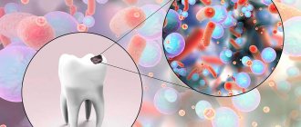
Hard lump-like formations are not uncommon in the mouth, especially on the roof of the mouth. Most of them are harmless, but sometimes a lump that appears is a harbinger of a serious illness. Scientists have found that there is practically no pathology that does not affect the condition of the oral mucosa.
The entire mucous membrane of the oral surface is covered with stratified squamous epithelium. In the area of the hard palate, keratinization of the epithelium occurs, where the stratum corneum is presented in the form of several rows of cells in which there are no nuclei. There is no submucosal layer in this place. Cones can develop in the epithelium, muscle fibers, and blood vessels.
Classification of palate cancer
Based on location, cancerous tumors are divided into 2 types of malignant tumors of the palate. In case of cancer of the hard palate, the malignant neoplasm is located at the border of the nasopharynx and the oral cavity, affects the bone structures and spreads to all layers of the oral mucosa. In the case of cancer of the soft palate, the tumor is localized in the mucous layer and muscles of the vault of the oral cavity.
Based on the histological structure of the tumor, there are 3 types of malignant neoplasms of the palate:
- Cylinder;
- Adenocarcinoma;
- Squamous cell carcinoma.
Cylindroma (adenocystic carcinoma) is formed from glandular tissue. It is characterized by rapid, uncontrolled growth of pathologically altered cells and quickly metastasizes. Adenocarcinoma develops from the epithelium of the oral cavity. The tumor can be localized in all parts of the hard and soft palate. Squamous cell carcinoma is the most common type of oral malignancy. The tumor affects the mucous membrane.
Causes and symptoms of a painful tubercle that has jumped up on the palate
Factors influencing the appearance of bumps on the palate have different etiologies.
Possible external reasons:
- trauma after tooth extraction, jaw surgery;
- burn;
- smoking;
- congenital anomaly in the form of a bony protrusion on the palate;
Internal reasons:
- oncology;
- benign tumors;
- viral diseases (herpes);
- allergy;
- hyperkeratosis of the mucous membrane, leading to the appearance of growths;
- ulcers due to lack of nutrients, gastrointestinal infections;
- dental abscess.
Initially, a painful tubercle is perceived as a pimple that appears on the mucous membrane. Over time, it may disappear, but it can also continue to grow, become inflamed and cause discomfort. In this case, it is necessary to determine the cause of its development.
Cyst
A lump has appeared on the palate and it hurts - this is quite possibly a cyst that has formed, which appeared as a result of a malfunction of the salivary glands. It develops slowly and is a round, painless, lump-like formation.
Mucous cysts, or mucoceles, can be single or group, and the disorder lasts from several days to weeks. They rupture on their own, often while eating, and do not require treatment. They pose a danger if they are a source of infection.
A cyst can develop in the area behind the two front teeth; such a growth is called a palatal papilla cyst. It is painless, often goes unnoticed, and if it becomes infected, it is removed surgically.
Pemphigus
The disease, which belongs to the group of bullous dermatitis, is observed in genetically predisposed people.
Can develop under the influence of various factors:
- taking medications;
- stress;
- consumption of certain foods;
- physical factors
In medicine, several clinical forms of pemphigus have been identified. They are characterized by a long course, a process of remissions and relapses. The most common form of pemphigus is vulgar. The first foci of blistering rashes of various sizes appear in the mucous membrane of the mouth, nose, and pharynx, causing pain during eating and talking.
The thin coating of the blisters quickly breaks open, forming erosions that soon become the cause of bad breath. Subsequently, rashes appear on the body. Symptoms of medicinal pemphigus are similar to vulgaris.
Myxoma
A benign formation manifests itself in the form of a myxoma, which looks like a bumpy lump on the roof of the mouth. This is a rare type of tumor that arises from mucous tissue. It can appear at any age in both men and women.
The macroscopic tumor is similar to a node consisting of whitish-yellow tissue, has no clearly defined boundaries, and does not form metastases. In many cases, no treatment will be needed, but the lumps, if they are causing problems, can be removed surgically.
Cancer
Morphological changes in the oral mucosa often lead to the formation of tumors. Cancerous tumors are characterized by rapid growth and a tendency to metastasize. An ulcerative form of cancer often forms on the palate.
At the beginning of the disease, a dense tubercle is noticeable, which later develops into an ulcer. With a light touch, the bottom of the ulcer may bleed, subsequently the bone is exposed, tooth mobility develops, and metastases quickly appear in the lymph nodes.
Additional symptoms that are present:
- feeling of a lump in the throat;
- non-healing wound at the site of tubercle formation;
- bleeding;
- jaw pain, stiffness;
- a sore throat.
Treatment depends entirely on the location and stage of the cancer. People who smoke a lot should especially monitor the formations that appear in the area of the palate.
https://www.youtube.com/watch?v=0j8O9ADV3MA
Solution
The muscular structure of the palate is covered on top with a mucous membrane, which is subject to the development of an inflammatory process. Inflammation often occurs under the influence of such factors:
- burn of the mucous membrane as a result of consuming excessively hot food and drinks;
- damage to the palate due to dental diseases - periodontitis, stomatitis, caries, pulpitis;
- disruption of the oral environment due to the action of metals - when wearing braces or installing crowns;
- smoking;
- allergic reactions to medications;
- neurological diseases affecting the joints of the upper or lower jaw;
- osteomyelitis - an infectious lesion of the bone tissue of the jaw;
- malignant neoplasms;
- infectious diseases of the upper respiratory tract - sore throat, tonsillitis, pharyngitis, rhinitis.
The development of the inflammatory process of the palate may be facilitated by some of these causes in their entirety or separately from each other.
White plaque on the mucous membrane is a classic sign of a developing inflammatory process.
Symptoms of inflammation of the gums and palate:
- plaque on the mucous membrane (white, yellowish, brown);
- hyperemia;
- burning, itching;
- multiple small ulcers;
- swelling.
It happens that the signs of the inflammatory process in the palate are so pronounced that the patient experiences discomfort with every meal (not only when chewing, but also during swallowing). It is noteworthy that similar manifestations also occur with angina (inflammation of the tonsils). With the development of abnormal dental phenomena, the symptoms of the inflammatory process also affect the periodontal tissues; in this case, multiple white ulcers may appear on the palate.
Under the influence of galvanic current when installing braces (metal crowns), the condition of the mucous membrane of the palate in the mouth also changes, it can become inflamed. Smokers also often suffer from unpleasant sensations in the corresponding focus - tobacco smoke injures the delicate mucous membrane and it becomes more susceptible to “attacks” of pathogenic microflora.
Important! Benign and malignant tumors of the throat, inflammatory processes in the salivary glands of any location also lead to pathological changes in the structure of the palatal soft tissues. Soreness of the soft palate during eating may indicate the presence of scratches, cracks in the mucosa, or the absence of muscle adhesions after the installation of permanent dentures
Soreness of the soft palate during eating may indicate the presence of scratches, cracks in the mucosa, or the absence of muscle adhesions after the installation of permanent dentures
The main causes of pain in the palate are:
- violation of the integrity of the mucous membrane;
- metabolic failure;
- activation of the body’s defenses in response to “attacks” of a bacterial or fungal infection.
It is necessary to treat an inflamed palate based on the cause of the abnormal process. As a rule, medications are supplemented with traditional methods. Inflammation of the nerves and tonsils requires antibiotics (only as prescribed by a doctor). For stomatitis, treatment with local and systemic antifungal agents is indicated.
Regardless of the origin of the inflammatory process in the palate, it is impossible to cure it without a special diet. So, until the mucous membrane calms down and heals, doctors recommend that patients avoid rough, solid foods, excessively hot, cold foods (drinks). Smokers should at least temporarily give up their addiction.
Important! Given the secondary nature of the inflammatory process in the palate, any treatment will be ineffective without eliminating the root cause of the anomaly (benign, malignant tumor, inflammation of the salivary glands, any dental problems)
In what case and which doctor should I contact?
A lump has appeared on the roof of your mouth and it hurts - this is not yet a reason to visit a doctor.
You can contact the clinic if other symptoms appear:
- Increasing pain when chewing and swallowing.
- Increase in volume of the tubercle.
- The appearance of bad breath.
- The thickening does not go away within several weeks.
First of all, you should contact your dentist. Subsequent examinations can be carried out by doctors: a surgeon, a therapist, an otolaryngologist.
Complications
When any formation appears on a child’s palate, two threats arise: obstruction of the airway and the addition of an infection.
In the first case, the main role is played by the size of the cone. The larger it is, the more the lumen of the pharynx narrows, which can lead to breathing difficulties. Particular attention should be paid to this aspect in children with completely or partially absent breathing through the nose (in children with adenoids, polyps, sinusitis, vasomotor rhinitis, etc.). For them, the “alternative” method of breathing, through the mouth, is especially important.
Infection can occur at any time. The location of the lump on the palate makes it vulnerable to any mechanical or thermal irritants - a hard lump of food or hot tea easily injures the mucous membrane, which creates pathways for bacteria to penetrate into it. In this case, a purulent process develops.
Less commonly, necrotization of the neoplasm may occur. This is especially true for angiomas that are located on a stalk - a small “column” of mucosa that makes the neoplasm mobile. In rare cases, such a leg is significantly long, which allows it to twist around its axis. This stops the flow and outflow of blood, and after complete tissue necrosis, a black lump is observed on the child’s upper palate.
Diagnostic measures
Diagnosis of cancer is difficult in the initial period of tumor appearance. It is during this period that a thorough examination of the oral cavity and tissue biopsy are necessary.
| Type of seal | State Definition | Type of study | Symptoms |
| Hemangioma | palpation of the tubercle, during which the lump is first reduced in size and then restored to its original shape | taking a smear for bacteriological analysis | turns pale when pressed and may bleed |
| Myxoma | visual | smear for histology, general blood analysis, X-ray (if necessary), puncture | difficult to see, especially during the initial stage, myxoma grows slowly |
| Cyst | visual | clinical blood test, biopsy sample, x-ray | no pain is felt, the growth does not interfere with eating or talking |
| Pemphigus | visual | clinical blood test, test for Nikolsky syndrome | caries as a concomitant symptom |
| Angioma | oral examination | blood test, smear | healing is long-term, bleeds, interferes with eating and talking. |
A lump has appeared on the palate and the pain may be a hemangioma.
In most cases, when making a diagnosis, two main types of examination are implied: examination of the mucous membrane and examination using X-rays. In cases where the doctor is in doubt, a puncture is prescribed.
Symptoms
Ulcers, or in scientific terms aphthae, cause a lot of inconvenience, so they cannot be asymptomatic and a person will immediately understand that the mucous membrane of the palate is not in order:
- Due to the appearance of ulcers, the mucosal tissues swell, and this can make speech difficult.
- It is impossible to eat hot, spicy, sour and salty foods, as they will irritate the damaged surface of the palate.
- A person, even with his mouth closed at rest, may experience pain, and in advanced cases, the ulcers bleed.
Not all canker sores are harmless. Of course, if they were formed due to mechanical damage to the mucous membrane, they will go away on their own in the shortest possible time.
If the ulcers do not go away, you need to track the accompanying symptoms in time to begin treating systemic diseases:
- When the herpes virus gets on the mucous membrane of the mouth, a person’s condition generally worsens, body temperature may rise, lymph nodes become enlarged, salivation increases, and then small wounds form on the palate.
- Often, with a decrease in immunity or long-term use of antibiotics, it is possible to develop a disease such as candidiasis or thrush of the oral mucosa. In this case, a burning sensation is felt, taste sensations are lost, and a characteristic white coating of a cheesy appearance forms on the entire mucous membrane; eventually, aphthae can form on the palate, which sometimes bleed.
- When infected with syphilis at the beginning in the oral cavity, a rash may occur, which then develops into ulcers on the soft tissue of the palate and cheeks.
- Aphthous stomatitis. A chronic disease characterized by inflammation of the oral mucosa and the appearance of ulcers on the palate. Besides. such stomatitis can be a symptom of generalized aphthosis, a disease that affects all mucous tissues in humans (gastrointestinal tract, genitals, eyes). Ulcers form gray-yellow in size from 2 to 10 mm, depending on the course and stage of the disease. They are quite deep, painful, do not heal for a long time and often recur.
- With pulmonary tuberculosis, initially small compactions in the form of reddened tubercles may form on the palate, which then transform into shallow round wounds.
Principles of treatment
Depending on the type of formation on the palate, the doctor will suggest treatment methods. The basis of treatment for pemphigus is glucocorticosteroid drugs, prescribed according to a confirmed diagnosis and vital indications.
Treatment of cancer depends on the location and degree of development of the tumor, and includes surgical methods, including cryodestruction, and radiation techniques. Cysts are removed surgically if they become infected.
Sclerosis
Neoplasms that arise from changes in the vascular system are removed during sclerotherapy. To do this, a drug is injected into the lumen of the vessel, causing gluing of the vascular walls. The most popular agent used in sclerotherapy is 70% ethyl alcohol. An injection is made into the thickening in the oral cavity, which leads to a decrease in the size of the tumor.
Cryotherapy
In medical practice, local cryotherapy has found wide application. It is used to treat precancerous conditions of the oral cavity, as well as primary prevention of malignant tumors.
The procedure is carried out using applicators made of porous titanium nickelide. The applicator is immersed in a vessel with liquid nitrogen and after it is saturated, cryotherapy is carried out.
The cauterization procedure lasts no more than 60 seconds. Subsequently, preventive procedures should be carried out with a cryoapplicator at a frequency of 2-3 times per second. The total duration of cauterization is 1-2 seconds daily, for 7-10 sessions. Wound healing after the procedures is successful, leaving no traces of scarring.
Cryotherapy stimulates blood flow and does not affect the general condition of the patient; observation of patients showed no relapses.
Electrocoagulation
The procedure is performed under local anesthesia; after removal, a crust forms at the site of the tumor. The essence of the method is that tissue is excised using an electric knife and coagulation of blood vessels is carried out at the same time.
After the procedure, it is recommended to protect the wound from infections until new epithelium is formed by rinsing the mouth with antiseptics. Surgery is often performed to remove angiomas in the oral cavity. In addition to this procedure, laser therapy is often prescribed.
Laser
A tubercle that has popped up on the roof of the mouth, causing anxiety and pain, can be removed using laser therapy. Under the influence of rays, the lump on the hard palate is removed layer by layer.
The surrounding tissues are little injured, the mucous membrane recovers quite quickly after surgery.
Galvanoacoustic loop
If the formation that has arisen on the hard palate has a stalk, which is typical for many types of tumors, then a galvanoacoustic loop is an effective method.
Before the procedure, the oral cavity is treated with an anesthetic, then the stem is tightened using a special loop, and a current is passed through the loop. The heated loop anneals the tumor. After surgery, you should rinse your mouth with antiseptics, and anti-inflammatory drugs are prescribed.
Radium therapy
This type of therapy is used to remove angiomas formed from small capillaries. This method of excision of formations within healthy tissue is considered the simplest. With the help of certain drugs, the radium isotope is delivered directly to the tumor, thereby affecting the tissue, damaging the tumor.
Chemotherapy
Treatment of malignant neoplasms is carried out using intravenous drugs that kill not only the tumor, but also the spread of metastases.
Therapy has side effects:
- severe headache;
- vomiting;
- diarrhea;
- hair loss;
- nausea;
- general weakening of the body and loss of immunity.
The number of sessions is determined depending on the stage of the lesion.
Prerequisites for the occurrence of a lump
Modern medicine has not created a complete list of reasons why bumps may appear. Doctors' reasoning is based on hypothetical cause-and-effect relationships. The appearance of a lump on the palate is caused by:
- Bad habits (smoking, alcohol, oral drugs);
- The presence of micro and macro injuries to the oral cavity (surgeries, scratches of the upper parts of the oral cavity);
- Availability of dentures;
- Viral infections;
- Intrauterine disorders (hemangioma is a congenital disease and acquired from the mother);
- Violation of the activity and integrity of the mucous membrane (typical of angina);
- Congenital and acquired dysfunction of the glands (the main cause of cysts).
There are two more hypothetical reasons for the appearance of malignant tumors:
- Eating too hot and spicy foods, constantly disrupting the structure of the cells of the oral cavity;
- The presence of papillomatosis or leukoplakia - diseases that are precancerous lumps that can develop into an oncological disease.
Folk remedies
A lump has appeared on the roof of the mouth and it hurts - this is the reason for many people to turn to traditional medicine. It should be borne in mind that in the presence of malignant tumors, traditional methods should only complement the main therapy. Before resorting to home remedies, you should consult an oncologist.
Bumps on the roof of your mouth that are not cause for concern can be treated at home. For this, plant components are used; spotted hemlock, celandine, borax or aconite. After undergoing chemotherapy sessions, the following will help restore strength: lady's slipper, cat's claw root. Tinctures are prepared from these herbs and taken over a long period of time.
- Daily 1 tsp. cat's claw is poured into a glass of boiling water, infused and taken for 3 months, 3 glasses a day.
- The same infusion is made from the herb spotted slipper.
- Tincture of fragrant laurel leaves is taken 1 tbsp. l. 5 times a day, in small sips. To do this, take 250 g of leaves, add 0.5 liters of alcohol and infuse for 2 weeks in a dark place.
Aloe juice helps a lot. The pulp from the leaves can be applied in the form of applications to the thickening or rinsed with juice in the mouth. A soda solution prepared with warm water helps speed up healing if you rinse your mouth with it 2-3 times a day.
You can make a paste from baking soda:
- Mix 1 tsp. soda with a small amount of water, bring to a paste.
- Apply the mixture to the thickening of the mouth and leave for a few minutes.
Honey procedures performed 3-4 times a day will help get rid of the growth. To do this, you need to lubricate the palatal cone with warm honey.
To reduce pain and relieve inflammation, mouth rinses based on witch hazel are used.
A tubercle that appears on the roof of the mouth does not cause many problems if there are no alarming accompanying symptoms. To prevent infections, you should follow basic hygiene rules, and in case of severe pain, seek help from a doctor.
Article design: Vladimir the Great
Diseases that cause a pain response on the inside of the upper area of the mouth
Pain in the palate occurs due to external and internal factors. External causes include irritation of the oral mucosa by chemical and thermal factors. Minor microdamages lead to inflammation of the palate, as they create an ideal environment for the proliferation of pathogenic flora. Dentists identify several main causes of the problem.
Infections
The provoking factors of the problem include pathogens of fungal, viral and bacterial origin. There are pathogenic agents in the oral cavity of any person, but they are activated only under favorable conditions: a decrease in general and local immunity; metabolic disorders; chronic diseases.
The herpes virus affects not only the membranes of the palate, but also the mucous membranes of the entire oral cavity, including the lips. At the initial stage of the disease, the mucous membranes of the mouth become covered with red spots.
Fungal infection affects not only children, as many mistakenly believe, but also adult patients. The main symptoms of infection with yeast-like fungi are swelling of the soft tissues of the mouth and the appearance of a white coating on them.
The disease can develop secondarily as a result of infections of the ENT organs:
- tonsillitis;
- tonsillitis;
- inflammation of the tonsils.
The mucous membranes of the mouth are very thin, so hard and rough foods easily damage them. The same applies to cold and hot foods. The problem is often observed in adults and children who have the bad habit of biting their nails or putting foreign objects into their mouths. Violation of tissue integrity leads to the development of edema. The situation is complicated by poor oral hygiene.
Dental diseases that are not treated in a timely manner negatively affect the microflora of the mouth. In this case, gum inflammation is often accompanied by severe pain and swelling of the cheeks. The problem is caused by stomatitis, caries, dental cyst, periodontitis, osteomyelitis, pulpitis, periodontal disease.
Osteomyelitis is associated with inflammation of the deep layers of the gums and jaw structures, causing pain in the roof of the mouth
Why does the sky hurt? Other causes of palate damage include:
- Leukoplakia. The condition is considered an early stage of the oncological process. Leukoplakia occurs due to temperature irritants and injury to the mucous membranes.
- Smoking. Under the influence of nicotine and tobacco smoke, the structures of the mouth undergo changes. As a result, a person experiences pain in the upper palate of the mouth.
- Poor quality treatment. Complications occur with improper prosthetics or after the removal of teeth damaged by caries.
- Degenerative processes of bone structures. In this case, unpleasant sensations spread to the joints and jaw.
- Sialometaplasia is a benign tumor that affects the mucous membranes of the mouth. The tumor gradually enlarges and then ruptures, leaving behind a large bleeding wound.
In cases where the palate hurts, what to do can be determined after determining the cause of this phenomenon. The structure of the tissues of the palate has such a feature that minor damage, a scratch, or a burn from hot food can cause symptoms of pain. They open the way for infection to enter.
Pathogens in the form of bacteria and viruses can activate the process of local inflammation without wounds, just like fungal microorganisms. If the palate in the mouth hurts, the causes may be different, and the treatment tactics depend on their identification. It is determined by symptoms and test material (mucosal smear).
- burns: thermal, chemical;
- injury;
- herpes;
- a benign formation or oncological process, in which gray spots gradually become noticeable;
- pharyngitis, tonsillitis;
- problems with teeth (caries, pulpitis) and their supporting structures (periodontal disease, periodontitis, osteomyelitis);
- unsuccessful prosthetics;
- leukoplakia;
- stomatitis;
- rhinitis (atrophic);
- neuralgia.
Redness on the soft tissue of the upper septum of the mouth from a thermal burn
The cause may be surgery. The pain is localized mainly in one place, on the side of complex surgical manipulation.
Namely after:
- fistula removal;
- cutting gums;
- penetration into the bone tissue of the jaw;
- root extraction.
There are times when I wake up in the morning and feel that my palate hurts. This also has its preconditions. These are diseases of the articular zone between the upper and lower jaw.
Diseases in which a lump on the gum does not hurt
There are diseases in which the resulting compaction does not bother the patient with pain. It is because of this that people are in no hurry to see a doctor, which can subsequently complicate treatment. In what diseases does a lump that appears on dental tissue not hurt?
Fistula
If a lump with pus appears in the gum, for the flow of which there is a hole in the gum, we are talking about a fistula. The formation is a growth 3-5 mm in size with a white tip. When the outlet is not blocked by compacted exudate and pus flows freely, such compaction, as a rule, does not hurt. The reason for the formation of a fistula is a complication of periodontitis, in which the tissue grows and becomes infected by accumulated bacteria. Despite the fact that pus periodically flows out and does not accumulate in large quantities, if left untreated, an acute fistula becomes chronic. If the fistula is not treated, there is a risk of infection spreading through the periodontium and tooth loss.
Exostosis
Exostosis is a type of jaw abnormality when the bone protrudes slightly to the surface. Such a lump is hard and painless to the touch, since it is, in fact, a bone or osteocartilaginous growth of a benign nature. The causes of occurrence are hereditary predisposition, congenital pathology or consequences of injury. With such an anomaly, prosthetics are difficult or even impossible: the prosthesis will rub the skin on the bone growth, which can cause injury and infection.
Important! Exostosis is determined by x-rays. The decision to remove is made by the patient, but it is worth considering that such growths can become malignant over time.
Epulis
If a lump appears above the gum, shaped like a mushroom on a stalk, pale pink or red in color, we are talking about epulis - a pathological formation, common mainly in women, and also occurs in children when their first teeth erupt. The reasons for the appearance of epulis are often post-traumatic in nature (large filling, tartar, chips), however, a pathological growth can also appear due to malocclusion, hormonal disorders or wearing low-quality dentures. The symptoms of epulis are similar to those of gingivitis, so for an accurate diagnosis, the doctor uses radiography or a histological test.
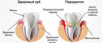
Periodontitis
You should think about periodontitis if a lump has formed on the gum - hard to the touch, up to 10 mm in diameter. The causes of inflammation of the periodontal tissues are usually unsealed dental canals and their infection. An abscess forms at the root apex. A characteristic sign of the disease is that when you press vertically on a tooth, pain appears. If the pus has already drained, there may be no pain, but the disease cannot be ignored, as it can lead to tooth loss and further spread of infection.
Attention!
If a seal appears on the gum, under no circumstances should you apply a warm compress or hot rinses: this can aggravate the course of the disease!
Hematoma
A soft-to-touch swelling with watery contents – a hematoma – is formed as a result of tissue injury during tooth extraction. This bump, as a rule, does not require special treatment, dissolving a few days after its appearance.
Do not ignore preventive visits to the dentist.
It is enough to visit a specialist 1 – 2 times a year, which will allow you to promptly identify any dental problem at an early stage of development. This means that its elimination will be quick, easy and without complications.
By clicking the “request a call” button you agree to the personal data processing policy.