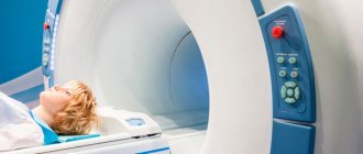
Image of the brain on MRI results Magnetic resonance imaging is one of the best methods for diagnosing the brain. It is painless and has a small list of contraindications.
Neurosurgeons prescribe a study for an accurate diagnosis after injuries and when pathologies are suspected, manifested by complaints of acute or persistent headache, tinnitus, confusion, weakness, memory impairment, etc.
Along with computed tomography, MRI plays an important role in recognizing life-threatening diseases. When a doctor refers patients for a magnetic wave scan, many people wonder if they need to prepare for an MRI of the brain. The answers are in this article.
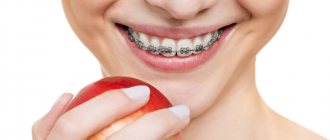
Is it possible to do an MRI of the head with metal crowns on the teeth?
Magnetic resonance imaging (MRI) is a highly informative examination method. It allows you to identify defects in tissues and organs as small as 0.2 mm - such pathologies are not detected by other diagnostic devices. But doctors warn that MRI and metal are a dangerous combination.
A stalemate is developing - most adults have metal structures in their mouths: dentures, pins, braces, etc. And they still undergo MRI. So are metal products really contraindications for examination?
To give an answer, you need to explain the principle of operation of a magnetic resonance imaging scanner. It sends out radio waves and magnetizes the nuclei of hydrogen atoms located throughout the body. The latter “respond” to a powerful magnet or, in other words, enter into resonance. Based on the arrangement of hydrogen atoms, the program creates a picture and displays it on the monitor.
But not only the nuclei of hydrogen atoms, but also metals can “respond” to the magnet of the device. However, not all: there are materials that cannot be affected. These are paramagnetic materials:
- titanium; (titanium crowns for teeth and their characteristics)
- platinum;
- aluminum;
- tantalum.
Cases of displacement of metal products are very rare
In addition to paramagnets, there are 2 more groups of metals:
- Ferromagnets. These include iron, chromium, nickel, cobalt, and steel. They react strongly to the magnet: they heat up, move and can even “break out” of the body. There were cases when a part of the body with an installed ferromagnetic structure (arm, leg) “stuck” to the tomograph and the device had to be completely demagnetized - there was no other way to free the patient.
- Diamagnets. These are hydrogen, copper, silver, gold (gold crowns on teeth, frills). They are not significantly magnetized and do not heat up or move during MRI. However, they create interference and distort the picture.
Where can metal be found in the body?
Our body naturally does not contain pure metal. But doctors are actively using this durable material to make implants and various prostheses. Metal “spare parts” can be found in almost any part of the body: in the mouth, arm, leg, head, ear. Moreover, sometimes the patient himself does not even remember that something was replaced with a metal prosthesis, and only remembers it before the procedure. And the other, on the contrary, does not know whether he can conduct this research. What to do if you have been sent for MRI diagnostics in Moscow, and you are phoning in front of every metal detector?
The main metal “spare parts” that doctors use:
- In dentistry: implants, pins, braces, metal crowns, bridges, removable dentures with a metal base, inlays.
- In traumatology and orthopedics: pins, knitting needles, plates (for example, for fixing bones after a fracture), joint endoprostheses (usually have metal elements).
- In neurosurgery: hemostatic clips on cerebral vessels.
- In cardiology: artificial aortic and mitral heart valves.
- In otorhinolaryngology: cochlear implants.
As you can see, the list is quite impressive. Moreover, these are only the most common options; in fact, there are many more of them. What should such a patient do if he needs MRI diagnostics?
Should you tell your doctor that you have dental implants?
Dental replacement implants are made primarily from titanium. This is a paramagnetic material that does not resonate with a magnetic tomograph. But this does not mean that there are no restrictions on the examination.
To avoid having to undergo an MRI twice, notify your doctor about the implant so that he can change the machine settings
The diagnostician must be informed about artificial teeth for two reasons:
- Titanium is capable of distorting the picture. Yes, the metal does not heat up and does not move. But it can create minor interference. In order not to undergo an expensive procedure twice, it is better to immediately notify the doctor about the implant - he will change the settings of the device.
- The titanium pin is not the only element of the implant. In addition to the rod itself, there is also an abutment (gum former) and a crown. They are made from ceramics or various types of metal and then lined with porcelain.
Therefore, patients are always asked about the presence of implants at least twice: when giving a referral and immediately before diagnosis.
Indications for use
An MRI during pregnancy may be prescribed if there are no other ways to accurately diagnose. Most often it is prescribed:
- If you suspect the presence of pathologies during pregnancy, fetal development;
- If you suspect the presence of malignant tumors that cannot be detected otherwise;
- If there is an urgent need to examine the condition of the bones or blood vessels of the expectant mother;
- When diagnosing injuries and preparing for emergency surgery.
Myths for patients with implants
Before undergoing an MRI, people with artificial teeth are frightened by their acquaintances with stories: they say that the implants are torn out, tearing the gums and breaking the jaws. But these are just rumors with little truth.
There are 3 misconceptions about undergoing MRI with implants:
- The implants will be pulled out of the jaw. 90% of the rods are made of titanium, which does not react to magnetic fields. In addition, the metal pins are embedded in the bone, so they physically cannot move.
- The implant will distort the picture. The rod itself is made of paramagnetic material and does not affect the data. But the crown on the implant can be made of di- or ferromagnetic alloys. And it already affects the results of the examination.
- The implant will heat up, causing tissue burns. Again, titanium does not resonate with a magnetic field. Only a crown with a cobalt-nickel or chromium frame can heat up.
Implants are not an absolute contraindication to examination. There are limitations, but they are related to the capabilities of the device - some models cannot be configured so that they do not react to metal parts. In this case, they simply select another medical center with more powerful and modern equipment.
Implants are not an absolute contraindication to MRI if it is possible to change the device settings
Is it possible to do MRI with endoprostheses and other implants?
What to do if the patient has various types of implants right in his body? First of all, this must be reported to the specialist conducting the examination, since many metals are ferromagnetic and can shift in the body under the influence of a magnetic field.
For an MRI with a steel wire in the body, everything is not so simple. Iron causes the magnetic field to deviate from a given direction, which leads to distortion of the resulting images and the appearance of artifacts (defects) on them. In addition, the needle can heat up, creating unpleasant sensations for the patient.
Also, the answer to the question whether it is possible to do an MRI with an endoprosthesis depends on what material it is made of. If it is titanium, then there are no restrictions. If it is made of ferromagnetic materials, then this is a contraindication for research. You can find out exactly what metal the implant is made of in the design passport, which is issued to the patient after prosthetics.
Other problems with crowns
In addition to affecting MRI results, metal crowns cause other problems: they break, chip, and the root canals and gums underneath may become inflamed, or the tooth stump may collapse. But most often, fixed dentures become loose or fly off.
What to do if the crown of a tooth is loose or has fallen off?
Crowns or bridges made of diamagnetic or ferromagnetic metals may begin to wobble after MRI. This is due to the displacement of the prostheses during the examination and subsequent decementation. In the future, the structures will fall out.
The presence of metal structures does not affect the results of a CT examination
However, the causes of poor fixation are not always related to magnetic resonance imaging. Sometimes dentures become loose and fly off due to:
- washing out the cement from under the crown - if its edge does not fit tightly to the neck of the tooth, the bond (adhesive composition) is gradually destroyed under the influence of saliva and drinks;
- poorly made design - it will not fit tightly if it does not follow the shape of the tooth in the smallest detail;
- small volume of the stump or its destruction - the remains do not hold the prosthesis, and it flies off.
Regardless of the reason, contact an orthopedic dentist. He will correct a loose or fallen crown and re-fix it. If the prosthesis cannot be repaired, a new one will be made and installed.
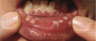
Swallowed a crown...
When a crown falls out, it is often swallowed along with saliva, food or drinks. There is no reason to worry - it will come out on its own along with the feces in a few hours or 1-2 days.
The only dangerous situation is when the prosthesis splits. In this case, an x-ray is taken, the location of the bridge or crown is determined, and then the stomach is washed out or an enema is given.
If you swallow a crown, it will be passed out in your stool within a maximum of 2 days.
But, in any case, the swallowed crown is reported to a gastroenterologist, surgeon or traumatologist. Magnetic resonance imaging is postponed until the structure is removed from the body.
Where to get an MRI in Moscow?
What should you do if doctors in your city refuse to examine you at the mere mention of prostheses and plates? Don't worry about it. On the website MRT-kliniki.ru you can choose any clinic in Moscow with good specialists who know all the nuances of performing tomography for “problem” patients. This site will help you not only choose the best diagnostic center, but also find out the cost of services. This is also an important point, because the price of an MRI in Moscow in one clinic can be 2-3 times higher than another. Therefore, it is worth comparing the cost and choosing a medical institution with a loyal pricing policy.
How long does a crown stay on a tooth?
The service life of entire crowns primarily depends on the material. So:
- metal ones last an average of 5 years;
- metal-ceramic – 8-10 years;
- ceramic and zirconium dioxide - 10-15 years and above.
The duration of wearing dentures is also affected by: how well the supporting tooth was treated and ground, the skill of the dental technician and the quality of oral care. The dentist is visited every six months to remove plaque from products and adjust them. And after 7-10 years, most crowns become unusable and are replaced with new ones.
But loose and broken dentures will not last long: from several days to a couple of weeks. Therefore, people contact the clinic immediately: the faster the doctor adjusts or replaces the bridge or crown, the greater the chance of saving the abutment tooth.
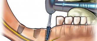
This is how a crown is sawed
What to take for an MRI of the brain?
In the medical department, MRIs are performed upon the direction of a doctor and upon self-referral. To obtain informative photos, it is recommended to take to the clinic previous examination results, an extract from the outpatient card, papers from the hospital and other documents related to the disease.
Before an MRI of the brain, tell your doctor if you have a fixed denture, an orthodontic structure to correct your bite, or tattoos that contain metal in their ink.
By adhering to the recommendations, the patient has a better chance of receiving comprehensive information about the pathologies of cerebral structures and the dynamics of changes during treatment.
How are crowns removed from teeth, indications?
In cases where the crown or bridge has moved during the MRI, they are removed and installed again or replaced with new prostheses. They do this in the following ways:
- sawing - the prosthesis is drilled out using drills with a cooling system; after removal, the structure is unsuitable for further use;
- ultrasound – ultrasonic waves destroy the cement, and the crown is easily removed from the stump;
- Koch apparatus - a special drill breaks the bond with gentle pushes, then the structure is lifted and removed;
- Coronaflex apparatus - compressed air is supplied under the edge of the prosthesis, this is the only method that eliminates damage to the structure and supporting teeth;
- special tools or sliding crown removers - used after destruction of the adhesive base using one of the above methods.
Orthopedic structures are also removed in case of resorption of cement, chips of ceramic lining, damage to the frame, inflammatory processes in the root canals, or the patient’s desire to replace the prosthesis with a better one.
It doesn't hurt to remove crowns
Is it possible to shoot on your own?
Sometimes the bridge or crown is so loose that the patient can tear it off himself. However, this should not be done, because... possible:
- damage to the prosthesis;
- fracture of the stump or root system of the tooth;
- traumatization of the mucous membrane or adjacent units.
If the denture becomes loose, the only correct option is to contact a dental clinic.
Does it hurt?
Removing dentures does not hurt at all, unless, of course, the patient rips them off himself. Because crowns and bridges are placed on already decayed teeth; in most units the pulp has been removed. Therefore, unpleasant sensations are excluded. There may only be a feeling of pressure on the jaw.
If the tooth under the crown is not pulpless or its roots are inflamed, dentures are not removed. The doctor administers anesthesia to eliminate pain.
How long does the procedure take?
The duration of prosthesis removal depends on the method. So, using a cut, the structure is removed in 10-15 minutes. But preliminary decementing of crowns using ultrasound, a Koch or Coronaflex apparatus can take several visits.
Artificial teeth are not an obstacle to magnetic resonance imaging. Modern designs are made from bioinert materials that do not harm the patient and do not affect the examination. If there is one of the items prohibited for examination in the mouth, it is removed and then fixed again.
Complications after the procedure
If a pregnant woman has properly prepared for the procedure and told the doctor all the features of the pregnancy, the presence of chronic diseases and other possible problems, then the procedure should be completely painless and absolutely safe for both the expectant mother and her child without any special complications. However, there may be a possibility that after an MRI, a woman will feel some discomfort caused by the need to remain in a horizontal, motionless position for a long time. That is why after the procedure you should take your time and also monitor your blood pressure, especially if you have problems with the cardiovascular system.
No. 7. How is the procedure done? It does not hurt?
The patient is positioned in the tomograph. The procedure takes about half an hour and does not cause pain. During this time, the patient with suspected coxarthrosis should remain motionless. This is the main difficulty for many. You may feel a slight warmth at the site of exposure.
During the procedure, the laboratory assistant is in the adjacent room, controls the tomograph, but constantly communicates with the patient through the communication system. The results will be ready the next day.
This is what a tomograph for MRI of the hip joint looks like
No. 10. What are the health risks after undergoing an MRI?
If all conditions are met, the risks are minimal. If there are implants in the body that the patient has not informed the doctor about, they may change location and lose functionality. This condition is life-threatening. There is also always the possibility of developing allergic reactions to contrast. Otherwise, the risks are not high if the examination is carried out by a qualified specialist.
What diseases can be seen in the hip joint using MRI? About this and much more in the video below:
For hip diseases, MRI is an excellent alternative to a painful biopsy. In terms of performance, she has no equal. The examination does not cause discomfort to the patient and does not have a negative effect on the body, since it does not use ionizing radiation. Magnetic resonance therapy has wide diagnostic capabilities, allowing the doctor to make the diagnosis as accurately as possible and prescribe effective treatment.