Modern technologies in medicine make it possible to detect the slightest disturbances in the human body, but for this it is important to comply with all indications and contraindications for a specific procedure. Magnetic resonance imaging is one of these tests that can give an accurate picture of your condition. Before carrying out it, the specialist is obliged to collect a complete medical history of the patient, including regarding the presence of any metal structures in organs and teeth. This is required in order to obtain accurate data after the procedure. In some cases, the presence of metal becomes a contraindication to examination. You can find out below whether they are metal-ceramic crowns.
When you can and cannot do an MRI of the brain with dental implants and metal crowns
Article navigation
- How does MRI work?
- When and who should not undergo the study
- MRI in the presence of an implant
- Do metal dental implants move?
- MRI of the head, brain and neck with implants
- Risks of MRI diagnostics with titanium dentures
- Should I tell my doctor about implants?
- Admission to tomography in the presence of crowns
- MRI of the brain in the presence of braces
- Myths for patients with implants
question to a specialist
In the age of modern technology, only the lazy have not heard of such a wonderful diagnostic device called a tomograph. There are several types, but the most accurate is the magnetic resonance machine. Doctors of various specialties refer for MRI. But a number of patients have a question: is it possible to do an MRI with implants and metal crowns on teeth? The answer to this question will surprise many - read further in our article about diagnostic methods for people with metal dentures and contraindications.
How metal products influence research
As a rule, employees of diagnostic centers answer positively the question of whether it is possible to do an MRI with dental crowns. But you need to understand that prostheses with the presence of magnetizable metal can cause distortions in the image. These are the so-called field artifacts. However, for modern tomographs this is not a problem. The patient only needs to notify the center administrator about the metal crown in advance or indicate this information in the questionnaire (it is filled out before the study). And the device will be adjusted in such a way as to minimize distortion or eliminate it altogether.
If the patient forgot to report metal crowns on his teeth, then it is possible that an MRI of the head or brain will have to be taken again - wasting time and money. However, if, for example, a metal prosthesis is in the mouth, and the area below the neck (chest, pelvis, arms or legs) was examined, then the image will not be spoiled in any case.
How does MRI work?
Let's figure out what a magnetic resonance imaging scanner is and how it works. This device is designed for targeted examination of tissues and blood vessels of the body. Modern devices with a power of 1.5-3 Tesla are capable of identifying problem areas smaller than 0.2 mm. The principle of operation is based on radio waves and the magnetization properties of the nuclei of hydrogen atoms, which are located everywhere in the body (and we remember that a person is 70% water).
The nuclei of hydrogen atoms “respond” to a powerful magnet (enter into resonance), and a special program records a picture and displays it on a computer. The diagnostic result consists of many virtual sections or sections of the desired organ - the brain or spinal cord, liver, vascular system, etc. And the radiologist makes a written report on the data obtained.
New teeth in 1 day - All-ON-4 - 180,000 rub.
All inclusive!
3D modeling of the structure with a prosthesis, implantation of 4 Osstem implants, installation of a fixed prosthesis on the same day. Free consultation with an implantologist +7 (495) 215-52-31 or write to us
In general, there are two types of tomographs that are installed in specialized institutions - a cone-beam tomograph (CT) and an MRI machine itself. The first, even with implants, does not distort the image, but is also not suitable for studying small areas (for example, brain tumors). During an MRI, a magnet may cause some image distortion in the area of metal products in the body - but whether implants are included in this list will be discussed later.
On a note! Tomographs are also available in dentistry - they are less powerful and give a much worse picture (however, for the needs of the clinic this is enough). Neither the presence of implants nor the presence of metal crowns is a contraindication to this study. However, such devices are cone-beam and do not contain magnets.
Is it possible to do an MRI if you have dental implants?
Many patients: “Can I do MRI with dental implants?” After all, today every third person has pins installed.
Indications for use
The reason for prescribing this procedure may be a suspicion of a tumor, disruption of vascular activity or blood supply to tissues. Most often, MRI is prescribed as the main examination of the brain, spine or spinal cord to diagnose diseases of the osteoarticular system and abdominal organs. The doctor can prescribe diagnostics for ligament injuries, gynecological diseases, kidney diseases, suspected tumors and metastases.
Features of the procedure
This procedure can be carried out both for the patient’s entire body and for a separate part. The patient lies down on the couch with the head fixed. The couch is then moved inside the MRI machine. The procedure takes from 30 to 90 minutes. The patient spends this time in a relaxed and motionless state. A special feature of the procedure is the loud noise of the device, so the patient is provided with either headphones or earplugs. Throughout the procedure, the patient has access to an emergency interrupt button. Before the procedure, all metal objects must be removed. In some cases, the procedure is performed under anesthesia. The images are usually ready immediately after the examination or the next day.
When and who should not undergo the study
In response to the question: “Is it possible to perform MRI with dental implants?”, doctors answer in the affirmative. But a number of conditions must be observed - they do not relate to the implants themselves, but are of a general nature. First, let's systematize the contraindications to this study. They are divided into 2 groups – relative and absolute. The following are considered absolute contraindications:
- any large ferromagnets in the human body: large metal implants on bones, middle ear prostheses, metal fragments in soft tissues. But dental implants are not included here,
- electronic stimulators: on the heart, middle ear prosthesis.
Ferromagnets are substances and alloys containing nickel, chromium, steel, cobalt, and any compounds with a large percentage of iron. Highly susceptible to magnetism. In a powerful magnetic field, ferromagnets can move if they are located in soft tissues. The consequences of such a movement are almost always unpredictable and can cause harm.
Relative contraindications - this term hides restrictions on MRI that can be circumvented. For example, undergo diagnostics later or on a different device. Let's consider the relative contraindications to MRI:
- inability to lie still during the examination: this may include small children and patients with nervous disorders. They are put into a short medicated sleep,
- early pregnancy: due to the lack of confirmed data on the harmful effects of the magnetic field on the fetus,
- hemostatic clamps in blood vessels,
- tattoos: if metal is present among the components of the pigment, then it will heat up in the device, even causing burns,
- braces, crowns or other prosthetic devices that contain ferromagnetic metals - primarily steel. But there are some peculiarities - if the prosthesis is in the mouth, and you do an MRI of the leg, then there are no risks.
Contraindications mainly relate specifically to MRI, and the closed type - when the patient is completely placed in the machine. There are also open-type tomographs - they have fewer restrictions for diagnosis, but their accuracy is lower.
Absolute contraindications to CT
Contraindications to computed tomography are associated with radiation exposure to the body. They are usually divided into absolute and relative.
- The main and absolute contraindication to CT is pregnancy due to the negative effects of X-rays on the fetus.
- Children under 14 years of age are not recommended to have a CT scan without a referral from a treating specialist due to the risk of developing cancer from radiation.
Frequent X-ray examinations may be a limitation for computed tomography. The recommended radiation dose for medical purposes per year is 25 mSv. Exceeding this radiation exposure is dangerous for the patient's health.
Magnetic resonance imaging in the presence of a dental implant
So why wouldn't a dental implant be an absolute limitation to MRI? Here you need to understand that implants are made from metals endowed with diamagnetic and paramagnetic properties. What it is? The operation of an MRI scanner is based on the magnetizing properties of certain substances. So, para- and diamagnets react weakly to powerful magnets, such as those installed in the tomograph.
Paramagnetic materials are almost not subject to the action of a magnetic field and do not shift in it. These are the following materials:
- titanium: the main metal from which modern implants are made,
- aluminum: medical gurneys made of this metal can be brought into the room where MRI is performed,
- platinum: softer and more expensive than titanium.
Diamagnets - magnetization is not significant, it occurs in the direction of the applied field:
- hydrogen: it was the presence of this element in the human body that made magnetic resonance imaging possible,
- copper: oxidizes quickly, not suitable for implantation into the body,
- gold and silver: very soft metals.
Magnetic resonance imaging can be performed on any part of the body. If the organ being studied is located in the lower half of the body, then the implant will not affect the result in any way. And if the upper part is examined, then there may be some distortions - however, the implant itself will not move, that is, it will not be magnetized and will not break out of the body.
Features of MRI in human treatment
Magnetic resonance imaging is a relatively new method for diagnosing serious pathologies in the human body. Its essence lies in the effect of a special magnetic field on enzymes that are found in human tissues and mucous membranes. When conducting an examination, you must be extremely careful, since each organ is diagnosed using its own method.
This is due to the fact that each tissue of the system has its own number of hydrogen atoms. It is they, under the influence of a magnetic source, that make it possible to obtain information about the presence or absence of a problem. The tomograph picks up the received signals, and the doctor deciphers them. During diagnostics, it is important to remove all possible interference, which can be even small metal and metal-ceramic elements.
Attention! The procedure itself can take quite a long time, the average diagnostic time is 40 minutes. It all depends on the tissues that need to be assessed. If the patient is afraid of closed spaces, the diagnosis is carried out in an open apparatus.
Do metal dental implants move during examination?
To understand whether an implant can move under the influence of a magnet, you need to know what it consists of. Dental implants are a modern replacement for your own lost teeth. It consists of a metal implant (reliably fixed in the jaw bone and replaces the tooth root), an abutment (connecting part) and a crown - an analogue of the upper part of the tooth. If the prosthesis is installed efficiently, then it is not at all perceived as a foreign body. It looks like it's your own tooth. These sensations are achieved thanks to the special structure of the implant - it contains micropores, and bone grows into them over time. Thus, it fully replaces the tooth root and does not wobble.
Why the implant does not move during MRI:
- rigid fixation in the jaw: the bone securely holds the tooth root prosthesis in place,
- consists of titanium: i.e. does not magnetize and does not heat up.
How long does a crown stay on a tooth?
The service life of entire crowns primarily depends on the material. So:
- metal ones last an average of 5 years;
- metal-ceramic – 8-10 years;
- ceramic and zirconium dioxide - 10-15 years and above.
The duration of wearing dentures is also affected by: how well the supporting tooth was treated and ground, the skill of the dental technician and the quality of oral care. The dentist is visited every six months to remove plaque from products and adjust them. And after 7-10 years, most crowns become unusable and are replaced with new ones.
But loose and broken dentures will not last long: from several days to a couple of weeks. Therefore, people contact the clinic immediately: the faster the doctor adjusts or replaces the bridge or crown, the greater the chance of saving the abutment tooth.
This is how a crown is sawed
MRI of the head, brain and neck for patients with dental implants – can it be done or not?
The brain is the control center for all organs of the body, and its proper functioning is vital. And nature could not leave such a necessary organ without protection, i.e. skulls But it is precisely this that creates a serious obstacle in the study of diseases of the brain and its blood vessels. X-rays and ultrasound cannot “enlighten” the skull, and computed tomography is low-power and will not be able to show pathological areas smaller than 1 mm in size. For this reason, MRI is prescribed to study the soft tissues of the head - the brain and blood vessels - as the most informative method.
MRI of the head with titanium dental implants has no contraindications and can be done. Just first you need to check with your dentist what material the implant is made of (although you should also have this information on hand), but the prosthesis. If you have metal-ceramic crowns installed, they may contain steel impurities - then it is better to refuse magnetic resonance imaging and undergo PET (positron emission tomography) or CT (computed tomography).
On a note! If one center refuses to give you an MRI, go to another - modern devices can muffle the “noise” from metal implants. But, naturally, these are the latest generation devices and you need to look for them.
Before undergoing an MRI, you must indicate in the questionnaire that you have a dental implant. Tell the specialist about this immediately before diagnosis.
Contraindications to MRI with crowns, bridges, removable dentures
MRI in the presence of metal crowns may be refused if the prostheses are massive and made of ferromagnetic materials - steel, iron, nickel or cobalt-chromium alloy. For example, if it is a fixed extended bridge or a removable clasp prosthesis (but the latter can be removed if necessary). In the case of single orthopedic structures, it is better to clarify in advance whether it is possible to undergo an MRI with metal crowns from a radiologist at the diagnostic center. If a specialist says that there is a danger of the prosthesis shifting or heating, then it is recommended to turn to alternative research methods (more on them below).
Risks of MRI diagnostics with titanium dentures
Due to the fact that prostheses implanted into the jaw bone are mostly made of paramagnetic titanium, magnetic resonance imaging with dental implants is a completely feasible and completely safe procedure. Titanium is a biologically inert material, i.e. it is not able to oxidize and release harmful substances during contact with bones, muscles and blood vessels of the human body. Of course, in medicine it is not pure titanium that is used, but its alloys with a small amount of other elements. This is done to make the material durable and light. But they also do not have any negative impact when undergoing an MRI.
By the way, titanium is comparable in strength to steel, and in lightness - to aluminum. Therefore, this metal has received recognition not only from dentists, but also from traumatologists and orthopedic surgeons.
Titanium is also used to produce abutments, gum formers, and metal arches (bases) for dentures.
Other problems with crowns
In addition to affecting MRI results, metal crowns cause other problems: they break, chip, and the root canals and gums underneath may become inflamed, or the tooth stump may collapse. But most often, fixed dentures become loose or fly off.
What to do if the crown of a tooth is loose or has fallen off?
Crowns or bridges made of diamagnetic or ferromagnetic metals may begin to wobble after MRI. This is due to the displacement of the prostheses during the examination and subsequent decementation. In the future, the structures will fall out.
The presence of metal structures does not affect the results of a CT examination
However, the causes of poor fixation are not always related to magnetic resonance imaging. Sometimes dentures become loose and fly off due to:
- washing out the cement from under the crown - if its edge does not fit tightly to the neck of the tooth, the bond (adhesive composition) is gradually destroyed under the influence of saliva and drinks;
- poorly made design - it will not fit tightly if it does not follow the shape of the tooth in the smallest detail;
- small volume of the stump or its destruction - the remains do not hold the prosthesis, and it flies off.
Regardless of the reason, contact an orthopedic dentist. He will correct a loose or fallen crown and re-fix it. If the prosthesis cannot be repaired, a new one will be made and installed.
Swallowed a crown...
When a crown falls out, it is often swallowed along with saliva, food or drinks. There is no reason to worry - it will come out on its own along with the feces in a few hours or 1-2 days.
The only dangerous situation is when the prosthesis splits. In this case, an x-ray is taken, the location of the bridge or crown is determined, and then the stomach is washed out or an enema is given.
If you swallow a crown, it will be passed out in your stool within a maximum of 2 days.
But, in any case, the swallowed crown is reported to a gastroenterologist, surgeon or traumatologist. Magnetic resonance imaging is postponed until the structure is removed from the body.
Should you tell your doctor that you have dental implants?
A patient with installed dental implants must notify the doctor about this. Then the specialist makes the necessary adjustments, taking into account the location and composition of the prostheses. These settings eliminate the appearance of blurred distortions (also called field artifacts). And then the results of the examination will be clear and precise.
But if a person decides to keep silent about “non-native” teeth, then the picture will turn out blurry, because implants, although slightly, do affect the quality of the pictures. And such a careless patient will have to pay for the procedure and undergo diagnostics again. Why does the blame in this case fall on the patient? Because before the MRI, he will definitely be asked about the presence of implants and what kind of metal they are made of.
“Mom 2 years ago had an MRI of the head and blood vessels, using a high-field tomograph. And she’s had implants installed in her upper jaw for about 5 years now. And nothing, everything went fine. There are no special sensations during the procedure. Just be sure to tell the girls at the reception that there are implants before starting the procedure. They warn the doctor themselves, and then the device is reconfigured.”
Vitalina, from correspondence on the woman.ru forum.
Is it possible to do an MRI if there is a pin in the tooth?
Severe destruction of the tooth crown, which prevents reliable fixation of the filling, requires the installation of a dental pin (rod). Sometimes a stump pin tab is used instead.
Some patients confuse an implant with a dental post. It's not the same thing. The pin, just like the stump inlay, is installed in the root canal of the tooth, and the implant is implanted into the bone tissue, sometimes extended.
A rod-shaped pin can be made of titanium, palladium, gold alloys, platinum, stainless steel, fiberglass, and ceramics. Gutta-percha rods are also used. The same materials are used for the pin stump insert. Titanium structures are the most durable and popular.
The metal of the pin can affect the quality of the MRI, so it is important to know what material it is made of. Titanium and gutta-percha rods do not react to the magnetic field of the tomograph and are not a contraindication for MRI. Other metals, especially precious metals, significantly distort MRI results when examining the head and chest.
It is necessary to warn the radiologist about the presence of dental pins so that the doctor can decide whether to conduct the study. New MRI scanners make it possible to eliminate the effect on the ferromagnetic alloy when changing the device settings.
Is it possible to have access to tomography if I have crowns?
Are there any special features in MRI diagnostics for patients with dental crowns? This question also worries many. Some people worry that under the influence of magnets, metal, metal-ceramic crowns and pins can fly out of their places and damage something in the mouth. Experts note that metal crowns and pins have absolutely no effect on MRI results, but only if they are made of pure ceramics, zirconium dioxide or high-quality alloys. If the crown is made of metal-ceramics, then it is worth clarifying the metal content in the base - it is likely that you will not be allowed to conduct research.
When can you do an MRI with metal-ceramics?
About 5 years ago, any metal-ceramic crowns ceased to be a hindrance during magnetic resonance therapy. This is due to the fact that they are made from inert alloys. Such components do not interact in any way with the magnetic field, and therefore cannot come into resonance. This allows you to examine any organ of the body, including the head and jaw. There will not be even the slightest glare in the pictures.
But at the same time, it is necessary to pay attention to the alloys from which the fasteners for the crowns are made. In exceptional cases, they are made from alloys that give false results on MRI. But since the fasteners are small, they will not heat up much during diagnostics, which may make it possible to carry out the required examination. The problem can only arise when diagnosing the tissues of the jaw and teeth.
Attention! Since the patient cannot independently answer what metals are used in his design, the doctor will refer the patient to an orthodontist. The dentist must give an opinion on what alloys were used in the manufacture of the crowns. Based on the information received, a decision will be made on a possible examination.
MRI of the brain in the presence of braces
At the beginning of the article, we talked about relative contraindications to undergoing magnetic resonance imaging. These include various bracket systems. Such structures rarely contain titanium (the structure is still bulky, and titanium is not a cheap metal), so they are made from inexpensive ferromagnetic materials (mostly medical steel). These materials, in turn, are magnetized in an MRI scanner. Therefore, braces, retainers and clasp dentures are prohibited from MRI scanning of the head and neck.
But you can undergo diagnostics of the lower part of the body or in an open-type apparatus.
But if the clasp prosthesis is easy to remove and undergo diagnostics without problems, then difficulties may arise with braces. There are 3 ways here:
- Braces and retainers can be removed: if urgent examination is required,
- undergo PET or computed tomography: there are no contraindications for them,
- undergo an examination after the end of the period of wearing braces or retainers: if the diagnosis is not urgent.
DENTAL PROSTHESIS WITH 4 OSSTEM IMPLANTS FOR 1 JAW - RUB 130,000.
Implantation even with bone tissue atrophy. Work guarantee! Save RUR 20,000.
on promotion >> Free consultation with an implantologist +7 (495) 215-52-31 or write to us
When is examination completely prohibited?
As mentioned above, regardless of when you had a metal-ceramic crown installed, a specialist will refer you to an orthodontist. This is required in order to find out what metal the teeth were made of. If the structure contains alloys such as iron, cobalt or nickel, the procedure is strictly prohibited. They interact very actively with the magnetic field, which can cause very sad consequences.
You can find them below.
- Due to resonance, the alloys identified above become very hot under the influence of magnets, which can lead to burns of the oral mucosa.
- When examining the heart and head, crowns may fly off, especially if there is poor contact with the main structure. The magnet will simply pull them out of their places, which can lead to painful shock and, accordingly, the need for urgent hospitalization.
- The crowns may move slightly out of place, which will require them to be re-fixed, which is quite expensive.
- An incorrect diagnosis will be made, which is especially dangerous if the presence of an oncological tumor is suspected. The initial stages of cancer are difficult to detect with other equipment.
If it is necessary to conduct an MRI examination, and the patient has complete contraindications, it is necessary to remove all metal-ceramic crowns that contain prohibited metal. Most often, such manipulations have to be undertaken if the patient suffers from severe pain in the jaw or teeth. During such examinations, it is necessary to remove the entire structure so that it allows identifying pathology.
Attention! Metal ceramics containing iron, cobalt and nickel are not a contraindication if you want to check the condition of the spinal region, abdominal systems and pelvic organs. In this case, they do not come into contact with the magnetic field in any way and do not distort the result. The procedure is absolutely safe.
Myths for patients with implants
The most common myth about magnetic resonance imaging scares most patients. An incompetent interlocutor - a work colleague or a neighbor next door - may say in horror: “What, you can’t do a tomography with implants - the prosthesis will be pulled out of the jaw by a magnet and it will stick somewhere, or the device will be damaged.” And the person will believe and refuse MRI in favor of a less informative, but more harmful examination (for example, computed tomography).
Every patient referred for magnetic resonance imaging should remember that dental implants are not a contraindication. And if you are denied such an examination, then you need to look for a clinic with modern equipment and a qualified radiologist.
Author: Vasiliev A. A. (Thank you for your help in writing the article and the information provided)
How does the type of prosthesis relate to the quality and possibility of performing MRI?
MRI in the presence of dental crowns is most often performed as a routine procedure; less often, the laboratory assistant has to adjust the equipment, and in exceptional cases the patient is denied examination. Next, we will talk about which crowns and other orthopedic elements can be used for magnetic resonance imaging without fear, and which ones can cause harm.
Plastic and metal-plastic
Synthetic materials, in particular dental plastics and acrylic, are not prone to magnetization. And in metal-plastic ones, the thickness of the metal is very small. Therefore, as a rule, there are no contraindications for magnetic resonance imaging.
Ceramics, composite and metal ceramics
Ceramics and composite materials, as well as ceramic composites, are not attracted to or repelled by a magnet. Therefore, if you have ceramic (porcelain), ceramic-composite, glass-ceramic, zirconium dioxide and aluminum dioxide crowns, bridges or veneers, there is absolutely nothing to fear. Are MRIs done with metal-ceramic crowns or bridges? Yes, there are practically no contraindications here either. But it is optimal if the patient knows what metal the metal base of the prosthesis is made of and will not hide information before diagnosis.
Metal crowns
Is it possible to do an MRI with metal crowns? Here the answer will depend on what metal the prosthesis is made of[1]. Gold or titanium are not magnetized, therefore they are safe during the examination process and do not cause distortion. Cobalt-chromium or nickel alloys contain ferromagnets, but despite this fact, magnetic resonance imaging is also often performed on such patients. For example, with single crowns. But if there is a long bridge made of an inexpensive alloy in your mouth, then you need to consult in advance with a specialist at the clinic where the tomography is planned.
“My mother underwent an MRI of the brain in 1995, and she had 2 gold “Soviet” bridges in her mouth. Nobody asked anything about crowns then. The examination went well, without any problems at all. So there is no need to be afraid. Just don’t forget to take off all rings and earrings, hairpins, and a belt with a buckle before the MRI. They can cause problems."
Autumn_19, review from gidpozubam.ru
Pins, stump inlays
The presence of metal pins and stump inlays also does not become an obstacle to MRI. But again, you need to know what they are made of, whether these metal alloys are magnetized or not. On the other hand, the size of these orthopedic elements is very small, and the pins and inlays themselves are firmly “seated” on dental cement, which should protect them from displacement.
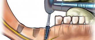
How are crowns removed from teeth, indications?
In cases where the crown or bridge has moved during the MRI, they are removed and installed again or replaced with new prostheses. They do this in the following ways:
- sawing - the prosthesis is drilled out using drills with a cooling system; after removal, the structure is unsuitable for further use;
- ultrasound – ultrasonic waves destroy the cement, and the crown is easily removed from the stump;
- Koch apparatus - a special drill breaks the bond with gentle pushes, then the structure is lifted and removed;
- Coronaflex apparatus - compressed air is supplied under the edge of the prosthesis, this is the only method that eliminates damage to the structure and supporting teeth;
- special tools or sliding crown removers - used after destruction of the adhesive base using one of the above methods.
Orthopedic structures are also removed in case of resorption of cement, chips of ceramic lining, damage to the frame, inflammatory processes in the root canals, or the patient’s desire to replace the prosthesis with a better one.
It doesn't hurt to remove crowns
Is it possible to shoot on your own?
Sometimes the bridge or crown is so loose that the patient can tear it off himself. However, this should not be done, because... possible:
- damage to the prosthesis;
- fracture of the stump or root system of the tooth;
- traumatization of the mucous membrane or adjacent units.
If the denture becomes loose, the only correct option is to contact a dental clinic.
Does it hurt?
Removing dentures does not hurt at all, unless, of course, the patient rips them off himself. Because crowns and bridges are placed on already decayed teeth; in most units the pulp has been removed. Therefore, unpleasant sensations are excluded. There may only be a feeling of pressure on the jaw.
If the tooth under the crown is not pulpless or its roots are inflamed, dentures are not removed. The doctor administers anesthesia to eliminate pain.
How long does the procedure take?
The duration of prosthesis removal depends on the method. So, using a cut, the structure is removed in 10-15 minutes. But preliminary decementing of crowns using ultrasound, a Koch or Coronaflex apparatus can take several visits.
Artificial teeth are not an obstacle to magnetic resonance imaging. Modern designs are made from bioinert materials that do not harm the patient and do not affect the examination. If there is one of the items prohibited for examination in the mouth, it is removed and then fixed again or replaced with a new one.
How MRI works
The final result of an MRI examination is an image of the human body “from the inside”, created using magnetic waves. A magnetic field is formed in the device, in which the patient is placed, and the computer records the return signal emanating from the molecules of his body.
The resulting image is not just a photograph. Magnetic pulses create a three-dimensional layer-by-layer image, that is, the body can be viewed at different depths and from different angles. The method allows you to obtain accurate information:
- about the location of neoplasms, even small ones;
- about micro-strokes and hemorrhages;
- vascular deformation, aneurysm and multiple sclerosis, invisible to other types of scanning;
- intervertebral hernias not detected on other devices.