An MRI machine, by generating a strong magnetic field around a person, is capable of examining many disorders and pathologies of organs and tissues, but it can have a negative effect on the body of patients who have metal foreign bodies. One of the contraindications for the MRI procedure is the presence of implants made of various metals and alloys. Implants are considered to be pins in bone tissue, joints, permanent structures, pacemakers, and dentures. Why do doctors recommend choosing a different examination method in the presence of metal implants? Is their presence an absolute contraindication for the procedure? If there are metal objects in the body, in particular titanium, is it possible to undergo an MRI or not?
MRI and metal plates
Depending on the relationship of any metal to the influence of a magnetic field, they are divided into diagmagnets (in the field they are subject to weak repulsion), paramagnetic (weakly attracted by the magnetic field) and ferromagnetic (strongly susceptible to the influence of the field).
In exceptional situations, the doctor may prescribe an MRI if the patient has metal plates. If there is metal in the body, the examination can only be carried out if its immediate location is outside the range of the magnetic field, or the diagnosis will be performed using low-field equipment. However, in the vast majority of cases, metal prostheses are a contraindication to the procedure.
In the presence of titanium plates in the leg and other parts of the body, diagnostics are carried out without restrictions, since titanium is paramagnetic and is not characterized by strong attraction in a magnetic field. MRI with a titanium prosthesis is as informative and harmless as without it.
Is it possible to do an MRI if there is a pin in the tooth or if you wear braces - MEDSI
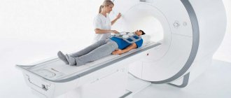
Magnetic resonance imaging (MRI) is a type of examination of the body that is carried out using magnetic waves using a tomograph. The impulses sent by this device are reflected differently from each organ of the human body due to magnetic resonance. The result is a series of images of the organ under study from different sides and in different planes. Their detail helps the doctor make the diagnosis most accurately.
In what cases is this analysis used?
This study is necessary to identify problems with the spine, joints, brain, cardiovascular system, gastrointestinal tract and other organs. Thanks to it, it is possible to identify tumors, inflammation, and post-traumatic changes.
A doctor may prescribe an MRI in cases such as:
- Chronic headache, cramps, back pain, dizziness
- Determination of the state of the brain and the presence of pathologies in it
- Examination before or after surgery to monitor the patient's condition
- More accurate diagnosis after other tests
Application of tomograph in dentistry
In some cases, this testing may be required for problems such as:
- Pain and spasms when chewing
- Involuntary jaw movements
- Unusual sounds in the jaw
Is it possible to do an MRI of the brain if you have metal teeth?
Implants are placed in place of extracted teeth. They consist of a metal support and an artificial crown. Many patients who have undergone such surgery wonder whether the CT scan will harm them.
In this case, much depends on the metal from which the implant is made. Materials such as titanium and tantalum are inert and do not interact with the magnetic field and therefore do not distort the analysis result.
For implants made of ferromagnetic materials (alloys of gold, steel, etc.) there is a loose limitation for this analysis. It is due to the fact that these metals enter into a magnetic interaction with the tomograph. It will not harm the patient in any way. The problem is the image distortion that occurs due to this interaction.
What to do if you have metal teeth?
In the presence of non-inert metal implants, the use of magnetic resonance imaging of the head and chest is not recommended. For other organs, this analysis can be carried out - the tomogram is not distorted. If a head examination is needed and the patient has metal teeth, the doctor may order a CT scan or other test.
To prevent the patient from having to undergo an examination, the results of which may be useless or inaccurate, he should inform the doctor about the presence of metal implants and find out from the dentist exactly what material they are made of.
Is it possible to do the procedure with metal crowns?
Before performing an MRI of the brain, you need to remember about dental crowns. They are placed if the teeth are severely damaged and filling no longer helps. For their manufacture, metal, metal-ceramics and various alloys are used. Crowns made of copper-gold alloy or steel greatly influence the analysis results.
More modern crowns, for the manufacture of which inert metals and ceramic coating were used, do not affect the tomogram. Fasteners are made of cobalt, nickel or iron. They interact with the magnetic field, but do not heat up much and, due to their small size, do not affect the result of the study.
If the patient does not know what material his crowns are made of, he can contact the dentist to get a description of them.
Modern MEDSI equipment allows you to change its settings to avoid distortion of the tomogram. But only a doctor can determine how much this will help. If it is impossible to conduct an MRI of the brain due to metal crowns, he may prescribe another study.
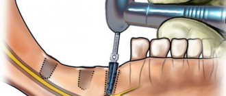
Is it possible to do the procedure with a pin?
The pins are installed to securely fix the filling. Some patients are afraid to have an MRI of the brain if they have a pin in their tooth. They fear that the metal will heat up or be attracted to the magnet, destroying the jaw and other oral tissues.
Even if the pin is not made of inert materials (alloys of gold, steel, etc.), it does not deform the tissue around it and does not heat up much. But it will distort the tomogram of the head. Pins made of precious metals have the greatest impact on the results.
If a patient who needs an MRI of the brain has titanium, ceramic or gutta-percha pins in his teeth, then no problems will arise during the procedure.
To avoid distortion of the analysis results, before the procedure it is necessary to warn the doctor about the presence of the pin and make an appointment with the dentist to clarify the issue of its material.
How to do an MRI of the brain and keep braces?
Braces are placed to correct the bite and straighten the teeth. They can influence the results of the study only if they are made of non-inert materials. If they are titanium or polymer, then no reaction with the magnetic field will occur and there will be no interference on the tomogram.
Braces will have to be removed only if they are made of ferromagnetic materials (precious metals, steel alloys), and it is vitally important to conduct a tomography examination rather than a CT scan or other tests.
What do you need to get a procedure at MEDSI?
You must take one of two steps:
- Call and schedule a consultation or
- Contact one of twenty medical centers throughout Moscow
Here you will be met:
- Experienced radiologists of the highest qualification category
- Modern premium equipment that allows you to regulate voltage and neutralize interference
- Possibility of making an urgent tomogram in case of injury or sudden illness
MRI after stenting
After stenting, an MR examination is not only allowed, but also prescribed. Therefore, the answer to the question of whether it is possible to do an MRI after stenting is positive. But the specialist conducting magnetic resonance imaging must know exactly what material the stents are made of.
You can absolutely safely carry out examinations with bioabsorbable stents, since they consist of a biopolymer - after a given time they are absorbed, but the lumen of the vessel is preserved.
In other cases, stents are made of inert metal alloys: stainless steel, cobalt alloys, etc. Note that the patient is required to strictly follow the instructions for the stent, i.e. if it states that MRI should not be done in the first few weeks after stenting, then this applies not only to the area where the stent is installed, but to the entire body. Even if it is not directly located in the apparatus tunnel, the magnetic field works equally strongly in the room where the tomograph is installed.
Sometimes immediate diagnosis is necessary when the presence of a stent is not known before MRI is performed because the patient does not have time to report them. Practice has confirmed that the materials currently used for the manufacture of stents are not ferromagnetic and do not respond to external field influences, and, therefore, are MRI-compatible.
Sign up for an MRI
If you have pins in your teeth, a brain MRI can be easily performed at our center. We use modern equipment, which allows us to obtain the most informative data based on the results.
Among the important advantages of contacting us:
- Providing the most detailed and comprehensive conclusion on the results.
- High speed of research.
- Affordable prices and special discounts for different categories of buyers.
- Convenient location.
To make an appointment to visit the center at a convenient time, leave a request on the website or call us. We will answer all your questions and check if there are any contraindications. We provide many options for examinations using magnetic resonance imaging.
Is it possible to do an MRI with iron crowns?
If you have old-style crowns made of iron, you cannot screen the brain and heart. The metal heats up significantly, which causes severe pain in the patient, deformation of the metal structure - the integrity of the implants may be compromised or they may fly off the teeth.
With crowns and dentures with metal-ceramics, screening of the brain and heart area is allowed, but there is a high probability of an unreliable result due to a distortion of the response to magnetic field signals.
Regardless of the type of alloys of crowns and prostheses, it is allowed to conduct MRI of the lumbar spine, abdominal organs and retroperitoneal space, pelvic region and extremities in closed-type devices.
When installing pins, high-strength titanium implants are often used. Their presence does not affect the reliability of the examination results; moreover, the size of the pins is so small that the magnetic field does not have any significant effect on them.
Metal crowns made of polymer alloys also do not distort magnetic field signals, however, you should check with your dentist about the possibility of conducting an MRI. Some structures heat up, so the procedure will cause significant discomfort to the patient.
If a patient has dental bridges installed, then they probably contain separate parts - pins, plates, screws of various sizes. For their manufacture, diamagnetic, ferromagnetic and paramagnetic materials are used - cobalt, iron alloy and nickel, which react differently to magnetic field signals. Therefore, you should check with your dentist what materials were used to make the prosthesis, and notify the tomography specialist - he will decide on the possibility of conducting an MRI.
Dental implants and brain MRI
Implantation of artificial teeth is a fairly common orthodontic procedure. This is a convenient and aesthetic way to replace lost teeth. Dental technologies for manufacturing implants are constantly being improved: outdated metal prostheses (made of iron and nickel) have been replaced by new materials. Titanium is the most commonly used metal in the manufacturing of implant mounts. It is inert to the magnetic field (does not heat up, is not attracted, does not magnetize), is not rejected by the body, and does not oxidize.
But, despite the relative “adequacy” of the reaction of paramagnetic substances to the effects of magnetic radiation, patients with dentures are not recommended to have an MRI of the head.
Is it possible to do an MRI with braces?
Modern brace systems are made of expensive and durable alloys that do not deform under the influence of magnetic-nuclear radiation and cannot move or injure the patient’s oral mucosa.
Small structures do not distort tomograph signals and do not heat up; their response to a magnetic field is very weak.
You cannot do an MRI if a fairly voluminous structure - more than 20 cm - is secured with ferromagnetic retainers. In this case, the bracket may become hot.
Is it necessary to do an MRI if a bracket is swallowed to accurately determine its position in the intestine? It is impossible to swallow a large brace, but a small one will come out naturally. To speed up this process, you need to eat more viscous porridge and drink fluids.
Braces do not affect the patient's condition during MRI, but they can result in insufficiently reliable results when scanning the brain, heart area, thoracic or cervical spine.
In cases where it is urgently necessary to screen the brain and cardiac system, and doctors do not see an alternative to MRI, you should contact orthodontists and remove dental implants. After tomography, they are reinstalled in the required volume.
Composition of modern endoprostheses
All plates, pins and endoprostheses, which are used in modern traumatology and orthopedics, consist of a variety of alloys. Note that different implants contain different amounts of paramagnetic and ferromagnetic materials. The properties of each endoprosthesis, pin or plate depend on the composition.
Not all prostheses are 100% metal. Most of them contain ceramics or polyethylene. The latter does not interact with the magnetic field, therefore, it does not in any way affect the MRI results and the course of the procedure. However, ceramics most often contain aluminum oxide, which still has a certain magnetic susceptibility.
Destroyed components of the hip joint implant.
Possible combinations of materials in endoprostheses:
- ceramics + ceramics;
- ceramics + polyethylene;
- metal + polyethylene;
- metal + ceramics;
- metal + metal.
Fact! Plates and pins for fixing bone fragments are made of metal alloys. The same applies to external fixation devices (Illizarov type) and clips that are placed on vessels.
Composition of artificial joints:
- cobalt;
- chromium;
- molybdenum;
- titanium;
- zirconium;
- tantalum;
- niobium.
Having familiarized yourself with the composition, you can understand how it will behave in a resonant tomograph. The magnetic properties of each endoprosthesis are determined not only by the material from which it is made, but also by its shape and size. Steel pins and plates longer than 20 cm can heat up above the permissible limit.
Fact! Products containing large amounts of nickel and cobalt interact especially actively with a magnetic field. This means that diagnostics with such endoprostheses should be performed with extreme caution.
Is it possible to do MRI with endoprostheses and other implants?
What to do if the patient has various types of implants right in his body? First of all, this must be reported to the specialist conducting the examination, since many metals are ferromagnetic and can shift in the body under the influence of a magnetic field.
For an MRI with a steel wire in the body, everything is not so simple. Iron causes the magnetic field to deviate from a given direction, which leads to distortion of the resulting images and the appearance of artifacts (defects) on them. In addition, the needle can heat up, creating unpleasant sensations for the patient.
Also, the answer to the question whether it is possible to do an MRI with an endoprosthesis depends on what material it is made of. If it is titanium, then there are no restrictions. If it is made of ferromagnetic materials, then this is a contraindication for research. You can find out exactly what metal the implant is made of in the design passport, which is issued to the patient after prosthetics.
What implants can be used for MRI?
MRI is allowed for people who have undergone hip or knee replacement. It is important that the endoprosthesis or fixation for osteosynthesis is made of metals or ceramics with low magnetic susceptibility. This avoids displacement or overheating of the structure during the examination.
Knee endoprosthesis.
People with hernia mesh, dental, breast and joint replacements are also allowed to have an MRI. All these implants are made from materials that do not interact with the magnetic field. This makes the study safe. However, you should consult a specialist before an MRI. The doctor will assess the possible risks and recommend the necessary precautions.
Minimally invasive endoprosthetics in the Czech Republic: doctors, rehabilitation, terms and prices.
Find out more
How do iron parts of implants affect different areas during MRI?
Regardless of the area of examination, before the procedure it is necessary to remove all metal objects:
- buttons,
- rivets,
- hooks,
- lightning,
- buckles,
- keys,
- coins,
- keychains,
- decorations,
- watch,
- Cell phones,
- magnetic media (cassettes, floppy disks),
- credit cards.
In addition, it is necessary to warn medical personnel about the presence of implants or prostheses in the body.
, especially those containing metals. During an MRI of the head and cervical spine, the iron alloys in the denture fixtures can heat up and move. This can lead not only to distorted diagnostic results, but also to discomfort or possible injury to the subject. However, such an outcome is only possible if there are large fragments of metal in the body, and the mass of the pin or plate is negligible. Therefore, even with a large number of implants, the occurrence of such a situation is almost impossible. And certainly, the presence of an implant in the body is not a reason to refuse an MRI.
The presence of metal fasteners in dental implants does not have any effect on other types of MRI - bones, joints, thoracic and lumbar spine. Direct MRI diagnostics of teeth is rarely prescribed; it is difficult to “get close” to the jaw using this tomograph.
And MRIs of the limbs, lower back and spine are without consequences. Dental diagnostics using this method is rarely prescribed - it is difficult to “get close” to the jaw using MRI.