Studies using a magnetic resonance imaging scanner are informative. A distinctive feature of MRI is a targeted examination of a selected area and the ability to detect pathology at the very beginning of development. But patients are prevented from undergoing this procedure by the presence of contraindications. Many people are particularly interested in whether it is possible to do MRI of the pituitary gland or other parts of the body with dental implants and metal crowns. More details later in the article.
How MRI works
The final result of an MRI examination is an image of the human body “from the inside”, created using magnetic waves. A magnetic field is formed in the device, in which the patient is placed, and the computer records the return signal emanating from the molecules of his body.
The resulting image is not just a photograph. Magnetic pulses create a three-dimensional layer-by-layer image, that is, the body can be viewed at different depths and from different angles. The method allows you to obtain accurate information:
- about the location of neoplasms, even small ones;
- about micro-strokes and hemorrhages;
- vascular deformation, aneurysm and multiple sclerosis, invisible to other types of scanning;
- intervertebral hernias not detected on other devices.
How an MRI scanner works - the essence of the method
Before understanding how MRI machines and dental crowns interact with each other, you need to understand the essence of the analysis itself. So, magnetic resonance imaging is a method of obtaining layer-by-layer images of internal organs, based on nuclear magnetic resonance.
The magnetic field of the tomograph and electromagnetic waves interact with the nuclei of hydrogen atoms - and this element is found in large quantities in tissues (water, that is, hydrogen plus oxygen, makes up about 70% of the human body). The computer records information received from hydrogen nuclei and converts it into layer-by-layer images - these are virtual sections of organs, blood vessels, and nerves. The thickness of one such slice is only 0.2 mm on modern tomographs with a power of 1.5-3 Tesla. And there are a lot of cuts themselves. That is, as a result, you can see very tiny pathologies inside the body without surgery or a microscope.
Only until 30.09 South Korean implant Osstem - 19,900 rubles.
Hurry up to sign up for a free consultation and lock in promotional prices.
Call now or request a call
Opening hours: 24 hours a day - seven days a week
The MRI technique is used primarily to study soft tissues and organs of the body. Whereas CT (computed tomography using X-rays) better reflects the condition of hard tissues - bones.
Effect of MRI procedure on implants
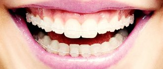
Since the examination is based on the use of magnetic waves, many patients have a logical question: is it possible to do MRI with dental implants? These concerns are understandable, but not always justified.
In dentistry, as in any other area of medicine, prosthetic technology is constantly evolving, new materials and techniques are appearing. So, if previously dental crowns were made of an alloy of copper and gold, today you will be offered metal ceramics or titanium alloy.
What is the difference? Old copper-gold technology produced distortions in MRI images
, but did not affect the health or condition of the patient during the procedure. Today in dentistry, titanium with small admixtures of other elements is widely used, which makes the prosthesis lighter and more durable. By its nature, titanium is chemically inert, therefore it does not oxidize, does not emit harmful substances and does not react to magnetic waves, which means it does not distort images. All this applies not only to teeth, but also to prosthetic bones and joints.
Metal-ceramics during MRI also does not affect either the state of health or the diagnostic result.
But, in addition to the implant itself, there are also pins, plates and screws on which it is attached. They may be the problem. Implant fastening parts are made from various ferromagnets and special iron alloys
, which have positive magnetic susceptibility. Exposure to a magnetic field can produce distorted results and affect the patient’s well-being during the procedure.
Interaction of metals with a tomograph
Taking an MRI if you have crowns can, in principle, lead to displacement and/or heating of metal prostheses. But only under a combination of several conditions - a relatively large mass of the metal and the presence of magnetization properties. Here you should refer to the following classification of metals and their alloys:
- ferromagnetic materials (iron, steel, nickel, cobalt, chromium): these substances are highly susceptible to the action of a magnetic field. It is they that can shift or heat up during MRI, but if their concentration is large enough,
- Diamagnets (hydrogen, silver, copper, gold): the ability to magnetize is very weak, i.e. There is no harm to the body during the examination,
- paramagnetic materials (titanium, tantalum, platinum, aluminum): do not interact with the magnetic field, i.e. absolutely safe for research - they will not heat up and will not move. For example, gurneys that can be brought into the room with a tomograph are made of aluminum.
How do iron parts of implants affect different areas during MRI?
Regardless of the area of examination, before the procedure it is necessary to remove all metal objects:
- buttons,
- rivets,
- hooks,
- lightning,
- buckles,
- keys,
- coins,
- keychains,
- decorations,
- watch,
- Cell phones,
- magnetic media (cassettes, floppy disks),
- credit cards.
In addition, it is necessary to warn medical personnel about the presence of implants or prostheses in the body.
, especially those containing metals. During an MRI of the head and cervical spine, the iron alloys in the denture fixtures can heat up and move. This can lead not only to distorted diagnostic results, but also to discomfort or possible injury to the subject. However, such an outcome is only possible if there are large fragments of metal in the body, and the mass of the pin or plate is negligible. Therefore, even with a large number of implants, the occurrence of such a situation is almost impossible. And certainly, the presence of an implant in the body is not a reason to refuse an MRI.
The presence of metal fasteners in dental implants does not have any effect on other types of MRI - bones, joints, thoracic and lumbar spine. Direct MRI diagnostics of teeth is rarely prescribed; it is difficult to “get close” to the jaw using this tomograph.
And MRIs of the limbs, lower back and spine are without consequences. Dental diagnostics using this method is rarely prescribed - it is difficult to “get close” to the jaw using MRI.
Price
The main pricing factors in computed tomography:
- size of the study area;
- printing on film or recording on digital media;
- the need for a written report from a radiologist.
Prices for 3D diagnostics in our Center are included in the consultation before surgery:
- 3D image for diagnostic and advisory purposes - RUB 3,000. ;
- 3D image with recording on digital media for third-party organizations - RUB 5,200.
A list and explanation of cases for installing implants with an indication of cost are listed here.
Myths about the MRI procedure
What not to believe
when going for an MRI:
During the procedure, the implants heat up and move.
The specific weight of the metal part of the implant fastening is negligible, which means there will be no defects in the images or discomfort for the patient. But it is still worth warning medical personnel if it consists of zirconium dioxide, pure ceramics or expensive alloys, there are no contraindications for MRI, but if it is metal ceramics, it is necessary to clarify what metals and impurities the implant consists of. If the composition contains a lot of ferromagnetic metals, the image quality will most likely be reduced and it will be impossible to evaluate the structures behind the structure.
You should not do an MRI during pregnancy.
This is only partially true. In the first trimester of pregnancy, the laying of tissues and vital organs of the child occurs, so any physical, chemical and biological effects should be excluded. Therefore, MRI during this period is performed only for emergency indications. However, in the later stages, diagnosis makes it possible to determine the position of the child, identify a narrow pelvis in a pregnant woman and enable the obstetrician to prepare for the correct delivery.
Going through an MRI with prosthetics is painful.
No, this is an absolutely painless and safe procedure. You can undergo MRI even with titanium plates; they are not affected by the magnetic field and cannot cause harm. The procedure can only be denied if a steel plate is implanted in the patient's body, since it heats up and moves under the influence of a magnetic field, which can injure the patient.
Do I need to tell my doctor about the presence of metal prostheses and implants?
Definitely necessary! After all, the tomograph needs fine tuning for an informative image. And as for dental implants, you should know that the bulk of them are created from the bioinert paramagnetic titanium. There is a small percentage of dental implants made from tantalum, which is also used to make surgical tantalum staples and “sutures” - tantalum is also paramagnetic, i.e. does not pose a danger. Another option is a combination of zirconium dioxide and titanium (Roxolid alloy), which is also not magnetized. In any case, a patient with dental implants should have a service book on hand, which indicates the model and brand of implants - it is easy to check from them what the structures are made of.
But if the implant was placed a long time ago - more than 20 years ago, or was made individually in a laboratory at the clinic, then most likely it is not made of titanium or tantalum, but from a cheaper alloy that can be magnetized. Here it is better to abandon MRI in favor of alternative diagnostics.
What could be the real reason for refusing an MRI?
- Claustrophobia. The patient is placed in a closed apparatus for a short time, which can provoke a panic attack. But here it all depends on the degree of claustrophobia and the type of examination. In addition, each device has an “alarm” button, when pressed, the procedure immediately stops and medical personnel approach the patient.
- An installed pacemaker may come into dangerous resonance with magnetic waves and stop working.
- Before starting the examination, the hearing aid must be removed, since the magnet in the hearing aid may be damaged under the influence of a magnetic field. And the presence of middle and inner ear implants is an absolute contraindication, since they often contain metal alloys with strong magnetic properties.
- An insulin pump is an absolute contraindication for MRI.
Before the procedure, you must tell your doctor
about everything that may affect your health and the diagnostic result. You can’t hush something up for fear that an MRI won’t be done. As a rule, any problem can be solved, and in the case of metal prostheses and implants in the patient’s body, the doctor will simply adjust the tomograph so that, taking into account the location and composition of the prosthesis, the device will produce the highest quality clear image. If there are absolute contraindications to MRI, the doctor will select the most informative and permitted type of examination in your case.
If the patient “forgets” about the fake ideal smile, the pictures will turn out blurry, and he will have to undergo an MRI again, having honestly admitted all the interventions in the body.
If you are planning to undergo MRI diagnostics from qualified specialists, using modern expert-class equipment in comfortable conditions and in the shortest possible time, call the phone number listed on the website, or leave a request in the feedback form. Medical specialists will answer your questions and conduct all necessary examinations within one business day. Let's take care of your health together!
When not to use
Despite its many benefits, there are cases when CT scanning cannot be used. Contraindications include:
- the patient is allergic to the contrast agent that is administered before the procedure;
- pregnancy (to eliminate the risk of negative effects on the developing fetus);
- breastfeeding (if a woman has been injected with a contrast agent, the child should be switched to an alternative diet for 48 hours);
- active children under 14 years of age - during diagnosis, sudden movements should not be made and an active child will not tolerate this even for a short period of time;
- claustrophobia - the tomograph is a closed space that can cause panic in a person with such a disease;
- diabetes mellitus, thyroid disease or kidney pathology - the introduction of contrast in such cases will provoke complications.
Computed tomography can be performed without the injection of a contrast agent. This significantly reduces the list of listed contraindications. Only a specialist can choose the most appropriate examination method, taking into account the patient’s existing problems.
Important! Experts do not recommend doing CT scans more than 2 times a year . After scheduled examinations, it is necessary to take a break for at least a year. It all depends on the radiation dose, diagnosis, circumstances surrounding the procedure, and individual characteristics of the body.
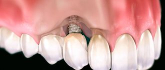
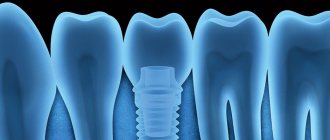
Why do you need a CT scan during implantation?
When undergoing dental implantation, a CT scan is considered desirable. Thanks to this innovative technology, implantation operations have become more predictable, carried out much faster and safer, ultimately reducing the patient’s rehabilitation period. After all, the process of osseointegration directly depends on the quality of their installation, and this can only be achieved after a thorough examination.
It is CT that allows the dentist to:
- preliminary study of the patient’s bone density;
- analyze existing defects and identify inflammatory processes;
- determine the location of the maxillary sinuses, the location of nerves and blood vessels;
- calculate the exact dimensions of the future product and the depth of its penetration into the bone;
- install the implant at the desired angle of inclination, in accordance with the dentition;
- plan the permissible physiological load on the implant.
That's why all of this is so important. After all, dental implantation involves the implantation of an artificial root into the gums and bone tissue, and if all the features of the jaw are not taken into account and it ends up in the wrong place, then the person will constantly experience discomfort or pain.
When installing prostheses on implants, it is also necessary to do a CT scan (other examination methods are allowed). This will help the dentist perform all the necessary manipulations with millimeter precision, and will save the patient time and money.
Application in individual cases
For a dental implant to last for many years, it must be completely surrounded by the jawbone. But its structure differs from person to person: in anatomical shape, height and width, density, presence of depressions and defects, etc. Therefore, for a dentist, the task of introducing an artificial root is not an easy one, and requires an individual approach:
- The implant must be placed where the bone is strong and thick.
- It should be positioned at such an angle that the tooth on it does not protrude from the natural row (aesthetic effect).
- It is important to correctly determine the depth of penetration into the bone so as not to touch the nerve or damage important anatomical structures.
Only computed tomography and preliminary 3D modeling will help you take into account all the nuances.
When implanting upper teeth
The most dangerous is the installation of implants in the lateral sections of the upper jaw. In these zones, deep in the bone, there is a maxillary sinus, which usually increases in size when teeth are lost. This situation will significantly complicate the operation if a CT scan is not performed in advance.
Before implanting teeth in the upper jaw, preliminary preparation and diagnosis of the maxillary sinus and nasal cavity are required. The fact is that this zone can complicate the procedure for a number of reasons:
- the presence of acute or chronic inflammation in the maxillary sinuses;
- tumors, polyps, cysts;
- increased size of sinuses due to tooth loss.
Chronic inflammation of the maxillary sinus will inevitably cause swelling of the mucous membrane. In addition, there may be exudate underneath, which will negate the dentist’s work and will not allow the implant to take root. The normal condition of the sinus is a rounded thickening of the mucosa in the alveolar bay, known as a mucocele.
To avoid complications after surgery or complete rejection of the implant, it is first necessary to cure all diseases that may become an obstacle to its installation (including sinusitis). Proper installation of an artificial root in the upper jaw should be carried out after joint consultations of two specialists: an ENT specialist (otolaryngologist) and a dentist. This will help predict the results of treatment and implantation in advance.
Dental indications for tomography
- Diseases of the temporomandibular joint;
- the presence of extraneous sounds when closing and opening the jaw, displacement of the lower jaw to the side;
- malocclusion;
- involuntary jaw movements;
- pain, discomfort when chewing food;
- spasms of muscle tissue when opening and closing the mouth;
- complications after treatment or tooth extraction;
- diseases of the pulp, gums;
- study of neoplasms in the maxillofacial region.