Increased tooth wear – This is a process of increased tissue loss: first, the enamel becomes thinner, and then the inside of the teeth. Typically, the process of abrasion is constant - teeth gradually wear out throughout human life: first, the tooth enamel becomes thinner, and in old age it comes to the inner layers. Increased abrasion is a similar process that occurs at an accelerated rate: a real problem, and we will tell you how to deal with it.
The pathological loss of dentin and enamel is different in that it occurs much faster than with normal physiological abrasion. In this case, we talk about abnormal tooth wear, which is a recognized dental problem that mainly affects men over the age of 30. In this case, the decrease in the dental layer begins gradually, and as it progresses, it is no longer possible to influence the disease or cure it. That is why it is extremely important not to miss the initial stage of destruction of tooth enamel - so that tooth extraction, treatment and prosthetics are not required.
How to tell if your teeth are really big
The size of human dental crowns has its own physiological limits. More often than others, the upper cutting elements, which are located in the center, seem excessively large. These two teeth in a mature person range from 9 to 13 mm in length. It usually depends on height. On each side of them, 2 teeth are usually shorter by a couple of millimeters. The ratio between the dimensions of the tooth crown in width and height is also important. Its ideal value is 1.25%. Long front teeth are counted when this number becomes smaller.
This condition has its own name – macrodentia. It is diagnosed only when a person’s teeth are one and a half times larger than the standard size.
On a note!
With a normal crown size, from under the lip you can see the edge of the upper teeth, not exceeding 3 mm. Large front teeth in a child or teenager are quite normal. Gradually, the enamel wears down and the size of large crowns decreases.
When should you see a doctor?
Dentist help is necessary if one of the incisors is in a high or low position. In children, this defect can disappear without any intervention when replacing milk teeth with molars. If different lengths of the front teeth persist despite a fully formed bite, you should consult a doctor.
You should also contact a specialist:
- when the enamel darkens,
- increased sensitivity of teeth,
- acute toothache,
- cutter mobility,
- formation of a sharp cutting edge.
The dentist will conduct an examination, determine the cause of the problem and help solve the problem.
Reasons for lengthening teeth
There are not very many reasons why a person can develop 2 large teeth. But they need to be clarified and taken into account if the issue is correction of the defect.
- People often have large teeth from birth, due to genetic predisposition.
- Over time, the front teeth can become large due to age-related weakening of the gums.
- Teeth can become larger than others due to inflammation and bone loss.
Causes of dental defects
Lengthening or shortening of one of the incisors is usually a congenital anomaly. This feature of dentition development is associated with genetic factors. Experts also include the following as possible causes of the problem:
- periodontitis,
- malocclusion,
- presence of supernumerary teeth,
- abnormal shape of the alveolar process,
- disruption of the process of resorption of the roots of baby teeth.
Changes in the length of the central incisor can also be a consequence of jaw trauma. If a tooth is mechanically damaged, its lower part may chip. Because of this, the appearance of the smile deteriorates, discomfort occurs when chewing, and the risk of developing various diseases increases.
What to do if your two front teeth are large
Before and after photos show how the appearance of your smile and face changes after correction of large incisors. There are several methods that help give a large dental element the required shape. All of these can help make the tooth smaller.
- Contour type plastic surgery. This is a method in which the shape is given by grinding with a special grinding machine.
- Composite restoration (contouring and bonding). After grinding the dental crown, additional composite materials are used. This coating not only improves the shape of the teeth, but also protects the enamel layer.
- Application of veneers. Such onlays can be installed on adjacent teeth so that the front ones do not stand out so much. This is especially true if for some reason turning is impossible. Another option is to install lumineers. These are more elegant plates that do not require much grinding of the teeth, and in some situations the teeth for them do not need to be ground at all.
- There are situations when enlarged teeth are a consequence of a crooked bite. Classic braces help correct this problem. This method is recommended most often in adolescence from 11 to 18 years.
- Implantation. It is used in particularly difficult cases when it is necessary to remove a large tooth. Subsequently, the removed elements are replaced with implants.
Before choosing the appropriate option for a particular case, the dentist will offer to undergo an examination, including an examination and x-ray of the jaws. Experts often recommend leaving dental crowns in their original form, but if it is decided to correct the defect, then it will be necessary to increase hygienic care. Use fluoridated toothpastes, dental floss and even an irrigator. Because the enamel layer will become more at risk of carious destruction.
Stages of dental destruction: when are consultation and diagnosis necessary?
By degree of destruction:
- Stage 1: there is a smooth smoothing of the tubercles on the enamel and the cutting edges of the teeth;
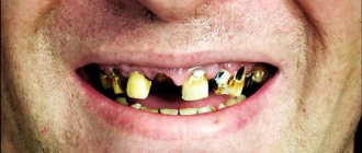
- Stage 2: tooth enamel is completely erased, the destruction process reaches dentin (the inner part of the tooth);
- Stage 3: the size of the part of the tooth located above the gum and clearly visible is only two-thirds of the norm;
- Stage 4: the crown of the tooth is completely destroyed and falls to the level of the gum.
Due to increased abrasion, the most unimaginable patterns may appear on the surface of tooth enamel, indicating damage to the enamel in the vertical, horizontal directions, in the form of dots, etc.
- Promotion
Dental treatment under anesthesia
Promotion for sedation in Moscow!
If you experience: — Severe stress during dental treatment — Fear of the sound of dental instruments — Time-limited visits to the dentist
old price
12 000 ₽
price
5 900 ₽
Free consultation RUB 5,900 for 30 minutes
Limited offer. Sign up for a free consultation. There are contraindications, consultation with a specialist is required.
- Promotion
Teeth whitening ZOOM
Promotion for teeth whitening in Moscow!
As a gift: - Prof. Oral hygiene – Whitening care set with individual trays
old price
40 000 ₽
price
19 900 ₽
Free consultation Sign up for a free consultation. There are contraindications, consultation with a specialist is required.
Possible consequences if left untreated
The front incisors, which are slightly longer than the others, do not cause any problems to a person and are even an object of fashion. This is due to the fact that enlarged crowns give the face a more youthful appearance and are not always an aesthetic flaw. Pathological enlargement of dental elements leads to unpleasant consequences.
- Very wide teeth can push, twist, and protrude beyond the row because there is not enough room for them. An abnormal bite develops, affecting not only the beauty of the smile, but also the digestive processes.
- If a child has a very large molar, it can become an obstacle to the eruption of neighboring elements.
- If the incisors have become larger over time, and were not this way from birth, then this indicates problems with the gums. In such a situation, before fixing the teeth, it is necessary to treat the gum tissue.
- Speech is distorted under the influence of pathologically large dental elements.
- A person with such a defect experiences psychological discomfort. He avoids communication and does not smile or does it very stiffly, controlling the process.
What types of diastema exist and why do they occur?
Diastemas, as well as tremata, come in different types. Experts divide them by etiology, location, and type of displacement. Doctors also distinguish between false and true diastemas separately. The latter are always a consequence of pathology, while false ones are found only in children and disappear as the teeth grow.
Symmetrical and asymmetrical diastema
Most often, the diastema is symmetrical, that is, the incisors are equidistant from the frenulum. An asymmetrical arrangement is much less common, and with it, only one incisor is displaced, the second is in the correct position.
Lateral deviation of crowns
With lateral deviation of the crowns, the roots are located correctly, the discrepancy begins only on the visible part of the teeth. Most often, this pathology is a consequence of supernumerary teeth. Also, a gap may appear due to bad habits.
Body lateral displacement
The peculiarity of this type of displacement is the complete deviation of the teeth along with the roots. It is provoked by congenital adentia, the lower attachment of the frenulum, and compaction of the middle suture on the bone tissue. In the latter case, the teeth cannot overcome the obstacle and take the correct position.
Medial inclination of central incisors
This type of diastema is the most difficult, since it simultaneously causes deviation of the teeth from the center and their rotation along the axis. The cause is almost always supernumerary teeth located between the first incisors or odontoma, a benign tumor growing in the jaw bone.
Celebrities with big teeth
Celebrities often have large front central incisors. They give the image something special and unusual. Such a smile makes a person stand out from the rest. The list of public figures with large incisors:
- Hilary Swank;
- Steve Tyler;
- Jon Heeder;
- Ann Hataway;
- Keira Knightley;
- Tom Cruise.
Is it possible to fix a hole between teeth at home?
It is impossible to remove diastema at home. To carry out effective correction, specialists use a whole range of diagnostic measures, as well as devices that are manufactured specifically for the patient. It is naive to believe that the problem can be eliminated simply by tying the two front teeth with floss. Moreover, such manipulations are dangerous, as they can damage the gums or the teeth themselves.
Sometimes you can hear stories about how a child's teeth were bandaged and the cleft disappeared. But, in this case, we are talking about a false diastema, which goes away on its own without any intervention.
What are the alternatives to teeth straightening?
Filing is a violation of the integrity of the tooth, so in the future it is possible to increase tissue sensitivity. If the patient has minor cosmetic defects (for example, incisors of different heights), orthodontic treatment can be done without drilling. Before straightening your teeth, you need to consider all correction options: mouthguards, trainers, braces, aligners, and also compare the prices of teeth filing and orthodontic bite correction.
Based on: 4 votes
Symptoms
It is difficult to miss such a fact when a tooth has crumbled. This usually occurs while eating, and a hard piece of enamel is felt in the mouth. Sometimes there are many small pieces at once. There is a feeling of panic. After all, we know from school that enamel is the hardest tissue in the body. Why does this happen, causing teeth to crumble and crack?
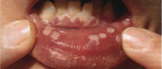
It happens that it all starts with stains on the enamel - this is a symptom that not everything is in order. And you need to make a visit to the dentist before the crown of the tooth collapses.
Anomalies in the shape and size of teeth - symptoms and treatment
Anomalies in the shape and size of teeth are variations in the development of teeth and the dental system that deviate from the norm.
Malformations of the dental system are divided into major (congenital) and minor. Congenital anomalies include facial clefts, such as cleft lip, cleft palate, etc.; to small ones - anomalies in the eruption, number, shape, size and structure of teeth. Unlike congenital malformations, minor anomalies are not accompanied by significant impairments and do not threaten the patient’s life, but they significantly affect the aesthetics of the tooth and treatment tactics, and can also lead to caries, malocclusion and other complications [1].
This article describes frequently occurring minor anomalies in the shape and size of teeth: macro- and microdentia, fusion of teeth, etc. Absolute and relative indicators of their frequency and prevalence are shown in the table below [11].
| Types of anomalies | Anomaly subtypes | Frequency of anomalies in the population (%) | Prevalence of anomalies (%) |
| Size anomalies | Macrodentia | 0,42 | 0,16 |
| Microdentia | 7,87 | 3,08 | |
| Shape anomalies | Fusion and fusion of teeth | 0,21 | 0,08 |
| Taurodontism | 11,27 | 4,41 |
Subtypes of anomalies and reasons for their development
All anomalies in the shape and development of teeth can be formed under the influence of negative factors that disrupt the development of fetal tissues and organs during pregnancy. These include:
- medications;
- nutritional supplements;
- viruses;
- industrial poisons;
- alcohol;
- tobacco smoke, etc.
Macrodentia is characterized by an increase in the size of the teeth - a large crown and a tapering root. It can affect both all and individual teeth [5]. The prevalence of macrodentia in permanent teeth is 0.03-1.9%. This pathology occurs more often in men. The large size of the crown causes problems with teething and leads to deformation of the dentition [9][10].
Microdentia is characterized by the reduction of one or all teeth [6]. According to epidemiological studies, the prevalence of the anomaly ranges from 1.5% to 2%, and is more common in women than in men [19]. Teeth with this pathology usually have a cone shape.
Macro- and microdentia can be hereditary. Since the formation of follicles (buds) of permanent teeth occurs at different times, starting from the 5th month of intrauterine development and up to 3 years, the number of abnormal teeth will depend on the impact of negative factors during this period.
Taurodontism (bull tooth) refers to abnormalities in tooth shape. It manifests itself in the form of an enlarged pulp chamber - the “core” of the tooth, consisting of connective tissue, nerves, blood and lymph vessels. The base of the pulp moves closer to the apex of the tooth, and there is no narrowing at the level of the cemento-enamel junction [12].
Taurodontism can occur at any age; it is most often found in patients 13-19 years old [11]. As a rule, the anomaly does not appear before this age, since the roots of permanent chewing teeth form before the age of 13.
Dental evagination (protrusion) affects the crown of the tooth. The anomaly occurs during the period of formation and formation of tooth buds. It appears in the form of an additional tubercle that protrudes from the crown of the tooth. This tubercle consists of enamel and dentin. Its size, structure and location vary widely. It can be horny, conical or pyramidal in shape. Otherwise, such an anomaly is called a tuberculate protrusion, a claw-shaped tubercle, a Leong premolar, and an occlusal enamel pearl.
The reasons for the development of evagination are not fully understood. The influence of X-linked and autosomal dominant inheritance is assumed [2]. With X-linked inheritance, the anomaly appears only in men, and women are only carriers of the mutant gene. With autosomal dominant inheritance, boys and girls with dental evagination are born equally often, and at least one of the child’s parents also has this anomaly.
Dental invagination causes the enamel to retreat into the dental crown. Otherwise, such an anomaly is called a “tooth in a tooth.” Intussusception occurs before calcification of dental tissues during fetal development [13]. It can affect any tooth, but most often occurs on the upper lateral incisors (front teeth).
How to prevent diastema in a child
If diastema is a common occurrence among relatives, the parents of a small child should inform the dentist about this when the child is 2-3 years old. The specialist will observe the dynamics and, if necessary, offer age-appropriate types of correction.
Among the general recommendations on how to prevent gaps between teeth in children, the main one is to ensure that the child does not develop bad habits, such as biting nails, pencils and pens, and sucking fingers.
How to get rid of the problem, solution methods
The dentist will decide how to stop tooth decay after identifying the cause. If treatment by other doctors is necessary, then in parallel it is already possible to begin the restoration of dental units.
Dental restoration methods:
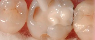
- filling.
Installing a filling in place of the missing piece of tissue. If the carious cavity is large enough and the walls are fragile, then an inlay with a composite material may be indicated; - veneers.
If there is a chip or crack in the front teeth of the frontal area, a thin ceramic overlay will correct the situation, hiding all the defects; - crowns
When the crown of a tooth is severely damaged, the doctor may recommend installing an artificial one from the selected material. Most often these are metal ceramics, ceramics, zirconium dioxide. It is important what condition the gums are in; - remineralization.
Restoring the normal balance of vitamins in the enamel structure. A special composition is applied to the surface of the teeth for several minutes. The tissue is saturated, enriched with calcium, fluoride, becomes stronger and can even remove caries at the chalk spot stage; - fluoridation.
Fluoride-containing pastes nourish the enamel. Fluorine is responsible for crystalline calcium compounds. Fluoride varnish seals dentinal tubules, strengthening teeth; - taking active
calcium and vitamin D
The ecological situation of the region of residence is one of the causes of dental health pathologies. The quality of air, water and food is far from ideal. Replenish the deficiency with complex supplements, spend your weekends in nature. Love organic farm products.