The first molars of the chewing group of teeth (6s) are large 2-3-root teeth, the main function of which is the primary mechanical processing of food. They are subject to high chewing loads. Due to their complex structure and location in the row, it is difficult to remove bacterial plaque around the molars, which leads to the development of caries and destruction of dental tissue. The loss of the first molars is fraught with impaired chewing function, displacement of the dentition, impaired occlusion, and other serious problems. Since the upper 6s are located almost close to the maxillary sinuses, the implantation of the 6s in the upper and lower jaws is different.
- When used:
Absence of 1 tooth - Type of anesthesia:
Local anesthesia - Procedure time:
From 15 minutes - Treatment period:
Up to 4 months - Healing period:
Up to 7 days - Age restrictions:
From 18 years old
Features of the structure of the root system
Human teeth have a purely individual structure, so the root system of the same element may differ from person to person, including the number of canals in it and their branches. The structure of the roots is directly related to the immediate purpose of the tooth:
- incisors of the upper and lower jaws - help to bite off food,
- canines and premolars - perform the primary chewing function,
- molars - take on the main chewing load and help crush solid food as thoroughly as possible.
The photo shows teeth with roots.
The last ones in the row, sixes and sevens, and sometimes eights, require more nutrients to remain strong and wear-resistant throughout a person’s life. Therefore, premolars and molars usually have a developed canal system. But before we move on to a more detailed consideration of the structural features of the root system, let’s look at what a tooth actually consists of.
Features of implantation of the 6th lower tooth
The six of the lower jaw bears more loads during chewing than the upper jaw. Therefore, implants are selected taking this fact into account - wide and of sufficient length. The nuances of implantation from below:
- easier to carry out than on the top;
- implants take root faster - in 2-4 months, since the jawbone is denser and anatomically higher;
- but there is a risk of damage to the trigeminal nerve if the protocol is not followed, which is complicated by numbness of the chin and lips.
If the bone is small in height, then a preliminary operation is performed to increase it. The gum is peeled off, bone material is added, and a resorbable membrane is applied to secure it. Recovery takes up to six months.
Without preliminary bone grafting, surgery is possible if the atrophy is moderate. In this case, our Center uses bone growth stimulants. They are fixed to the neck of the artificial root during its installation and trigger the processes of bone tissue regeneration. In this case, implant healing and bone tissue growth occur simultaneously.
What is a human tooth made of?
The visible part or crown is covered with a layer of enamel - the strongest tissue in the human body. Beneath it is dentin, and then the walls of the pulp chamber. The pulp (nerve) is a neurovascular bundle that smoothly passes into the root canals. At the very end of the root there is a small apical opening - nerve endings, vessels and capillaries pass through it.
The photo shows the structure of the tooth
Thus, if for some reason the pulp becomes inflamed, the pathological process spreads very quickly through the canals. Essentially, these are communicating vessels, so if one of the passages is missed during treatment and filling, the inflammatory process will continue to develop and ultimately lead to the need to completely remove the diseased element.
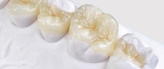
Installing a crown on the sixth tooth
The method of installing a crown depends on the degree of destruction of the crown part of the tooth. If, as a result of treatment, it is possible to save the tooth, but the filling area is large, then dentists recommend installing a crown. If the degree of destruction is small, but there are indications for installing a crown (chips, changes in enamel, abrasion of the tooth), then the tooth is given the desired shape, and then the crown is fixed.
If there is practically nothing left of the crown part of the tooth, then first a pin is installed in the treated root canal, then the tooth is built up on it, prepared, and only then a crown is installed.
Prosthetics consists of several stages
- Consultation and examination, referral for imaging, approval of a treatment plan.
- Sanitation of the oral cavity: professional hygiene, treatment of dental diseases and inflammatory processes.
- Preparing the tooth for crown installation: root canal treatment, pulp removal (if necessary), restoration of the coronal part with a filling, giving the tooth a cylindrical shape.
- Taking an impression, making a plaster model, from which the crown will then be cast. Our clinic has its own dental laboratory, which allows us to produce orthopedic structures on the same day. If it takes more than a day to create a crown, for example, when the clinic does not have its own dental laboratory, then a temporary onlay is placed on the tooth to protect it from germs and various damages.
- Trying on the structure and fixing it with dental cement.
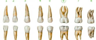
Differences in the structure of incisors, canines and molars on the upper and lower jaws - table
As mentioned above, the number of channels is largely determined by the range of the element. The essence is the same: the very last chewing teeth bear the maximum chewing load, so they must be strong and resilient, and for this they need abundant nutrition - this is provided by a developed internal system. The table below shows average data on the number of passages in the tooth roots in the upper and lower jaws.
| Tooth | Number of channels | |
| Fangs | upper | 1 |
| lower | 2 | |
| Incisors | upper | 1 |
| lower | 1-2 | |
| Premolars | upper | usually 2, but sometimes from 1 to 3 |
| lower | first – 1-2, second – 1 | |
| Molars | top first | 3 or 4 |
| second | 3, less often 4 | |
| third | 5 | |
| lower first | usually 3, but can be from 4 to 5 | |
| second | usually 3, but sometimes 4 | |
| third | 3 | |
On the upper jaw
The teeth on the upper jaw have their own structural characteristics, and therefore they are somewhat different from their antagonists on the lower jaw. Here are the main distinguishing features:
- second molars (sixes) most often have three canal passages, but in some cases there are 4,
- premolars usually have 2 canals, although in some situations their number can vary from 1 to 3.
The photo shows the teeth of the upper jaw.
It should be noted that the roots on the upper jaw usually have a more complex and branched structure. Therefore, upper molars are more difficult to treat, which explains the higher percentage of complications due to incompletely healed cavities.
On the lower jaw
The structure of the upper and lower jaws is different, which is partly due to the peculiarities in the distribution of chewing load. In the lower teeth, as a rule, there are fewer canals, but here much depends on the individual anatomical parameters of the jaw apparatus. Therefore, the patient must undergo an X-ray examination, and with the image in hand, the doctor will be able to begin treatment directly.
The following are the features in the internal structure of the incisors, canines and molars of the lower jaw:
- in the first molars (sixes) there can be from 2 to 4 moves - there is no exact number to focus on,
- the lower second premolar (five) most often has one canal, although in approximately 10% of cases specialists find two canals at once1,
- the first premolar usually has only one root, but in about a third of cases there are 2,
- Eights are the most unpredictable - the exact number of roots located inside can only be determined using radiography. As a rule, there are no more than 3 of them, but often during treatment specialists identify additional cavities. This is one of the reasons why wisdom teeth are difficult to treat therapeutically.
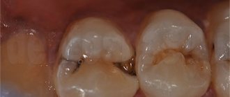
The photo shows the teeth of the lower jaw.
Regardless of the type and stage of the disease, competent dental treatment necessarily involves a preliminary x-ray diagnosis. Otherwise, any wrong move by the doctor can easily lead to serious complications.
Eights - third molars
A wisdom tooth usually has a complex and intricate root system - this is the only element in which the number of roots can reach 5. But this is extremely rare, and more often the figure eight has from 3 to 4 roots, while the lower third molar may not have any. more than 3.
The photo shows wisdom teeth
We are talking about a rudiment that we inherited from our ancient ancestors. Now we do not need to carefully crush very tough foods, such as raw meat. Therefore, there was no longer any urgent need for the figure eight, and due to the reduction in the size of the jaws, there was practically no free space left for it.
Because of this, the tooth almost always erupts with problems, since it initially begins to grow in the wrong position and often half remains under the gum - retention and dystopia. All this significantly complicates the treatment of pathological processes that are localized in this area. Therefore, most often the eight has to be deleted.
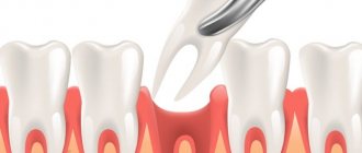
Preparing to remove the six
- Correct selection of tools. There are several types of forceps and elevators that can only be used when removing specific teeth. An error in choosing a tool leads to quite unpleasant consequences: for example, the removal of the 6th tooth of the upper or lower jaw cannot be carried out with narrow forceps - this will lead to the fact that it will either be crushed, or the patient will feel pain at the time of direct removal.
- Be sure to choose the right anesthesia before the procedure. Most use local anesthesia - an injection of lidocaine or ultracaine. But in some cases, such drugs are not suitable - for example, if the patient has an individual intolerance. Another point that dentists usually do not take into account is the fear of dentists, which amounts to dental phobia. To calm the patient down, you can sedate the patient and the doctor’s removal procedures will be absolutely calm.
- An examination of the oral cavity before tooth extraction is a mandatory procedure. This rule is due to the fact that after the removal of some teeth, problems with neighboring ones may begin. For example, removing a six implies some impact on the entire row.
Types of root canals
As noted above, the internal structure of each individual element has its own individual characteristics. But there is also a classification that makes it possible to group various options for the structure of dental canals into several categories:
- for one root there is one passage and one apical foramen,
- the root has several branches that connect closer to the apical foramen,
- two branched passages with one mouth and two apical openings,
- in one root, the cavities are connected and separated several times,
- three canals emerge from the same orifice but approach three different apical foramina.
The internal structure of each individual tooth has its own individual characteristics.
The number of roots and canals may be the same, but more often their number differs. In this case, cavities of various types may be present in one chewing premolar or molar.
Features of the structure of the root system in children
Each baby tooth has one nerve, just like the molars. In addition, they have a similar root system structure. In pathological processes, temporary elements are also opened, processed and filled, but the tactics largely depend on how long ago they erupted and how much time is left before the bite changes. Thus, the roots of baby teeth are usually filled with a special paste, which dissolves as the root system dissolves.
The process of root formation in permanent incisors, canines and molars takes about three years. If the root system is still at the stage of its development, special compounds are chosen for filling it - pastes with a high concentration of fluorine and calcium.
The photo shows the process of resorption of the roots of baby teeth
Why do sixth teeth deteriorate more often than others?
The sixth teeth (first molars according to dental classification) are molars. The rudiments are laid at the 5th month of fetal development, and at the 9th month their mineralization occurs, which continues throughout the first month after the birth of the child. Their final formation with enamel occurs only by the age of 2-3 years. Therefore, the condition of the root and crown depends on the course of pregnancy and childbirth.
Sixes do not erupt in early childhood, since the function of chewing is not yet needed. When they erupt, they do not have predecessors in the form of milk teeth, so they essentially grow from scratch. Of the permanent molars, they begin to erupt first at 5-6 years, so they last longer than others. Frequent diseases, removals and the need for implants are associated with this.
When inflammation begins - diseases and treatment
The internal structures of the tooth can become inflamed as a result of rapid development, caries, pulpitis or periodontitis. To determine the exact cause of the problem and the stage of development of the pathology, the patient must undergo an x-ray examination. The standard treatment regimen is as follows:
- the doctor removes an old filling or opens a carious cavity to open access to the insides of the teeth,
- if the nerve is still there, it is removed using a special instrument - depulpation is performed,
- Next, the doctor carefully cleans the canals and treats the walls with an antiseptic,
- fills the canals and fixes the filling on top. When treating periodontitis, to ensure the elimination of inflammatory processes, it is usually necessary to apply medication and install a temporary filling - and this procedure will have to be completed more than once.
“My root canals were cured in just 2 visits. It’s a pity, of course, that the nerve had to be removed. They say the tooth can darken. But the treatment was quite quick and easy, and there were no problems afterwards. I heard that sometimes people go for several months to clean out the roots, but at my second appointment they gave me a permanent filling. By the way, it was the first time they filled with gutta-percha, and it still has the same smell!”
KirillMigo, from correspondence on the forum www.32top.ru
The photo shows a treatment regimen for periodontitis.
The treatment regimen described above is a universal scenario that can be changed depending on the type, shape and stage of the pathological process. But the essence remains the same: it is important that all passages are thoroughly cleaned and sealed. Otherwise, relapses of inflammatory processes are possible, and then the risk of complete tooth loss will increase significantly.
How to care for a crown
The service life of crowns varies from 5 to 15 years, depending on the material and compliance with hygiene rules. Despite the fact that the crown is made of artificial material, plaque also accumulates on it. You should take care of it the same way you would take care of your teeth. Particular attention should be paid to the area at the edge of the gum. Dental plaque in these places can cause inflammation and other problems in the body.
Basic recommendations for crown care
- Brushing teeth 2 times a day
- Use of additional hygiene products: brush, dental floss, irrigator.
- Professional teeth cleaning every six months. If the dentist indicates other dates, follow his recommendations.
- Preventative visits to the dentist every six months.
- Monitor the condition of the joints between the tooth and the crown.
- Do not eat cold and hot food at the same time.
If you experience crown mobility, an unpleasant taste, or any other discomfort in the oral cavity, you should make an appointment with your doctor. Otherwise, it can lead to serious complications. You can get to our doctors by calling: 220-86-30
Why do we need to know the structural features of roots?
X-ray diagnostics and computed tomography in particular allow us to carefully study the root system and at the same time identify the narrowest and most intricate branches. And if the length of the canal is determined instrumentally, then the exact number of internal passages will be clearly visible on the x-ray image. Information regarding their cross-country ability is also extremely important:
- curvature up to 25 degrees – tool processing available,
- within the range from 25 to 50 degrees – the cavity is considered impassable,
- from 50 degrees - it is impossible to carry out instrumental treatment, but if the corner is in close proximity to the mouth, a specialist can try to improve patency.
An X-ray examination allows you to examine the canals and roots of the teeth.
Due to severe narrowing or even overgrowth as a result of an advanced pathological process, the doctor may not find the canal at all. Among other reasons for this situation, experts in the field of endodontics identify age-related changes and previous medical errors.
The complex anatomy of teeth makes endodontic treatment a complex and risky undertaking. But with the advent of modern technologies in the arsenal of specialists, the likelihood of medical errors has noticeably decreased. We are talking about treatment under a microscope, when during work the doctor has a clear picture of what is happening in real time before his eyes. The use of optics can significantly reduce the risk of poor-quality filling or root perforation, which means that treatment can be carried out efficiently and without complications
- Dmitrienko CB, Krayushkin A.I. Anatomy of human teeth, 2003.
Prosthetics of 6 teeth
What crowns to put
Crown for a chewing tooth
Installing a crown on the sixth tooth
How to care for a crown
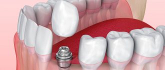
Implantation of 6 teeth
Many people believe that the first permanent teeth are the lower incisors. Actually this is not true. The first to appear are the molars—the sixth teeth, or “sixes.” They erupt behind the second primary molars and line up with the rest of the baby teeth. Due to the fact that the sixth teeth are “pioneers” among other molars, they bear a large chewing load. With poor hygiene or untimely treatment, these teeth begin to decay. Today there are many methods for restoring “sixes”; let’s dwell in more detail on prosthetics.