The significant prevalence of dental caries in children determines the relevance of its prevention. Dental care for children should be preventive. Only under this condition does a real reduction in major dental diseases become possible [1, 2, 6].
Today, local fluoride prophylaxis is recognized as more effective than systemic fluoride prophylaxis [9]. In this case, the factor of the constant presence of fluorides on the tooth surface is important [8, 10].
According to WHO, fluoride-containing toothpastes occupy approximately 95% of the European toothpaste market, which, when used regularly, reduce the increase in caries by 25% [7].
Under natural conditions, the source for the entry of the main structural elements of calcium and phosphorus into the enamel is the oral fluid. However, the remineralizing potential of saliva allows stopping caries at the spot stage only in 50% of cases [6]. Therefore, to increase the caries resistance of enamel, it is necessary to use products that include components that increase its mineral saturation.
Evidence has appeared in the literature that the remineralization process is supported enzymatically; in particular, the inclusion of phosphates and calcium in tooth enamel under the influence of phosphatases is significantly activated. The activity of these enzymes increases in the presence of chlorine and magnesium ions. The substrate for alkaline and acid phosphatases is calcium glycerophosphate. Studies using immobilized alkaline phosphatase have demonstrated that it is in the presence of glycerophosphate that the mineralization process of teeth is most successful. The reason for this may be a greater acceleration of the formation of apatite under the action of immobilized alkaline phosphatase than brushite or whitlockite [5]. In addition, to carry out the processes of mineralization and remineralization, the presence of biopolymers and hyaluronic acid in the interprismatic spaces of the enamel is necessary [3].
The purpose of the study was to increase the caries resistance of dental enamel in children and adolescents by using a complex remineralizing fluoride-containing coating with tricalcium phosphate.
What is caries resistance?
In dentistry, a condition called caries resistance is often encountered, but what does this mean? How is this condition characterized?
Attention! Caries resistance is considered to be increased resistance to carious lesions. Increased resistance to caries is usually observed in people who adhere to a healthy lifestyle. In addition, this condition is observed in people who are not burdened with any pathologies or severe complications and who follow a balanced diet. Tooth decay resistance properties are achieved by consuming essential vitamins and minerals to strengthen tooth tissue and bones.
Caries is a disease of our time; it occurs in almost every person in one form or another. But there are those who have innate immunity, this phenomenon is called caries resistance.
Medical Internet conferences
Introduction. Resistance to dental caries (DC) is ensured by the correct formation and formation of tooth buds and the physiological development of hard dental tissues [4, 12]. One of the conditions for tooth resistance to caries is the formation of a complete enamel structure, which begins with the formation of a protein matrix and ends with the mineralization of the enamel. The mineralization process is of great importance, the usefulness of which is ensured by a properly formed protein matrix [6, 8, 15]. Changes in the elemental composition of teeth make it possible to identify metabolic disorders during the development of teeth after eruption [13, 14], and therefore, the study of the chemical composition of the surface layers of enamel in these teeth is relevant.
The purpose of our study was to determine in vitro the degree of maturity of the enamel of permanent teeth and to study the content of microelements in its surface layers.
Object and methods of research. The study of dental surfaces using scanning electron microscopy was carried out at the Institute of Metal Physics named after G.V. Kurdyumov using equipment with the assistance of the Japanese company TOKYO BOEKI CIS LTD (manufactured by “Jeol”, Japan). To determine the microelement composition, the method of electron dispersive X-ray spectroscopy is indicative and accurate. The material for the study was the permanent third molars of children aged 16 to 18 years. Fifty-one unerupted intact permanent third molars, which were at the stage of root growth in length, were examined. The criteria for selecting permanent third molars were: similarity of developmental stages and morphology with permanent first molars in 6-year-old children, the possibility of obtaining material for research, namely extracted teeth for orthodontic reasons. The extracted teeth were washed with distilled water for three minutes. All samples were stored in tightly closed containers (10% streptomycin solution) at a temperature of (+2 ... + 4) ° C for two days.
Two days later, the studied samples were prepared for determination of trace element composition by electron dispersive X-ray spectroscopy. The content of chemical elements in the surface layer of permanent tooth enamel was determined in the equatorial, tubercular and cervical zones. The size of the areas of the enamel surface under study ranged from 50x50 µm to 250x250 µm (Fig. 1).
Rice.
Fig. 1. Area of study of immature enamel of a permanent tooth in an X-ray dispersive spectral analyzer INCA Energy 450. The content of chemical elements in the surface layer of enamel of a permanent tooth and the initial level of mineralization of each sample were determined using an X-ray characteristic spectrum (Fig. 2).
Rice. 2. X-ray characteristic spectrum of the surface layer of permanent tooth enamel.
When studying the surface of tooth enamel, the optimal magnification modes were determined (x100, x500, x1000, x3000) (Fig. 3-6).
Rice. 3. Immature enamel of a permanent tooth. Magnification x100.
Rice. 4. Immature enamel of a permanent tooth. Magnification x500.
Rice. 5. Immature enamel of a permanent tooth. Magnification x1000.
Rice. 6. Immature enamel of a permanent tooth. Magnification x3000.
Results and its discussion. When studying the chemical composition of tooth enamel using the EDS method, our studies determined similar indicators of the weight content of microelements (%): Ca2+ = 32.67 ± 8.07; Р5+=17.84±4.10; Na+ =0.84±1.23; Mg2+=0.15±0.11 (Table 1). In fact, we obtained results in which weight calcium and magnesium tended to decrease, and phosphorus and sodium were within normal limits [9].
By analyzing the atomic chemical composition of tooth enamel, we found that oxygen, sodium, chlorine, calcium, and phosphorus were determined in 100% of the samples. The elements Mg2+ were found in 63.89% of samples, F- in 30.56%, C4- in 13.89%, S2- in 8.33% of samples. The Ca2+ content was 18.87±6.28 (atom %), the P5+ content was 13.45±3.44 (atom %). The output level of mineralization by atomic (%) Ca/P ratio was 1.40 and was closer to the lower limit (1.33), after which irreversible changes in the enamel structure are observed [2]. A Ca/P ratio was found to be lower than the average values for human tooth enamel [1]. This shows that the enamel of teeth that have not erupted or have just erupted is immature.
At the same time, it has been established that the strength of dental tissue is influenced not only by the optimal ratio of basic microelements, such as calcium and phosphorus, but also by an increase in the amount of magnesium, sodium, potassium, silicon and a decrease in the content of sulfur and chlorine [3, 5].
Conclusion. The atomic chemical composition of the surface of tooth enamel made it possible to study electron dispersive spectral analysis. We found that 100% of the samples contained the following chemical elements: O2+, Na+, Cl-, Ca2+, P5+; Mg2+ was contained in 63.9% of the samples, F- in 30.6%, C4- in 13.9%, S2- in 8.3% of the samples. The Ca2+ content was 18.8±6.28 (atom%). The P5+ content is 13.45±3.44 (atom%). The output level of mineralization by atomic (%) ratio Ca/P was 1.40 and was closer to the lower limit (1.33). A lower Ca/P ratio was found than the average values for human tooth enamel. This shows that the enamel of teeth that just erupted was immature. The data we obtained coincide with the results of studying the content of fluorine, calcium and phosphorus in the enamel using X-ray photoelectron spectroscopy, which established an insufficient level of mineralization of the enamel of permanent teeth after eruption [10]. The presence of dental plaque on the teeth and the effect of other cariogenic factors, especially during this period, are very dangerous, which indicates the need to develop and carry out preventive measures, in particular exogenous ones, aimed at accelerating the mineralization of immature enamel of permanent teeth.
Determination of the chemical composition of immature enamel of permanent teeth using energy-dispersive X-ray spectroscopy is accurate and objective. Using this method, it is possible to reliably calculate the Ca/P coefficient of the enamel surface of permanent teeth, and thus determine the degree of maturity of the enamel of these teeth.
Modern concept of the etiology of caries
Based on historical theories , significant progress has now been made in studying the etiology and pathogenesis of dental caries.
The generally accepted mechanism for the occurrence of caries is the progressive demineralization of hard dental tissues under the influence of organic acids, the formation of which is associated with the activity of microorganisms.
Many etiological factors take part in the occurrence of the carious process, which allows us to consider caries a polyetiological disease.
The main etiological factors are:
· microflora of the oral cavity;
· nature and diet, fluoride content in water;
· quantity and quality of salivation;
general condition of the body;
· extreme effects on the body.
All of the above factors were called cariogenic and divided into general and local, which play an important role in the occurrence of caries.
General factors:
1. Poor diet and drinking water.
2. Somatic diseases, changes in the functional state of organs and systems during the period of formation and maturation of dental tissues.
3. Extreme effects on the body.
4. Heredity, which determines the usefulness of the structure and chemical composition of tooth tissue. Unfavorable genetic code.
Local factors:
1. Dental plaque and dental plaque, replete with microorganisms.
2. Violation of the composition and properties of oral fluid, which is an indicator of the condition of the body as a whole.
3. Carbohydrate sticky food residues in the mouth.
4. Resistance of dental tissues, due to the complete structure and chemical composition of the hard tissues of the tooth.
5. Deviations in the biochemical composition of hard dental tissues and defective structure of dental tissues.
6. Condition of the dental pulp.
7. The state of the dental system during the period of formation, development and eruption of teeth.
Cariogenic factors can be of varying intensity and nature, different options for their interaction contribute to the occurrence of caries, but the leading factor is the microflora of the oral cavity. It is now known that the carious process can develop in the presence of microorganisms in the oral cavity, excess amounts of carbohydrates in food and contact of carbohydrates and microorganisms with tooth enamel. It is well known that carbohydrate intake causes increased acid formation. Thus, taking 10 grams of sugar leads to an increase in lactic acid in saliva by 10-16 times [Leontyev V.K., 1978]. Studies have shown that at a pH more acidic than 6.2, saliva goes from being oversaturated with hydroxyapatite to undersaturated, and therefore turns from a mineralizing liquid to a demineralizing liquid (destroying the hard tissues of teeth). According to modern ideas, the cause of caries is prolonged exposure to acids on dental tissues. The formation of organic acids is associated with long-term enzymatic activity of microorganisms. Long-term exposure to organic acids on tissues is observed with poor oral hygiene, when a dental plaque forms on the enamel; it is under it that an acidic environment is created as a product of the enzymatic activity of a huge number of microorganisms that are capable of ideally absorbing carbohydrates retained in the oral cavity.
Thus, a carious cavity is formed in places of intense acid production, under the dental plaque, where the pH is more acidic 4 - 5. With good washing of the teeth with oral fluid, rare intake of sugar, the local pH shift is quickly leveled out. However, in areas of poor saliva access, with frequent intake of sugar, the demineralization process may prevail over the remineralization process. This means that carbohydrate consumption can be a decisive factor in the shift in pH and disruption of mineralization processes, which leads to the occurrence of caries.
It should be noted that the action of general factors is carried out, as a rule, through the action of local ones. That is, diet, the state of organs and systems, extreme situations can change the composition and properties of oral fluid, affect the microflora of dental plaque and dental plaque.
Sugar has a specific effect on metabolic processes in the oral cavity, causing a “metabolic explosion” after its consumption. This influence of simple carbohydrates is associated with their readiness to enter into metabolism (i.e. metabolism) already in the oral cavity, in contrast to proteins, fats and complex carbohydrates, which require preliminary hydrolysis: swelling and activation. The conditions for the absorption of carbohydrates by the microflora of the oral cavity are close to ideal. Which, naturally, affects the intensity and prevalence of caries.
So, according to modern views, the direct cause of progressive demineralization of hard dental tissues (caries) are organic acids, the formation of which is associated with long-term enzymatic activity of microorganisms. The occurrence of caries is the final stage of the effective interaction of a number of cariogenic factors.
It is known that at a young age the intensity of dental caries damage is higher than at an elderly age. This is due to insufficient mineralization of the tooth enamel immediately after its eruption. The maturation of enamel continues for more than two years, and only complete mineralization causes greater resistance of tooth enamel to the effects of acids, and vice versa, insufficient mineralization creates conditions for rapid demineralization and the occurrence of a carious process. After tooth eruption, the enamel initially matures in the area of the cutting edges and cusps of all teeth, so the carious process occurs precisely in immature fissures and the cervical region, which are risk areas. Today, maturation problems are central to the prevention and treatment of dental caries. Oral fluid plays a huge role in the formation of enamel; the remineralizing ability of the latter has been proven in a number of clinical and experimental studies [Aksamit L.A., 1978; Dubrovina L.A., 1989, Redinova T.L., 1989].
Normally, in the oral cavity, the processes of re- and demineralization are in a state of dynamic equilibrium, however, in the presence of cariogenic factors, a shift in equilibrium towards demineralization is observed.
The state of reduced resistance of dental tissues to cariogenic influences as a result of a violation of the nonspecific resistance of the body due to previous and existing somatic diseases, according to the definition of Professor V.K. Leontyev, is a cariogenic situation.
A cariogenic situation is created when any cariogenic factor or group of them, acting on a tooth, makes it susceptible to the effects of acids. Of course, the trigger is the microflora of the oral cavity, with the obligatory presence of carbohydrates and the contact of these two factors with dental tissues.
In conditions of reduced resistance of dental tissues, the cariogenic situation develops more easily and quickly.
Clinically, a cariogenic situation in the oral cavity is manifested by the following symptoms:
a. poor oral hygiene;
b. abundant plaque and tartar;
c. the presence of multiple chalky carious spots;
d. bleeding gums.
However, even in regions with a high prevalence of caries, there are people who do not have this disease, which made it possible to identify a group of caries-resistant individuals (resistant to caries). At the same time, there are people whose intensity of dental caries damage significantly exceeds the group average level; these were identified as caries-susceptible groups.
Caries resistance and caries susceptibility should be considered in terms of their relationship, just like cariogenic factors (general and local), they can be of varying strength. The occurrence of caries is possible with various options for their interaction. In caries-sensitive teeth, the pathological process occurs faster and more often, which depends on the general condition of the body in the past. Common diseases associated with caries during a given period of time cannot affect the structure and composition of mature teeth, however, disruption of the functional state of organs and systems of the body actively affects the occurrence and course of the carious process, changing the composition and properties of the oral fluid. Factors of resistance and susceptibility to caries are a consequence of certain relationships between the tooth surface and oral fluid. If, during progressive demineralization, cariogenic factors lose their strength or disappear, demineralization may be suspended. The occurrence of caries is determined by many factors, and in the presence of appropriate conditions they become the cause of the disease.
Teeth resistance to caries develops in individuals who are not burdened by previous and chronic concomitant diseases and their consequences, who eat nutritious food and water containing the necessary macro- and microelements, and are not exposed to any harmful influences. Each of the factors listed below depends on the general condition of the body, its reactivity and resistance.
Teeth resistance to caries, or caries resistance, is ensured by:
· chemical composition and structure of enamel and other tooth tissues;
· presence of pellicle;
· optimal chemical composition of saliva and its mineralizing activity;
· sufficient amount of oral fluid;
low level of tooth enamel permeability;
· good chewing load and self-cleaning of the tooth surface;
properties of dental plaque;
· good oral hygiene;
· diet features;
· correct formation of rudiments and development of dental tissues;
· timely and complete maturation of enamel after tooth eruption;
· specific and nonspecific factors for protecting the oral cavity.
The susceptibility of teeth to caries, or caries susceptibility, is promoted by:
· defective enamel maturation;
· a diet with a deficiency of proteins, macro- and microelements, and an excess of carbohydrates;
· water with insufficient fluoride;
absence of pellicle;
· composition of oral fluid, its concentration, viscosity, quantity and flow rate;
· the biochemical composition of the hard tissues of the tooth, which determines the course of caries, since a dense structure with minimal spaces in the crystal lattice slows down the course of caries and vice versa;
· condition of the neurovascular bundle;
· functional state of organs and systems of the body during the formation and maturation of dental tissues;
· abnormal tooth development due to common somatic diseases.
The carious process progresses if the rate of salivation decreases, the amount of saliva decreases, its viscosity increases and, conversely, the carious process slows down or stops at the stain stage with a sufficient amount of saliva and its normal viscosity. A high concentration of macro- and microelements in saliva also stops caries; with a low concentration of mineral elements and a high content of mucin, its progression is observed. Thick, smooth enamel, its dense structure and minimal spaces in the crystal lattice slow down the carious process. Pits, grooves, folds, depressions, thin enamel and loose structure contribute to the rapid progression of the pathological process. In many cases, dental caries occurs in immature fissures, which are risk areas, the latter also including the cervical areas of the teeth. V.K. Leontiev et al. [1984, 1989] in a clinical setting using electrometry showed that the process of enamel maturation is dynamic and depends on the anatomical identity of the tooth, its location, the topography of the tooth area and other factors. Rapid maturation of tooth enamel occurs in the area of the cutting edges and cusps within 4 - 6 months after their eruption. It is especially intense in the first days and weeks after tooth eruption. The enamel of the cutting edges of incisors and fangs matures 2 times faster than in the cervical area. The rate of maturation of the enamel of the fissures of teeth is much slower than that of the cusps and incisal edges, and largely depends on the degree of washing the teeth with saliva and covering the fissures with plaque. An important fact for practice has been established that in all cases, the complete maturation of fissures of premolars and molars varies within a period of up to 2 years. Moreover, in many cases, dental caries occurs in immature fissures and their destruction begins. The main sign of age-related changes in enamel is compaction and a decrease in structure variability due to a decrease in microporosity, which is consistent with the results of studies examining changes in calcium and phosphorus content during enamel maturation. The compaction of enamel is a consequence of the influx of macro- and microelements that change the chemical composition of the enamel, its structure, and properties (an increase in microhardness, a decrease in solubility and permeability occur simultaneously). These facts also explain the fact that at a young age the increase in the intensity of dental caries damage is higher than in the elderly.
V.V. Nedoseko et al. [1987] conducted clinical and laboratory studies to study the resistance of teeth to caries. The level of resistance was determined taking into account the intensity of damage to individual teeth (ITU), groups of teeth and their surfaces. 4 groups of resistance to caries were identified:
1. A high level of resistance was determined in caries-resistant individuals who do not have carious teeth and periodontal diseases. The rate of saliva secretion in such individuals is 2 times higher than in those susceptible to caries. The oral fluid sediment is characterized by low demineralizing activity, the pH of the oral fluid shifts to the alkaline side, the composition is characterized by a fairly high content of total and ionized calcium and a relatively low content of organic phosphate.
2. The average level of dental resistance to caries was detected in individuals whose foci of demineralization were localized on molars, premolars and sometimes canines, caries intensity (ICU) = 9.09 + 0.80 and a low oral hygiene index. The rate of saliva secretion is 2 times lower than in caries-resistant individuals, the pH of saliva is shifted to the alkaline side, it is oversaturated with hydroxyapatite by 16.4% more than the saliva of caries-resistant individuals. Oral fluid is characterized by a high content of inorganic phosphorus and an increased concentration of potassium ions. Saliva contains a large amount of sediment with increased utilization and demineralizing activity. The content of calcium, phosphorus and their ratio in enamel biopsies do not differ from those in caries-resistant individuals. This group is distinguished by the highest rate of remineralization of tooth enamel.
3. A low level of resistance was detected in individuals with caries intensity (CCI) = 17.65 + 1.27. Caries affected all groups of teeth except the lower incisors. The saliva reaction is neutral, it is oversaturated with calcium and phosphates, but the concentration of sodium and potassium is less than the saliva of persons with an average level of resistance. Dental plaque is highly cariogenic, and the hygiene index is low. The rate of enamel remineralization is quite high, but the rate of saliva secretion is 2 times lower than in caries-resistant patients.
4. A very low level of dental resistance to caries was found in individuals with the highest hygiene index and low saliva secretion rate. The oral fluid is undersaturated with hydroxyapatite by 10.3% compared to the saliva of persons resistant to caries. Intensity of the carious process (ICP) = 29.9 + 0.89, all groups of teeth are affected. The rate of enamel remineralization is sharply reduced. The cariogenicity of dental plaque is significantly higher compared to all other groups. The oral fluid contains significantly less total and ionized calcium and phosphates compared to other groups. The utilization and demineralizing activity of saliva is high.
With age, the number of individuals with a high level of resistance decreases among both men and women, mainly individuals with an average and low level of resistance predominate, however, there are significant group differences for each level of resistance in the hygiene index, salivary secretion rate, enamel remineralization rate and etc.
Elimination of the cariogenic situation is associated with remission of a general somatic disease, resumption of oral hygiene, change of place of residence, childbirth and completion of breastfeeding.
All of the above measures lead to the spontaneous disappearance of white carious spots without drug therapy.
The foci of demineralization receive calcium, phosphorus and fluorine from oral fluid, which has pronounced remineralizing activity and can normalize the permeability of enamel, which was increased as a result of exposure to organic acids. In turn, it should be noted that the course of caries in a cariogenic situation is characterized by rapidity, the presence of pigmented decay of dentin, chipping and sharp edges of the enamel. Such a caries clinic in a cariogenic situation is characterized by acute blooming or decompensated caries, that is, high activity of the course.
Article provided by JSC BIOMED
Relevance
. Currently known methods of preventing dental caries can effectively combat this disease. However, in order to select the most effective means of caries prevention, it is necessary first of all to clarify the schemes and indications for their use. This is closely related to the possibility of dynamically determining the individual resistance of dental hard tissues to caries, depending on the state of the body and treatment and preventive measures.
Purpose of the study
— to compare the effectiveness of existing methods for assessing caries resistance to improve control of enamel condition and the possibility of early diagnosis of caries and determining the effectiveness of prevention methods.
Material and methods.
The study included 40 patients aged 32 to 45 years, with an average age of 35 years. Patients were divided into 2 groups, randomized by gender, age and level of caries resistance. All patients underwent a clinical examination of the mouth with determination of the OHI-S index. To assess the condition of the enamel, the level of caries resistance was determined using the V.B. method. Nedoseko (V.B. Nedoseko, 1988) and performed an acid biopsy (V.K. Leontyev, V.A. Distel, 1975 modified by M.Yu. Zhitkova, 2000) with the calculation of the calcium-phosphorus ratio (Ca/P coefficient) , as well as the KOSRE test (V.K. Leontyev, T.L. Redinova, G.D. Ovrutsky, 1988), reflecting the rate of enamel mineralization. Patients in both groups received instruction in oral hygiene, dental treatment according to indications, and professional hygiene. Those observed in the main group were additionally prescribed the remineralizing gel Tus Mousse, which they rubbed into the enamel of their teeth 2 times a day, morning and evening, for 30 days. After 1 month, an acid biopsy was repeated in both groups to assess the condition of the enamel. To determine the correspondence between the two methods, assessing caries resistance (V.B. Nedoseko method, acid biopsy method), a correlation analysis of the obtained indicators was carried out.
Results.
The level of resistance in each group was not higher than moderate, the level of intensity of caries KPU was 15, the initial value of the Ca/P coefficient was 0.29.
The correlation coefficient between the level of caries resistance and the Ca/P ratio was quite high and amounted to 0.72. Recovery of the focus of demineralization after acid biopsy in the control group occurred after 25±5 days, in the main group - after 10±2 days ( p
<0.01), indicating a higher rate of remineralization with the drug used.
By the end of the month of observation, the Ca/P coefficient increased in the control group to 0.32, and in the main group against the background of the use of Tus Mouss gel - to 0.39 ( p
= 0.19 and
p
= 0.03 relative to the initial level, respectively). The Ca/P coefficient in the main group after remineralizing therapy was less correlated with the level of caries resistance. In the control group, the correlation coefficient was 0.63, in the main group - 0.32. This is explained by the fact that in the control group after a month the level of mineralization (according to V.B. Nedoseko) differed little from the initial one. In the main one, enamel resistance increased by 22% compared to the primary data.
Conclusion.
The acid biopsy method allows you to record changes in the condition of the enamel in a shorter time. Determination of the level of caries resistance using the V.B. method. Insufficient and indicators of acid biopsy of enamel are comparable, however, enamel biopsy allows not only to predict the development of caries, but also to quickly monitor the dynamics of caries resistance, which is necessary for studying different schemes and methods of prevention, choosing the most effective and economical, depending on the individual characteristics of patients.
How the disease develops
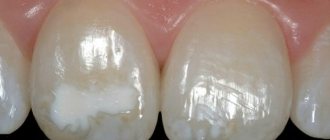
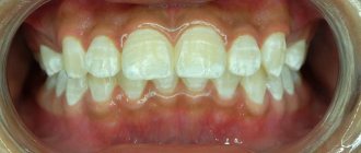
Spot stage. Tooth enamel is constantly exposed to acids that are formed during the fermentation of carbons. The top layer of enamel is more resistant to destruction processes, so the first signs of caries appear on the inner layers. They look like chalk spots visible on the surface. If the process occurs on the front teeth, then the patient can independently see these lesions. Only a dentist can notice the beginning of a lesion on the lateral chewing teeth.
The stage of intermediate caries occurs when the septum is completely destroyed and the carious cavity becomes visible. At this stage, brushing teeth and eating food become painful and cause serious discomfort.
The stage of deep caries occurs when the carious cavity reaches the pulp. If there is no treatment at this stage, the thin dentil layer disappears and the disease moves to the next level - pulpitis begins.
Treatment of caries
It is possible to use enamel remineralization, but only at the very first stage, when the manifestations of the disease look like spots.
At all other stages of caries development, the following algorithm is used:
- High-quality tooth cleaning is carried out;
- A filling material of the appropriate color is selected (a special scale is used for this);
- Anesthesia is used if necessary.
Modern equipment allows treatment to be carried out painlessly. If the caries condition is advanced or when the cervical area is affected, a decision is made to administer anesthetic injections.
Tissues affected by caries are drilled out.
A seal is installed.