Content
hide
1 Pulp - structure, functions, features
2 Why is a tooth without pulp considered dead?
3 How long does a tooth live without pulp?
4 What can lead to the need to remove a “dead” tooth: 4.1 There are several rules by adhering to which patients can prolong the use of the tooth:
Inflammation of the “nerve” of the tooth (pulpitis) is not always accompanied by severe toothache. A chronic infectious process can occur in a tooth unnoticed, leading to significant tooth destruction, the formation of a cyst on the root of the tooth, and even tooth loss. This pathology is an indication for tooth depulpation—removal of its “nerve.” Sometimes such news can come as a surprise to a patient if he did not suspect the presence of chronic pulpitis. At the same time, we are often asked - what will happen to the tooth later, after pulp removal? How long will the tooth live after this procedure, how will its condition change? We'll talk about this in the article.
Pulp - structure, functions, features
The pulp is also called the dental nerve. This is a soft connective tissue that contains a collection of nerve fibers, lymphatic and blood vessels. It is located in the tooth cavity (pulp chamber of the crown part of the tooth and in the root canals). Along the edge of the pulp there are cells, the processes of which are immersed in the dentin of the tooth. Thus, the pulp is closely connected with the hard tissues of the tooth. Through these connections, the trophic function is carried out; the pulp “nourishes” the walls of the tooth. Pain and mechanical sensitivity are also provided by the processes of pulp cells.
The blood supply to each tooth is provided by the common arteries and veins of the maxillofacial area. Small capillaries extend from them and enter the pulp, feeding the tooth. This structure protects the tooth from inflammation, proliferation of pathogens and other pathological processes.
The rich blood supply to the tooth also causes pain during inflammation. When the pulp becomes infected, a massive rush of blood occurs to its capillaries to provide a protective reaction, and swelling occurs inside the tooth. Enlarged vessels and soft tissues compress the nerve endings of the pulp, causing pain.
Reasons for changes in enamel color
The pulp is a neurovascular bundle that is responsible for feeding enamel and dentin with minerals, vitamins and other valuable elements. After depulpation, tissue metabolism is disrupted and the tooth ceases to receive the necessary nutrients. This is the main reason for darkening of the enamel after nerve removal. Changes in the color of a pulpless tooth are also provoked by:
- poor-quality endodontic treatment – incomplete pulp extraction, insufficient root canal treatment;
- the use of filling materials containing resorcinol-formalin, silver, tin;
- excessive consumption of tea, coffee, fluoridated water with high iron content;
- acid-base balance disorders;
- some diseases - pathologies of the urinary system and gastrointestinal tract, blood diseases caused by staphylococcus infections, calcium metabolism disorders.
Even with high-quality treatment, avoidance of coloring products and regular oral hygiene, sooner or later the pulpless tooth will darken. This can cause discomfort, especially if the tooth is located in the smile area. A visit to the dentist will solve the problem.
Why is a tooth without pulp considered dead?
The main thing that changes in the condition of the tooth after depulpation is that the trophism (nutrition) of the hard tissues is lost, the tooth becomes very fragile and reacts sharply to external factors.
The absence of nerve endings affects the protective function of the tooth: in the absence of a pain reaction, a person can damage the tooth by biting something hard on it and not notice it. The pulp ceases to protect the tooth from microbes, as a result of which it becomes more susceptible to caries.
As you can see, a pulpless tooth requires additional care, as it is more vulnerable than healthy ones.
Why does a tooth without a root darken?
Incisors, fangs, molars can darken due to abrasion of the enamel layer, from the occurrence of microcracks, as a result of the removal of nerve endings. The loss of nerve processes leads to a change in the shade of the enamel, leading to the appearance of dark brown spots. What factors provoke the darkening of pulpless masticatory organs?
- Lack of nutrients. In such a situation, moisture leaves the enamel, which causes it to darken.
- High degree of vulnerability. Without systematic feeding, porous tissue appears on the surface of the crowns, which allows coloring nutrients from tea, coffee, berries, and chocolate to pass through to the inner layers of the organ.
- The color of the crown is negatively affected by canal treatment. After such therapy, the tissues may turn blue.
- The corolla of the organ becomes dark after incorrect treatment of dental cavities. Small blood fragments, as well as pulp, may remain in the canals. Gradual hemolysis of the blood changes the color of the unit. It turns yellow, gray, brown.
A pulpless organ is no longer a tooth, it is only its shell. First, it weakens as the dentin dries out, as well as the enamel and mineral parts. Over time, the shade of the membrane differs from the tone of healthy organs. In such a situation, cosmetic teeth whitening is carried out, especially when the color of the upper incisors, threes or twos has changed.
There are more difficult problems when the tooth darkens from the inside due to bleeding. In such cases, typical lightening will not help, because it only affects the outer layers. Dentists offer a more complex method - intracanal. It's effective and safe. First, the old filling is removed. A bleaching agent is then placed into the cavity. Then a temporary filling is applied. If necessary, the clarifying reagent is introduced several times. The number of repetitions depends on the level of darkening. When the result is achieved, permanent filling material is applied.
How long does a tooth live without pulp?
This period varies greatly. In some patients, the tooth is destroyed within a year, and in others it can last for another ten years. What's the difference? Here are the factors that affect the condition of the tooth after depulpation:
- the degree of initial destruction of the crown of the tooth;
- pulp removal technique, quality of root canal filling;
- the method by which the tooth was restored after removal of the nerve (filling/inlay/crown);
- caring for the tooth after treatment, performing the necessary hygiene procedures.
After the tooth is depulped, treatment procedures are required that will improve its vitality. First of all, this is filling. Empty root canals are filled with special material. Complete and high-quality removal of the entire pulp, as well as hermetically filling the canals with filling material over the entire length, reduces the risk of inflammation of the tissues surrounding the tooth and the appearance of a cyst.
Depending on the degree of tooth decay and the specific clinical situation, the doctor may suggest restoring the tooth with either a filling, a ceramic inlay, or an artificial crown. An inlay and a crown are the optimal methods for restoring “dead” teeth, because they cover the tooth from above and fit hermetically to the tooth, thereby reducing the contact of the internal environment of the tooth with bacteria in the oral cavity, moisture and air, and protecting the tooth from chips and cracks. In addition, it is comfortable for the patient and has an aesthetic appearance.
Tooth weakening - after nerve removal
Because the tooth structure is slightly weakened after root canal treatment, the tooth loses some of its original capabilities. This translates into a higher risk of tooth fractures. Those patients who have had root canal treatment should avoid eating hard foods, such as nuts, or using other teeth to chew on them. To avoid the risk of tooth fracture after root canal treatment, you should consider installing a dental crown.
Failed root canal treatment
Root canal treatment may fail in approximately 5% of patients. This often leads to tooth extraction. Such cases occur for various reasons, sometimes even independent of the quality of canal filling.
What can lead to the need to remove a “dead” tooth:
- damage to the tooth root by caries;
- the occurrence of longitudinal fractures and root cracks (below the gum level);
- the presence of a cyst or granuloma on the root of the tooth, inflammation of the tissues surrounding the tooth, the treatment of which is not possible.
There are several rules that patients can follow to prolong the life of their teeth:
- cover the tooth with a ceramic inlay or crown;
- visit the dentist once every 6 months for a check-up, hygienic cleaning and, if necessary, a control X-ray of the tooth;
- do not chew hard food (nuts, seeds, crackers, etc.);
- Avoid temperature changes when eating (for example, hot coffee with cold ice cream).
As we see, a patient can do a lot for his health. But in order for the tooth to last longer, it is much more important to choose a specialized clinic that uses modern technologies, high-quality equipment and materials. An experienced and knowledgeable doctor will ensure the vital activity of a pulpless tooth for a long time.
Whitening methods
There are several ways to whiten a tooth after nerve removal. Effective technologies:
- Endobleaching;
- Restoration of units using artificial crowns;
- Veneers;
- Lightening with gels;
Gels based on hydrogen peroxide are used. It is not recommended to carry out the procedure at home, as there is a risk of damage to healthy organs, as well as burns on the mucous membranes of the oral cavity. In the dentist's office, the pathological organ must be isolated. In a professional substance for lightening the organ membrane, the concentration of peroxide is minimal. This is necessary in order not to damage the weakened enamel.
Before the procedure, the patient undergoes an x-ray, where he is given a picture of the oral cavity. The jaw is fixed using an expander. A special varnish is applied to healthy mucous tissues and healthy units to protect them from the ingress of the gel. Then the affected chewing organ is covered with a gel substance. A laser is used to enhance the reaction. During internal bleaching, the root is hermetically protected with medical cement. Thanks to this, the substance does not irritate the tissue in the area of the top of the hole. However, complete lightening from the inside is not always possible, so dark spots remain in some areas. Manipulation is not carried out if the units are highly vulnerable, with inflammation of the oral cavity, with periodontitis, or caries. Dentists do not offer lightening to teenagers and children. It is contraindicated for pregnant and lactating women.
The non-living organ itself slowly collapses and darkens again, because interference with the corolla weakens the tooth and increases the degree of damage. In such cases, in order to give the shell a natural shade, dentists often recommend the installation of artificial crowns and veneers.
Veneers
If intracanal tooth whitening has already been carried out several times, and it is not possible to repeat the procedure, dentists advise using thin plates made of composite materials or ceramics, that is, veneers. They are worn over crowns. The method is most often used in the restoration of masticatory organs. This technology gives damaged units a natural whiteness, restores their shape, and protects them from damage. Ceramic, zirconium, and porcelain plates are stronger than composite overlays. They have high strength, are aesthetically pleasing, and have a service life of several decades. Installation rules:
- First, the size and shade of the plates are selected.
- The top enamel layer of the crown is ground (0.5 to 1.5 millimeters of tissue is removed).
- Then the prepared crown is removed and an image is taken.
- Then, based on the plaster casts, the onlays are made in the dental laboratory.
- Plastic plates are temporarily placed on the crowns. They protect units from aggressive environments and damage.
- The fitted veneers are fixed with medical cream.
Contraindications include occlusal defects, a high degree of crown abrasion, bruxism, and severe organ destruction.
Crowns
To whiten a tooth without a nerve, artificial crowns are also used. They are installed on chewing organs that require restoration. They must preserve not only the roots, but also half of the outer part of the organ. In case of severe damage, dentists advise installing titanium rods and then putting the crown part on them. Advantages of artificial crowns:
- Elimination of color and shape defects that cannot be corrected using other technologies;
- Durable protection of organs from mechanical damage;
- Protection of the enamel layer from fractures in pulpless units;
- Distribution of the load when chewing evenly;
- Preservation of roots;
Prostheses are made of metal (titanium, gold, stainless steel). They have increased strength, but their appearance is unaesthetic. A durable material is metal ceramics. Sometimes the metal can be visible through the ceramic layer. Caps made of zirconium, aluminum oxide, and porcelain are used more often than others in modern dentistry. Zirconium dioxide has durability, high strength, excellent optical and aesthetic properties.
Endobleaching
If the unit has darkened from the inside, superficial lightening methods will not have an effect. Dentists advise using endobleaching, that is, lightening tissue from the inside. Using this technology, the dentist opens the root canals, cleans them, and also expands them. The old filling material is removed. The brightening agent is introduced into pre-prepared holes. It contains less than 20 percent hydrogen peroxide. Since the gel concentration is low, lightening occurs slowly and can take up to 15 days. When the enamel becomes lighter, permanent filling material is placed. Popular whitening system Opalescence. It is suitable for intra-canal clarification, as well as for external clarification.
The bleaching agent often contains sodium perbonate, and hydrogen peroxide is also added. You should be careful when consuming hard foods to prevent the tooth from chipping due to the fragility of the enamel layer. Endo-bleaching is not performed during lactation, pregnancy, gum disease, carious lesions, or allergies.
Prevention of dead teeth
The main measures aimed at preventing the appearance of dead teeth are:
- regular visits to dental clinics (at least twice a year);
- daily brushing of teeth (the procedure should be carried out at least twice a day, the duration of brushing should be more than 3 minutes);
- prevention of pulpitis, caries, periodontitis;
- using toothpicks and dental floss after meals;
- regular rinsing of the mouth with special solutions;
- competent selection of toothpastes and brushes;
- thorough cleaning of the inner surface of the cheeks, palate, tongue and other areas of the oral cavity that serve as places where pathogenic microbes accumulate;
- timely treatment of caries and other dental diseases.
Compliance with the above recommendations will not only prevent the appearance of dead teeth, but also preserve a beautiful, healthy, snow-white smile for a long time.
What to choose?
The lightening procedure is carried out at will. Often the frontal incisors and canines are lightened, because they are visible. If their shade differs by one or two shades, it is already noticeable to others. Lightening is a serious manipulation, as it involves chemical exposure. Therefore, it must be carried out by a dentist so that the result is long-lasting and the shade does not differ from the color of healthy teeth. To prevent the crowns from being destroyed or crumbling, the manipulation must be carried out by a specialist. Only a qualified dentist will protect the gum tissue from irritation by protecting it with special means. Also, the doctor will choose a safe method of action.
- An alternative to lightening from the inside are inlays and crowns, if the organ is not severely damaged. This solves the problem of color, as well as strengthening the unit, and reduces the risk of further destruction.
- The technology of restoration with veneers is suitable for patients with crooked, spotted teeth, the crown layer of which is slightly destroyed.
- Lightening from the inside is necessary when dentin darkens when other methods do not give results.
To avoid unpleasant situations, you should carefully care for your oral cavity, regularly visit the dental office, carry out timely sanitation and treatment of carious cavities. Taking care of the oral cavity is the key to the health of the whole body.
Associated symptoms
In addition to blackening of the enamel, other symptoms may develop:
- The appearance of erosions, gouges, black spots or spots on the enamel.
- Unpleasant odor from the mouth.
- Mobility of the filling.
- Pain when pressing, tapping or chewing.
- Inflammatory process in the gums around the filling, bleeding of the mucous membrane, swelling. The appearance of bumps or gumboil in an advanced stage.
Changes in tooth color should not be ignored. This is a clear sign of a pathological process.
Restoration of Darkened Teeth in the Frontal Region
Treatment of discolored teeth in the anterior region can be straightforward if the approach is planned according to the type of cause of discoloration the patient has.
Author: Giulio Pavolucci
The patient is a 20-year-old girl who complains of a chipped discolored central incisor. According to her, the tooth was endodontically treated about 7 years ago, without using a rubber dam, and became darker over time. It was also restored with composite and the filling took a couple of years to fall out. This problem is often associated with filtration phenomena or gutta-percha staining. Dark yellow and brown shades are associated with filtration and blood, while dark gray is associated with gutta-percha. The second option does not often respond well to bleaching.
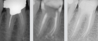
Photo 1. Initial situation: assessment of the degree of discoloration of the tooth 2.1 along with a class IV cavity.
Photo 2. Preoperative X-ray showed an incompletely filled root canal: repeated endodontic treatment is necessary.
Photo 3: A rubber dam should be used during endodontic treatment.
Photo 4. Intraoral x-ray.
Figure 5: Postoperative X-ray: Self-etching adhesive and a thin layer of flowable composite are applied over gutta-percha to seal the root canal system. This is really important before introducing bleaching agents into the pulp chamber to prevent root resorption.
Tooth whitening
The situation after tooth whitening with hydrogen peroxide (White Dental Beauty); The discoloration of tooth 2.1 has completely disappeared.
Photo 6.
Photo 7. Avoiding the use of bleaching agents for 20 days is necessary to remove all oxygen so that it cannot interfere with adhesion procedures. A new appointment was scheduled and a wax-up was done using a composite (no adhesive) and a silicone key was made directly into the patient's mouth.
Photo 8. Rubber dam is required when performing adhesion procedures.
Pin installation
The space for the installation of the pin was prepared, avoiding unnecessary grinding of the tooth tissue.
Photo 9.
Figure 10: A short bevel was created from the buccal surface along the fracture line.
Photo 11. Tooth preparation is completed.
Photo 12. The space under the pin was washed to clear it of gutta-percha and endodontic cement debris.
Photo 13. The correct pin was selected and the required length was measured.
Photo 14: The post was trimmed to ensure complete retention within the restoration.
Photo 15. The silicone key is very helpful in controlling the correct length of the pin.
Photo 16: Self-adhesive cement (Rely-X Unicem) was applied to the post space.
Photo 17. The fiberglass pin was inserted into the canal using forceps.
Photo 18. After photocuring, the pin is fixed in the canal.
Recreating the restoration
Although adhesion procedures are not necessary for cementing a post with self-adhesive cement, they are mandatory when reconstructing the restoration.
Photo 19.
Photo 20. To simplify the procedure, a layer-by-layer technique of one shade was chosen. A thin layer of A2B (Filtek Supreme XTE) was applied directly to the silicone key and photocured.
Photo 21. First, the palatal frame.
Figure 22: The posterior sectional matrix is very useful for creating the interproximal wall in anterior straight restorations because it provides the correct curvature for the composite.
Photo 23. Frame completed; With the technique of layering with one composite, it is very easy to complete the reconstruction of the restoration by filling the entire structure with it.
Photo 24. The restoration was completed.
Photo 25: Sanding and polishing procedures are extremely important to achieve proper aesthetic integration.
Photo 26. View of the restoration immediately after removal of the rubber dam.
Photo 27. Control image after 3 weeks: the patient is very satisfied with the restoration. The translation was carried out by S. Filanovich specifically for the OHI-S.COM website. Please, when copying material, do not forget to provide a link to the current page.
https://www.styleitaliano.org/
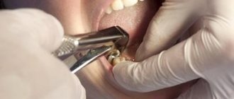
Removing the pulp of the affected tooth
Pulp removal is a complex dental procedure that requires a highly qualified doctor.
Stage 1 – local anesthesia or general anesthesia
At the first stage of treatment, local anesthesia is administered in the area of the affected tooth. In exceptional cases, the patient may require general anesthesia. The use of anesthetics allows one to avoid preliminary killing of the nerve with arsenic.
Stage 2 – x-ray
At the next stage, the doctor conducts an X-ray examination to determine the length of the root canals.
Stage 3 – pulp removal and tooth filling
All video presentations
After reviewing the x-rays, the dentist:
- gently removes pulp,
- cleans, rinses with antiseptic preparations and fills root canals,
- and then restores the natural anatomical shape of the tooth.
If all procedures are carried out competently, and the patient carefully and impeccably observes personal hygiene, then the service life of a pulpless tooth can reach several decades.
Severe toothache is a consequence of unprofessional dentists
Evidence that the dental operation was performed incorrectly is a sharp toothache accompanied by an increase in temperature. If depulpation is of poor quality, the patient may need repeated surgery. At the same time, aching, mild pain after removal of the dental nerve is considered normal.