Detection of root caries
Diagnostics is carried out according to a standard scheme, which is used when examining dental patients. This may include:
- collection of complaints, as well as information about life, concomitant diseases (history);
- examination and examination of the oral cavity using a dental mirror and probe;
- primary visual assessment of the condition of the periodontium and visible root surfaces;
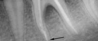
- X-ray examination with obtaining an intraoral targeted image, which provides information about the location, size and depth of the carious lesion.
When examining the oral cavity, the doctor evaluates the color of the tooth and the condition of the edges of the carious cavity. Probing makes it possible to assess the structure and density of root tissues and establishes the presence of painful areas. In the process of diagnosing root caries, special tests can also be used. They help determine the condition of the hard tissues of the tooth and periodontium, the oral hygiene index, and the status of the oral fluid.
Features of root caries
The clinical features of this type of caries include the spread of the pathological process mainly along the superficial part of the root. The carious cavity remains shallow for a long time. But at the same time, complications such as pulpitis and apical periodontitis often develop. This is due to the small distance from the root surface to the pulp.
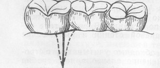
Types of caries localizations
Caries (lat. caries rotting) is a slow and hidden pathological process occurring in the hard tissues of the tooth. It is characterized first by focal demineralization of the enamel, then by the destruction of hard tooth tissues with the appearance of a cavity in the dentin. If left untreated, the tooth is lost or complications arise - pulpitis and periodontitis.
Cervical (circular)
The structure of a tooth is divided into a root, a neck and a crown - the latter is located above the gum. The neck and root are located in the gum and are protected by soft mucosal tissue.
When the carious process begins to destroy the tooth in the area of contact of the crown with the mucous membrane, we can talk about the cervical form of caries.
This category is the most difficult to diagnose and treat. It most often affects the incisors. When biting food with incisors, the food is not only “cut off”, but also gets clogged into the gum pockets, moving towards the gums.
Crown caries
Caries has the ability to affect not only healthy, but also already treated teeth - under a filling or crown. If there is a crown, it is difficult to detect, because the tooth does not have a nerve and pain is not felt. There are no external signs for a long time.
The causes of this type of caries are:
- defect of the dental crown with the appearance of access to the tooth tissue underneath it
- poorly treated tooth before prosthetics;
- gum disease;
- poor oral hygiene.
The problem is that the process spreads into the deep layers and spreads to the bone structures, as a result of which the tooth is often completely removed. That is why preventive examinations and an orthopantomogram - a panoramic image of the entire jaw - are so important.
Radical caries
Basal and cervical caries are similar. Both incisors and chewing teeth are affected. Destruction begins with the enamel, gradually moving to the nerve in the root canal. When the process touches bone tissue, it threatens with osteomyelitis.
The main reason for the occurrence is a cariogenic situation in the oral cavity, when plaque accumulates especially quickly. The enamel in the root part of the tooth is much thinner and more easily destroyed. It is most often diagnosed between the ages of 30 and 60 years.
Root caries
Root or subgingival caries occurs hidden. It can be with or without a cavity. Along the flow - active, suspended, secondary. It most often affects molars.
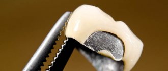
How to place a crown
If installation is required on the front teeth, the nerve must be removed, because when grinding the structure, the pulp can be burned. In premolars and molars, soft tissues are left if they are not involved in the pathological process.
After treatment, preparations begin for installation and production of the prosthesis. First, the doctor cleans the tooth surface and strengthens the remains of the tooth. If this is not possible and it is almost completely destroyed, a pin and a temporary crown are installed. After this, the laboratory prepares a permanent structure based on the casts. The specialist must accurately make the prosthesis not only in shape, but also in color, so that it does not differ in appearance from other teeth. Next, the temporary crown is replaced with a permanent one. Now we will find out whether caries can develop under it.
What is the danger of this disease?
Caries is dangerous because there are no symptoms for a number of years. It is detected more often in an advanced stage, when tooth extraction becomes the solution.
Visual diagnosis is difficult because all destructive processes occur inside the tooth. Pronounced plaque or tartar hides any stains on the enamel.
The root is hidden under the gum and external irritants do not affect it until a certain time. On the other hand, the root walls are thin, so they are destroyed quickly and with complications.
Causes of the disease
There are 3 main provoking factors that must act in a complex manner; none of them acts independently:
- Cariesogenic microflora - this includes Mutans streptococci, actinomycetes and certain types of lactobacilli. They should dominate the oral cavity. Bacteria produce organic acids from food carbohydrates, which cause demineralization of cement.
- Consumption of simple carbohydrates is the most cariogenic. Their breakdown produces glucan, which contributes to the appearance of plaque.
- Reduced caries resistance is a deterioration in the resistance of tooth tissue and the body as a whole. This is facilitated by a decrease in calcium content in hard root tissues, bad habits, and lack of saliva.
In addition, the anatomical features of the mouth can also have an effect - a small vestibule, a short frenulum, and bite pathology.
Gum disease, pocket formation
Gum pocket or otherwise periodontitis is a common occurrence in many patients. Many people do not treat it because it does not bother anyone for a long time.
The first manifestation of pathology is bleeding when brushing teeth, although outwardly they may look healthy. Dental pockets or depressions are normal, but their anatomical size should not exceed 3 mm.
Then the pocket is able to self-clean from food debris and epithelial particles. Reservoirs of bacterial accumulations form in deep gaps. This is accompanied by bad breath. Conventional treatment with antibiotics and brushing teeth with toothpaste have no effect.
Causes of periodontitis:
- insufficient hygiene;
- poor quality fillings;
- abnormal position of the dentition, malocclusion;
- hereditary predisposition;
- deficiency of vitamins and minerals;
- immunosuppression.
What should pregnant women do if they experience toothache under a crown?
It's no secret that during pregnancy a severe calcium deficiency occurs in the body. The teeth are the first to suffer from this. With proper nutrition, you can always replenish the calcium supply in a pregnant woman’s body. Alas, not all expectant mothers take care of themselves properly. It is not uncommon for pregnant women to visit the dentist.
If even the slightest toothache occurs, pregnant women are simply obliged to immediately consult a dentist. By the way, they are accepted everywhere out of turn. Know that any inflammation in the oral cavity, not to mention its purulent manifestation, necessarily affects the development of the fetus, and therefore the health of the newborn.
If it is not possible to quickly get to the dentist, pregnant women should at least slow down the further development of dental disease using traditional methods. To do this, you can rinse the mouth with soda, salt, chlorhexidine, chamomile or furatsilin. You can apply a piece of cotton wool soaked in a solution of dental drops to the area of pain. In particularly acute cases, you can take a paracetamol tablet. At the 5th month of pregnancy, you will already be able to withstand pain relief, so during this period, feel free to make an appointment with the dentist.
Lifestyle
If you have learned about the risk group for root caries (older age), this does not mean that you can relax. Caries has many causes and can occur at any age if basic precautions are not followed.
Those at risk include smokers, diabetics, pregnant women and even children. The provoking factors in this case are:
- smoking and lack of oral hygiene;
- infrequent brushing of teeth and lack of fluoride;
- poor nutrition with preference for desserts;
- frequent stress;
- alternating hot and cold;
- structural features of the oral cavity;
- alcohol abuse - alcohol breaks down into sugars and acids;
- abuse of coffee and strong tea, which creates an acidic environment in the oral cavity;
- lack of water intake, and therefore lack of saliva;
- gum injuries.
When a tooth hurts under the crown...
It is generally accepted that to treat a tooth, it is necessary to remove the crown. But high-quality dentures are not cheap, and they are not suitable for reuse. Therefore, it is doubly unpleasant when pain appears several months after prosthetics, and it becomes necessary to remove a crown that has not served its purpose.
In this case, patients often put off a visit to the dentist with all their might. For example, they take painkillers and antibiotics or experiment with very dubious “folk” methods. But by self-medicating, you will not solve, but will only aggravate the problem, since advanced inflammation is much more difficult to treat. If discomfort or pain occurs, you should immediately visit your dentist. Moreover, contrary to popular belief, modern treatment methods, in some cases, make it possible to do without removing the crown.
In some cases, treatment is possible without removing the crown.
Symptoms of the disease
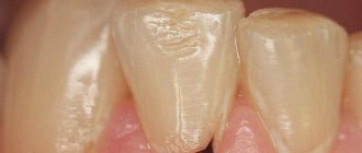
The process usually occurs without symptoms, but pain may occur when brushing a toothbrush, eating sour, salty, sweet, cold or hot foods.
After eliminating the irritant, everything goes away. The patient can see a doctor if a stain is found on the front surface of the incisors, but often it is hidden under plaque or tartar.
The ongoing carious process gradually reaches the dentin junction, penetrating first into its surface layers and then further. The cavity deepens with bacteria and food debris. There is a smell from the mouth. Irritants cause pain more and more frequently.
With cement caries, teeth become mobile and lose their support, and bleeding gums occur. These are already symptoms of periodontitis. Now, even when chewing food or brushing teeth, severe discomfort occurs.
Digestion begins to suffer. Teeth become hypersensitive to hot or cold.
Next, the process follows the Leus classification:
- Active lesion - the edges of the cavity are undermined. The cavity is filled with softened tissues and tends to grow.
- Suspended caries - no increase in the affected area is observed. The cavity is clean, the bottom is shiny and smooth, the edges are even and dense.
- Secondary caries - occurs under a filling.
Advantages of treating tooth root caries in our clinic
Working with affected subgingival areas of the tooth requires special practical skills. At Kariesu.net clinics, patients with complex dental diseases are treated by experienced specialists. The doctor considers each case individually. Explains in detail to the patient the essence of the identified pathology, informs about the features of the upcoming treatment. The main principle of our dentists’ work is minimal invasive intervention. Specialists try to preserve healthy tissue and the tooth itself.
Our clinics use traditional and new treatment technologies. Modern filling materials are used. An exceptionally polite and understanding attitude towards patients is practiced. Make an appointment with our doctor by phone or through the online form.
Diagnosis of the disease
The diagnosis is made in stages. Listening to complaints and visual examination make it possible to diagnose caries only in 13% of cases. Classic probing with a sharp probe and inspection with a mirror are informative. This allows you to examine the dentition.
Thermal diagnostics, electroodontometry and x-rays are also performed. If the patient has massive dental plaque, changes in the enamel will not be visible. In this case, professional teeth cleaning is first carried out to remove plaque and stone. All this is possible in a dental clinic.
At the dental clinic
The following types of studies can be performed at the clinic:
- Probing with a thin probe with a curved end - it is inserted under the gum, while the doctor has the opportunity to examine the structure of the tooth, its integrity, the presence of irregularities and chips. With rapidly progressing caries, the edges of the cavity are uneven and sharp. In the remission stage, the surface of the pathological focus is shiny, smooth, hard with smooth edges.
- Electroodontometry - can determine nerve damage and the depth of inflammation. The pulp reacts differently to the current strength.
- Thermal diagnostics - treatment of different areas of the tooth with a stream of water and heated wax. Unpleasant sensations that disappear after removing the irritant indicate caries.
- X-ray - a targeted photograph of one tooth or computed tomography. The presence and localization of obvious or hidden inflammation is determined with millimeter precision.
- Visiography is a special device - a visiograph scans the received data and transfers it to a computer, where the picture is studied from different angles and in detail, revealing the hidden process of inflammation.
Treatment or removal - what determines the outcome
The doctor makes a decision on treatment or tooth extraction after determining the depth of spread and the area of localization of the inflammatory process. It is mandatory to first carry out professional cleaning using an ultrasonic scaler, an AirFlow water-abrasive device and hand tools.
Initial caries is treated with conservative retherapy. For hard tissue defects, preparation and filling are performed. In severe cases, the tooth is removed.
Treatment of the initial stage of root caries
Conservative retherapy includes:
- fluoridation of teeth;
- covering teeth with protective agents
- training in proper oral hygiene
Cleaning in dentistry from stone and plaque
1243900247100
Professional teeth cleaning involves removing the accumulation of all pathogenic microflora around the root.
It involves not only removing plaque, but also stones. This elimination of negative factors protects teeth for a long time.
Antiseptic treatment of affected areas
Treatment of root caries involves drilling out all affected areas with a drill and treating the carious cavity with antiseptics and special preparations. A 2% chlorhexidine solution or a gel based on it is used as an antiseptic for the treatment of carious cavities.
Severe root damage
Modern dental methods make it possible to save a tooth even with extensive damage.
Depending on the specific situation, the doctor may prescribe:
- Resection - removal of the root apex.
- Hemisection - excision of the damaged part along with the crown that is adjacent to it.
- Amputation is the extraction of the affected part of multi-rooted teeth: one of them is removed.
- Separation is the separation of closely spaced teeth if the carious lesion is localized in the area of branching of the root system.
In rare cases, the doctor strengthens and restores the root not with a filling, but by installing a ceramic or metal stump that protects the tooth from fracture and infection. Of course, such treatment methods are expensive and not available to everyone. Most patients have to have their tooth removed.
What to do to protect your gums
If the gum is not treated, inflammation will remain and all treatment will become meaningless. The gums are protected by diathermocoagulation and retraction.
Diathermocoagulation
Diathermocoagulation – excision of excess areas of periodontitis with a coagulation knife. The knife immediately cauterizes the bleeding edges. The procedure is performed when there are severe changes in the gums. In such cases, filling is postponed until the soft tissue is completely regenerated.
Retraction
If the condition of the gums is advanced, treatment of caries becomes impossible - the mucous membrane bleeds and interferes with tooth treatment. This gum is excised or a special device is applied to move the gum back. But more often, retraction is performed - lowering the gingival contour or moving apart the overhanging edges of the gums using special hemostatic threads. A temporary filling is placed until complete healing.
What preventative measures are there?
To keep your teeth healthy for many years, you need to brush your teeth twice a day, use floss and a tongue scraper. After sweets, you should rinse your mouth and chew gum for 5-10 minutes.
Prevention also includes the removal of tartar. A sufficient amount of saliva is also important, so it is necessary to maintain a water regime.
Like any other pathology, caries is better prevented than treated. Therefore, it is important to visit the dentist twice a year for preventive examinations. People at risk should approach this especially responsibly.