Dentinal tubules.
They are pathways connecting the pulp and periodontium, the separating barrier between which is cement covering the tooth root. Normal, intact cement is an obstacle to the penetration of microorganisms and their metabolic products through these structures. Exposed dentinal tubules in areas of damaged cementum may serve as important communication routes between the dental cavity and the periodontium. Exposure of dentinal tubules can be a result of developmental defects, pathological processes, often inflammatory, or iatrogenic procedures. At the root of the tooth, dentinal tubules extend from the pulp to the dentin-cementum junction. They run in a relatively perpendicular direction to this boundary and vary in size from 1 to 3 mm in diameter. The diameter of dentinal tubules changes with age or in response to chronic low-intensity stimuli due to the formation of highly mineralized peritubular dentin. The number of dentinal tubules in the area of the dentinal-cementum junction is approximately 8000 per square millimeter. Simple arithmetic calculations show that the total area of dentinal tubules, providing communication between the tooth cavity and the periodontium, significantly exceeds the size of the apical foramen, around which inflammation most often occurs during pulp necrosis. There are approximately 15,000 tubules per square millimeter in the cervical root region, these tubules can be exposed by periodontitis or periodontal disease, as a result of iatrogenic interventions or developmental defects where cementum and enamel do not join at the enamel-cementum junction, thus leaving or creating areas exposed cement.
Another way of communication between the tooth cavity and the periodontium can be lateral and additional canals. They are located along the root of the tooth. Their prevalence and location have been well studied and documented in papers. 30-40% of teeth have additional canals and most of them are located in the apical third of the tooth root. 17% were shown to have additional canals in the apical portion, 9% in the middle third, and 2% in the upper third. However, there are relatively few destructive changes in the periodontium associated with the lateral canals. Studies show that out of 1000 patients examined, only 2% had the process located in the area of the lateral canals. Additional channels in the area of molar furcation are also pathways of communication between the pulp and periodontium. The prevalence of additional channels in this area is very high and reaches 76%. However, not all of these canals extend throughout the entire thickness of dentin to the bottom of the furcation. Seltzer showed that inflammation can spread to these areas, but the percentage of their involvement in pulp necrosis is relatively low.
Apical microsurgery as an opportunity to save a tooth
Starting from the standard therapeutic protocol to tooth-preserving surgery.
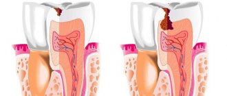
The anatomy of root canals is very complex and varied - there are branches, divergences and repeated closures of the canals, and specific bends. Also, the apex of the tooth itself (its apex) is practically a “tree” of small branches of the main canal.
All this helps to maintain minimal infection in the canals and its ability to continue to develop. In some cases, this infection is very minimal and the body comes to the aid of the doctor, completely blocking it. But sometimes, the infectious process is more active and, turning into a chronic form, slowly continues its development.
Several years after treatment, a focus of inflammation may be accidentally detected on control images, and the question of re-treatment or removal will arise. BUT since the doctor’s main task is to preserve the tooth, it is not always worth removing the tooth right away. After assessing its viability, doctors may suggest an alternative treatment - apical surgery.
Apical surgery is a complex of operations performed by resection of the upper quarter/third of the root.
The inflammatory focus is removed along with the root, and its place is filled with bone tissue.
To carry out these manipulations, an operating microscope or binoculars, ultrasound and microsurgical equipment are used, which allow retrograde filling and elimination of the consequences of periodontitis.
Indications for apical surgery:
- Lack of possibility of repeated endodontic treatment
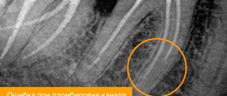
- Detection of instrument fragments in the extreme third of the canal, or excess material behind the root of the tooth (it is not possible to extract all this through the canal)
- Carrying out therapeutic intervention to no avail (sclerosis of the canals, impossibility of passing the canals due to cements or pins)
The planning of the operation is carried out jointly by the therapist and the surgeon. A CT scan is performed and the chances of a positive outcome are assessed. The surgeon evaluates the volume of bone tissue in the lesion area to understand the possibility of complete restoration of bone tissue.
A treatment plan is drawn up and all the nuances are discussed with the patient.
Let's consider the algorithm for apical surgery using a specific clinical example of patient Sergei, 39 years old. He is undergoing comprehensive rehabilitation at our clinic.
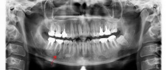
After all the examinations, a lesion was identified in the area of 2 central teeth 21,22.
Therapist Zapolskaya M.A. carried out repeated endodontic treatment in advance, the canals were filled with biodentine in the apical third and restored on fiberglass pins. On the day of canal filling, the patient immediately goes into the hands of a surgeon to perform resection of the root apexes (apical surgery).
The surgeon works with binoculars to better assess the surgical field. An incision is made to access the teeth, the flap is removed and the lesion is visualized.
Curettage of the lesion area is performed, treated with ultrasound and antiseptic solutions. The tips of the affected roots are partially resected and polished. The therapist controls the canal filling through the surgical field using a microscope.
The cavity is filled with bone material and hermetically sealed with a membrane. The membrane is fixed to the surface of the bone tissue with pins (surgical buttons)
The wound is sutured.
After surgery, the patient receives standard drug therapy and performs nursing procedures.
After 3 months, a control CT scan was performed to evaluate the results of the surgery. We are satisfied with the result and continue monitoring and monitoring every 3-6 months.
This clinical case went smoothly and pleased us with its result. However, I would like to note that not every tooth can be saved by apical surgery. To make a decision, the doctor always evaluates many factors - from the anatomy and location of the tooth, to the size of the lesion and the degree of destruction of the crown. The main thing here is to answer honestly the question of the appropriateness of such treatment. If the tooth cannot be restored or surgery can cause great trauma, then you should listen to the doctor and accept removal. If the doctor gives good prognosis and the tooth is strong and reliable, of course it’s worth trying all the ways to save it.
Be a team with your doctor, trust and listen to arguments, make a decision together and then the result will please everyone!
This article used the clinical case of G.V. Khokrishvili. and Zapolskaya M.A.
Apical foramen.
It is the main route of communication between the pulp and periodontium. Bacteria and their metabolic products can enter the periodontium and cause inflammation, with corresponding destruction of the bone and tooth root.
When the pulp is necrotic, microorganisms, inflammatory pathogens of various natures, lead to periodontal inflammation. However, it is extremely important and interesting that the same situation occurs when analyzing the opposite situation. It has been shown that in periodontal pathology the pulp is not involved in the process, at least until the pathological pocket reaches the apical foramen.
It appears that as long as the accessory and lateral canals are protected by intact cementum, inflammation does not spread to either side. This is confirmed by the preservation of the vital activity of the pulp in deep pathological pockets that do not reach the apical foramen. However, during surgical treatment of dental periodontium, when removing a layer of cement from the surface of the root, the pulp is often involved in the inflammatory process. Surgical intervention must be justified, and biological, mechanical and aesthetic feasibility must be taken into account.
Treatment of lateral tooth canals and apical branches
Endodontic treatment of teeth with lateral canals deserves special attention, since many misconceptions surrounding this topic have led to considerable confusion. In addition to the lateral canals, the following discussion includes various branches from the main canal, which also form communications between the canal system and the periodontium. Strictly speaking, branches include canal bifurcations, lateral canals and apical branches. Branches can occur throughout the root, but in most cases they are localized in the apical part of the root and in the chewing teeth.
According to research, 73.5% of branches are observed in the apical third of the root, 11% in the middle and 15% in the coronal third. Ricucci and Siqueira in their study found lateral canals and/or apical ramifications in almost 75% of the teeth examined, with the most common (80% or more) in molars and upper premolars.
Branches from the main root canal are formed after localized fragmentation of the epithelial lining of the root with the preservation of a small gap or with the preservation of blood vessels passing from the dental sac to the dental papilla. In such areas, dentinogenesis does not occur, and as a result, a canal is formed containing small blood vessels and sometimes nerves. The branches contain connective tissue and blood vessels that do not belong to the collateral blood supply system. Their participation in maintaining the vitality and function of the pulp is minimal, if at all, probably with the exception of branches localized in the most apical part of the root. Branching can serve as a route for the spread of microorganisms and their metabolic products from necrotic and infected tissues of the root canal into the periodontium, with the subsequent development of the pathological process in areas adjacent to the root. Likewise, pathogens from the periodontal pocket can penetrate into the pulp. The branches are poorly accessible for instrumentation, disinfection and filling. Next, the histological and microbiological status of tissues in the branches (in particular, in the lateral canals and apical branches) will be presented in various clinical conditions, including the development of periradicular pathology.
Side canal filling: what is the goal?
The need to fill lateral canals causes heated debate among dentists. The main goal of endodontic treatment should be to treat and fill the entire length of the root canal, including all lateral canals and apical branches. Some have even believed that those who use three-dimensional obturation techniques have historically established a technical and even moral superiority over those whose technique only fills the primary canals, so the ability to obturate the lateral canals was often considered an indicator of the clinician's skill, and unfilled lateral canals were seen as causes of apical periodontitis. In addition, some experts have suggested that inflamed and/or infected tissue in the lateral canals often leads to the development of pain. As a result, many clinicians and researchers have unjustifiably believed that filling these canals and apical branches is necessary for a successful outcome of endodontic therapy.
The misconception of mandatory filling of the lateral canals and apical branches as a quality criterion has led to many experimental studies aimed at assessing this possibility. However, most authors reported no significant differences between different techniques regarding the introduction of sealer into the lateral canals when using thermoplastic gutta-percha.
However, the need to seal the ramifications to improve treatment outcome has not been universally accepted. Weine showed that, despite the high prevalence, lateral canals are rarely detected radiographically after filling the main ones. In other words, in most cases, the branches are not diagnosed and remain unsealed, which, however, does not reduce the effectiveness of treatment, subject to adequate preparation, disinfection and filling of the root canal.
It should be noted that lateral canals and apical branches can lead to failure of endodontic dental treatment if their size is so large that they allow the accumulation of a significant number of microorganisms and provide wide access to periradicular tissues. Thus, disinfection of the lateral canals and apical branches in case of pulp necrosis and apical and (or) lateral periodontitis is one of the goals of treatment, despite the difficulty of achieving it even with the use of modern technology and materials.
It is not yet known whether the release of filling material into the ramifications increases the effectiveness of lateral sealing or disinfection in this area. The antimicrobial effect of such materials is very weak and short-lived, reaching its peak effectiveness before curing. Additionally, achieving a predictable seal when introducing material into a narrow and typically tortuous branch is unlikely and not supported by research. Based on the minimal antimicrobial activity and questionable sealing ability, we can conclude that the filling materials in the branches have no or weak influence on the treatment result.
Clinical Application
Ramifications are rarely diagnosed on pretreatment radiographs. The presence of lateral canals can only be assumed by the limited expansion of the periodontal fissure on the lateral surface of the root or a pronounced focus of lateral periodontitis. After obturation of the root canal, the lateral canals and apical branches can be visualized on radiographs when a sufficient amount of filling material (usually sealer, but also thermoplastic gutta-percha) is injected into them.
Radiologically, only lateral canals are determined to be of sufficiently large size and passing in the mesial-distal plane. Lateral canals and apical branches are more often identified after obturation of canals with necrotic tissues than during the treatment of teeth with vital pulp. This is probably explained by the operator’s greater alertness and the relatively high resistance of vital tissues in the branches. When describing filling material in a branch, the term “filling” should be replaced by the concepts of “removal”, “presence” or “penetration”, since targeted filling of branches in a clinical setting is practically impossible. This conclusion is confirmed by histological observations.
It is interesting to note the relatively low incidence of lateral periodontitis with pulp necrosis, despite the high prevalence of lateral branches. At present, there is no unambiguous explanation for this phenomenon, but it may depend on the size and patency of the branch, as well as the histological and microbiological characteristics of its contents. According to a morphological study, in 79 out of 100 permanent molars, the diameter of the openings of the lateral (additional) canals was 10-200 µm. In addition, the maximum diameter of the lateral canal was 2-3 times less than the average diameter of the main apical foramen. These differences in diameter between the main apical foramen and the lateral (additional) ones may explain the more frequent development of apical periodontitis compared to lateral.
Wide and open lateral branches can contain large arrays of microorganisms and their metabolic products, which increases the risk of developing lateral periodontitis. In turn, small branches contain fewer microbial irritants, which are often not enough to form a focus of pathology. Thus, a large focus of lateral periodontitis, as a rule, indicates the presence of a wide lateral canal with a significant infectious load that can cause inflammation in the lateral periodontium.
The pulp of the lateral canals and branches is characterized by a good blood supply from the periodontium and, therefore, significant resistance to necrosis and bacterial invasion. It is likely that this tissue becomes necrotic and becomes infected only with prolonged necrosis and an infectious process in the pulp of the main canal. The presence of inflamed but vital tissue in the branch also explains the possibility of healing of the lateral lesion without removal of materials into the lateral canal. Microorganisms in the main canal cause inflammation in the side canals. The inflammatory process can spread to the periodontium and is supported by waste products of microorganisms penetrating from the source of infection in the main canal through the lateral canal to the periodontal ligament. The developing lateral pathological focus, in turn, is not associated with infected necrotic branching tissue. In such cases, healing of the lateral lesion occurs provided that the infection in the main canal is effectively eliminated.
The emergence of new materials and technologies.
Allows you to significantly solve the aesthetic problem. Biological issues will be discussed below. The mechanical strength of the tooth is one of the most important factors ensuring the long-term preservation of the tooth as a functional unit in the dental arch. During surgical intervention, in all cases, it is necessary to find a reasonable compromise between the need to remove a significant amount of tooth tissue and ensure the mechanical stability of the remaining tooth structures. At the same time, in each specific case the problem is solved individually, taking into account the anatomy of the tooth crown, the topography of the orifices and the course of the root canals. Before any surgical intervention, a thorough x-ray examination is necessary, which makes it possible to determine the most important individual characteristics of the tooth.
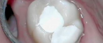
Often, endodontic intervention in the tooth cavity is attempted through a carious defect, without taking into account the anatomical features of the tooth. The general rule should be a clear understanding of the need to excise carious tissues before endodontic intervention itself, and sometimes to restore the tooth with permanent filling material. Only after this, ensuring rational access to the tooth cavity, which will be described in detail below.
Many problems that doctors unexpectedly encounter during endodontic treatment are often associated with insufficient knowledge of the morphology of the dental cavity. It is necessary to clearly understand not only the variants of the norm, but also deviations in the shape and directions of the canals, which are often encountered in a doctor’s practice. Clinical radiography can show the shape of roots and canals in only two projections. The third projection exists in the vestibulo-oral direction and is not visible on the radiograph.
Endodontic morphology of human teeth.
It has been studied by many researchers who have accumulated valuable observations about the shape, size and contours of the tooth canals. Modern literature has recently devoted enough space to the morphological features of teeth. Thus, a number of journal articles describe in sufficient detail the variants of the structure of dental cavities and root canals. At the same time, understanding the importance of presenting all aspects of endodontics in a single manual (and knowledge of dental morphology is one of the most important), the authors risk repeating themselves, but did not dare to ignore this section.
It is generally accepted that anatomically the dental cavity is divided into two parts:
- the pulp chamber, which is usually described as the coronal part;
- root canals, which are located inside the root.
Pulp chamber.
A single cavity, the dimensions of which follow the contours of the tooth crown. If the cusps are well defined on the crown, then the pulp horns are well defined in the pulp chamber. In multi-rooted teeth, the depth of the pulp chamber depends on the position of the furcation and can extend beyond the anatomical crown. In young teeth, the contours of the pulp chamber resemble the shape of the outer contours of dentin. With age, the tooth cavity decreases in size as a result of the deposition of secondary dentin, especially with caries, abrasion and other defects.
Root canals are an extension of the pulp chamber, and since the roots taper towards the apex, the canals also have a tapering shape and end in an apical foramen at the end of the root - foramen apicale, which rarely opens exactly at the anatomical apex of the root. During root formation, the pulp and periodontal tissues become partially separated, typically maintaining a major vascular and neural connection through the apical foramen.
It is necessary to immediately stipulate that the term “tooth canal” today does not quite reflect the complex formation with a large number of branches that exists in the roots of the teeth. Therefore, even for a single-rooted tooth, it is advisable to talk about the root canal system. Moreover, based on an analysis of the literature, we can assert that a single canal without branches and additional apical openings is practically never found.
Vertucci (1984).
Identified eight different channel configurations. In the case where the roots have one canal and one apical opening, he classifies them as type I. It should be emphasized that we are talking about any channel in a single root. The classification applies to each individual root and, at the same time, in multi-rooted teeth there can be any variations in different roots. It should also be emphasized that the classification provides only a rough, approximate diagram of possible options for root canal systems.
Types II - III, found especially often in the premolars of the upper and lower jaw, have branches at different levels of the tooth root, which then merge and end in one apical foramen. We emphasize once again that the division is very conditional and is more theoretical in nature. Therefore, we combined types II and III according to the Vertucci classification into one group.
Type IV (we are talking about each root separately) has two separate root canals at one mouth, ending in two separate apical openings. Possible variants can be represented by type VIII, where there are three canals with one orifice, ending in three apical foramina.
Types V, VI, and VII represent variations in root canal separation, fusion, and divergence that are common in the lower incisors.
The presented schemes can be variable, combined in different combinations and give a very complex topography of the root canal system. Clinicians should pay special attention to cases with multiple variations of the apical foramina, which is important for preventing possible complications during endodontic intervention.
Variations in dental pulp morphology are caused by genetic and environmental (phenotypic) influences. For example, the high frequency of single-rooted teeth with two canals suggests that they are the result of the fusion (joining together) of two separate roots in the relatively recent past.
It is very important to emphasize that since the roots of the teeth are wider in the vestubulo-oral direction than in the mesiodistal direction, the tooth canals follow the same pattern. The diameter of the root canal decreases towards the apical foramen and reaches the narrowest point 1-1.5 mm from the apical foramen. This point, the apical constriction or constriction, is located in the dentin just anterior to the first layers of cementum, and is the narrowest point in the tapered canal.
Treatment of Confluent Root Canals
When using nickel titanium rotary instruments, it is important to know the anatomy of the root canal system. One of the main risks when using rotary systems is instrument fracture. It is well known that the cause of instrument fracture is the insertion of a rotating instrument into the confluence of two canals.
Author: Calogero Bugea
Photo 1
Merging channels
Weine classified canal fusion into 4 types, depending on the configuration of the main root canal of the tooth, from the floor of the pulp chamber to the apical foramen. This classification is useful in everyday practice when we are dealing with two channels merging into one.
Photo 2 – TYPE 1: one orifice, one root canal, one apical foramen.
Photo 3 - TYPE 2: two orifices, two root canals, one apical foramen.
Photo 4- TYPE 3: two orifices, two root canals, two apical foramina.
Photo 5 - TYPE 4: one orifice, two root canals, two apical foramina.
Types 1 and 3 canals can be cleaned and shaped separately, as if they were in different roots. Due to the complexity of the anatomy, in all situations where we treat canals with possible fusions, we must consider each root separately. In this way, over-instrumentation and repositioning of the apical foramen, which can occur as a result of double peeling and shaping from two different directions, can be prevented. For example, the buccal-mesial canal of a lower molar in 50% of cases has a common apical foramen with the lingual-mesial canal. This one should be expanded less to reduce the risk of root weakening and slit perforation. With a type 2 configuration, which is easy to diagnose in the early stages, great care must be taken in the treatment of the canals. Type 4 is very difficult to diagnose, but if such a structure of the root canal system is suspected, several techniques can be used. Early diagnosis of common apical will prevent the risk of instrument fracture. Let's see in what situations it is really important to diagnose the 4th type of root canal system configuration:
Photo 6
Typically, in lower molars, the lingual mesial canal is straighter than the buccal canal. In upper molars, instead of two mesiobuccal canals, there is usually one. These canals are located closer to the center of the root.
Photo 7
Apical foramen
The image shows the typical change in the position of the apical foramen that occurs when the canal fusion is located at the apical part. Processing from two different directions leads to the formation of a teardrop-shaped hole and parallel walls. At different stages of endodontic treatment, early diagnosis can be performed using various methods. Traditional intraoral radiography does not provide information about the anatomical configuration of the root canal system. Sometimes patients have already had a CBCT scan (for previous procedures such as implantation or wisdom tooth extraction). In these cases, the doctor has the opportunity to visualize the anatomy before starting treatment.
Photo 8
Of course, the above scenario is common, but not as common as we would like. The best way to detect canal fusion, according to the author, is visualization using a gutta-percha point. When there is a possibility of merging canals, say, in a lower molar, in one canal (for example, lingual-mesial), the working length is determined with a small instrument, a gutta-percha pin is inserted into the already prepared canal, and then a small file (for example, K-file No. 10) is inserted into buccal mesial canal to measure its length. The file is inserted with short movements, while checking whether the pin moves in the second processed canal.
Photo 9
The K-file and gutta-percha pin are then removed from the canal. The post should be carefully examined, preferably with a magnifying lens, to look for any grooves, scratches, or folds left by noninvasive instrumentation of the buccal-mesial canal. This method is very effective because it only takes a few minutes to identify the additional channel.
Photo 10
On the other hand, this method may fail in the case of apical fusion or in a situation where the K-file in the fusion canal is unable to scratch the main pin.
Photo 11
Apex locator
Another method requires the use of an electronic apex locator. After preparing the first channel, the working length of the second channel is checked using an apex locator. Then the operation is repeated, leaving the last file inserted to the apical opening of the prepared canal. If the working length of the second canal is a few millimeters shorter this time, this will mean that the second canal shares a common apical foramen with the first, and their junction is the same distance from the common apex. The effectiveness of this method may be affected by the presence of irrigant or closed channels. Some doctors, especially those who use only the thermafil obturation system, prefer this method because it is simple and does not require the use of gutta-percha points. In the case of specific anatomy, such as the first premolar, which often has a type 3 canal configuration, the clinician can evaluate canal fusion during simultaneous insertion of the post and the Thermafil system carrier. As is the case with pin or Thermafil radiography during media selection.
Photo 12
Individually, the pins or carriers reach the desired maximum depth without hindrance.
Photo 13
When they are posted simultaneously in different channels, they are promoted alternately. For example, in the lower molar, if the instrument in the buccal canal goes to the working length, in the lingual it remains shorter and vice versa. The same test can be performed using Thermafil system media with similar results.
Photo 14
During irrigation, to avoid the formation of pores, it is recommended to frequently change the solution and aspirate the irrigation solution. This way, when the needle tip reaches the confluence, the solution will be drained from the two channels simultaneously. Aspiration can be done using a regular syringe or the Endovac system. This check is recommended not only to confirm the presence of channel fusion, but also to clean the fusion area that is not accessible to rotary instruments.
Photo 15
The image shows multiple connections between the two channels: this area is “inaccessible” to rotary instruments and only an effective irrigation protocol can clean and completely disinfect it. Diaphanization of Dr. A. Iandolo (Italy).
Photo 16
Even if there are several canals, the rules for their identification, formation, cleaning and obturation remain the same as for other structures of the root canal system.
Photo 17
Obturation of root canals
Fusion of canals can obscure a specific structure, so after completing the cleaning protocol, it is important to perform a three-dimensional obturation of this type of canal. During root canal obturation, it is recommended to insert gutta-percha points and sealer simultaneously and then condense the material in each canal. In obturation using a carrier, it is injected into one canal to block the passage of gutta-percha from the second canal, and then the confluent canal is obturated using the chosen method.
Conclusion
To maintain root canal configuration and prevent apical changes, slit perforations, and instrument fracture, it is important to understand root anatomy. Early diagnosis provides a safe and predictable solution to problems associated with confluent root canals.
The translation was made by Petrushchenko A. specifically for the OHI-S.COM website. Please, when copying material, do not forget to provide a link to the current page.
https://www.styleitaliano.org/
Literature
Weine FS, Healey HJ, Gerstein H, Evanson L. Canal configuration in the mesiobuccal root of the maxillary first molar and its endodontic significance. Oral Surg Oral Med Oral Pathol. 1969 Sep;28(3):419-25. Weine FS.Initiating endodontic therapy in posterior teeth. Part II. Maxillary molars. Compend Continue Educ Dent. 1982 Nov-Dec;3(6):455-64. Berutti E.Respecting apical foramina in the endodontic treatment of confluent canals. G Endodonzia. 1990;4(1):6-21. Hess W, Zürcher E. The anatomy of the root canals of the teeth of the permanent and deciduous dentitions, New York: William Wood & Co, 1925. Burns RC, Buchanan LS. Tooth Morphology and Access Openings. In Cohen S, Burns RC, editors: Pathways of the Pulp, 6th ed., Mosby Yearbook Co., 1994. Ruddle CJ. The Mesial-Buccal Root of the Maxillary First Molar: Treatment Considerations, The Endodontic Report, Fall/Winter, 1986. Ruddle JC. Endodontic Canal Preparation: Breakthrough cleaning and shaping strategies, Dentistry Today, February 1994
Location of the apical foramen.
Varies and can be located (depending on the amount of cement) ranging from 0.2 to 2 mm from the anatomical apex, and the apical constriction is from 0.5 to 1 mm from the apical foramen. Thus, the distance from the apical constriction to the anatomical apex can vary from 0.7 to 3 mm (according to Harty).
The dental pulp responds to damage by deposition of secondary dentin on the walls of the pulp chamber. With age, it decreases in size, the number of nerve fibers, blood vessels and connective tissue cells gradually decreases, and the fibrous component of the pulp increases. The pulp “ages” not only over time, but also under the influence of functional stimuli and chronic irritation. The rate at which the pulp deposits reparative dentin and decreases in volume varies from tooth to tooth and from patient to patient.
During root formation, the apical portion of the root pulp is described as “open” and funnel-shaped. As the tooth matures, the funnel-shaped opening closes and narrows to a normal root shape with a small apical opening. The location of the apical foramen may change in relation to the root apex due to the constant formation of cementum.
As a result of the deposition and mineralization of secondary dentin in the root canal, radiographically the pulp cavity may appear completely obliterated. However, such a canal, although not visible radiographically, may contain a significant amount of pulp or pulp decay, which can cause the development of inflammation in the periapical tissues.
Pulp and periodontal tissues.
They maintain communication not only through the main apical foramina, but also through accessory and lateral canals. Lateral (side) canals can be found in any root and are located approximately at right angles to the main root canal. Accessory canals usually branch off from the main canal at the apex (the so-called deltoid arborizations). Lowman et al. (1973) found that obvious lateral canals were present in the coronal or middle thirds of 59% of molars. Burch and Hulen (1974) found that 76% of all molars have an opening at the furcation. Kramer (1960), using intravascular injection techniques, found that these accessory canals often have an even larger diameter than the apical foramina, and the blood vessels passing through the lateral canals often have a larger diameter than the apical vessels and contribute more to blood supply to the pulp. It is very important to take into account the presence of such channels during endodontic treatment, since the presence of such channels allows the exchange of inflammatory products between the pulp and periodontium, and this can affect the outcome of endodontic therapy and the maintenance of periodontal health.
Most endodontists believe that endodontic instrumentation should be limited to apical constriction at the dentin-cementum junction. You cannot cross this point so as not to injure the periapical tissues during the preparation and obturation process.
There are quite a lot of works on determining the localization of the dentino-cemental junction, which is defined as an apical constriction, but studies on the size of this formation are rare in the available literature. Therefore, the work of Le Corn (2003) seems extremely interesting, since the author, based on the study of histological preparations, determined the dimensions of the apical foramen, apical constriction and their position in various groups of teeth. In this case, the author paid special attention to the level of distribution of cement into the root canal, measured from the most extreme border of the apical foramen. The apical foramen diameter and root canal diameter were measured at the point where the cement most extended into the root canal. It was found that the apical foramen was located on the medial side in 16.6%, on the distal side in 44.4%, on the palatine in 11.1%, and in only 27.7% on the apex of the tooth root. The average extent of cement into the root canal was 0.83 mm; the greatest extent of cement into the root canal was determined in the canines. The diameter of the apical foramen for various teeth was 0.45 mm for canines, 0.52 mm for lateral incisors, 0.35 mm for central incisors. The diameter of the root canal in the area of the dentin-cement junction (apical constriction) was 0.35 mm in the canines, 0.29 in the lateral incisors, 0.3 in the central incisors. Both the apical constriction and the root canal in this area had different shapes. The authors concluded that the dentin-cementum junction is where two tissues meet within the root canal and that the apical foramen cannot be the anatomical point for determining the apical limit of the preparation. Its use as an apical stop can result in damage to apical and periapical tissues due to wide variations in location. The study showed that the apical foramen rarely coincides with the anatomical apex of the root. According to radiographic and morphological data from the study of various teeth, the average distance between the apical foramen and the anatomical apex of the root usually ranges from 0.20 to 2.00 mm. Moreover, due to the deposition of cementum, the apical constriction tends to be located at a distance of about 0.5 - 1.00 mm from the apical foramen (Chapman, 1964). Ideally, the apical constriction should be used as a natural “stop” during endodontic therapy and the integrity of this constriction should be maintained throughout treatment to ensure long-term success. Having made a general overview of the morphological features relating to all teeth, we will then focus on the morphology of the intradental cavities of all teeth separately.
Crown of the tooth
The crown of a tooth (lat. corona dentis) is the part of the tooth protruding above the gum. The crown is covered with enamel - hard tissue, 95% consisting of inorganic substances and subject to the most powerful mechanical stress. There is a cavity in the crown of the tooth - dentin (hard tissue 2-6 mm thick) comes closer to the surface, then pulp, which fills both part of the crown and the root part of the tooth. The pulp contains the blood vessels and nerves of the tooth. Teeth cleaning and removal of dental deposits are carried out specifically from the crowns of the teeth.
Tooth neck
The neck of the tooth (lat. collum dentis) is the part of the tooth between the crown and root, covered by the gum.
Tooth roots
The root of the tooth (lat. radix dentis) is the part of the tooth located in the dental alveolus.
Fissure
On the chewing surface of the back teeth, between the cusps of the teeth there are grooves and grooves - fissures. The fissures can be narrow and very deep. The relief of the fissures is individual for each of us, but dental plaque gets stuck in the fissures of everyone. It is almost impossible to clean the fissures with a toothbrush. Bacteria in the oral cavity, processing plaque, form acid, which dissolves tooth tissue, forming caries. Even good oral hygiene is sometimes not enough. In this regard, fissure sealing has been successfully used throughout the world for 20 years.
Tooth enamel
Tooth enamel (or simply enamel, Latin enamelum) is the outer protective shell of the crown part of human teeth. Enamel is the hardest tissue in the human body, which is explained by the high content of inorganic substances - up to 97%. There is less water in tooth enamel than in other organs, 2-3%. Hardness reaches 397.6 kg/mm? (250-800 Vickers). The thickness of the enamel layer differs in different areas of the crown of the tooth and can reach 2.0 mm, and disappears at the neck of the tooth. Proper care of tooth enamel is one of the key aspects of human personal hygiene.
Dentine
Dentin (dentinum, LNH; lat. dens, dentis - tooth) is the hard tissue of the tooth, constituting its main part. The coronal part is covered with enamel, the root part of the dentin is covered with cement. Consists of 72% inorganic substances and 28% organic substances. Consists mainly of hydroxyapatite (70% by weight), organic material (20%) and water (10%), permeated with dentinal tubules and collagen fibers. Serves as the foundation of the tooth and supports tooth enamel. The thickness of the dentin layer ranges from 2 to 6 mm. Dentin hardness reaches 58.9 kgf/mm?. There are peripulpal (internal) and mantle (external) dentin. In peripulpal dentin, collagen fibers are located predominantly condensally and are called Ebner fibers. In mantle dentin, collagen fibers are arranged radially and are called Korff fibers. Dentin is divided into primary, secondary (replacement) and tertiary (irregular). Primary dentin is formed during the development of the tooth, before its eruption. Secondary (replacement) dentin is formed throughout a person’s life. It differs from the primary by a slower pace of development, a less systemic arrangement of dentinal tubules, a larger number of erythroglobular spaces, a larger amount of organic substances, higher permeability and less mineralization. Tertiary dentin (irregular) is formed during tooth trauma, tooth preparation, caries and other pathological processes, as a response to external irritation.
Dental pulp
Pulp (lat. pulpis dentis) is loose fibrous connective tissue that fills the tooth cavity, with a large number of nerve endings, blood and lymphatic vessels. Along the periphery of the pulp, odontoblasts are located in several layers, the processes of which are located in the dentinal tubules throughout the entire thickness of the dentin, performing a trophic function. The processes of odontoblasts include nerve formations that conduct pain sensations during mechanical, physical and chemical influences on dentin. Blood circulation and innervation of the pulp are carried out thanks to dental arterioles and venules, the nerve branches of the corresponding arteries and nerves of the jaws. Penetrating into the dental cavity through the apical opening of the tooth root canal, the neurovascular bundle breaks up into smaller branches of capillaries and nerves. The pulp helps stimulate regenerative processes, which manifest themselves in the formation of replacement dentin during the carious process. In addition, the pulp is a biological barrier that prevents the penetration of microorganisms from the carious cavity through the root canal beyond the tooth into the periodontium. The nerve formations of the pulp regulate the nutrition of the tooth, as well as the tooth’s perception of various irritations, including pain. The narrow apical opening and the abundance of vessels and nerve formations contribute to the rapid increase in inflammatory edema in acute pulpitis and compression of the nerve formations by the edema, which causes severe pain.
Tooth cavity
(lat. cavitas dentis) The space inside the tooth formed from the cavity of the crown and root canals. This cavity is filled with pulp.
Cavity of the tooth crown
(lat. cavitas coronae) Part of the tooth cavity, located under the crown and repeating its internal contours.
Tooth root canals
The root canal of a tooth (lat. canalis radicis dentis) is an anatomical space inside the root of a tooth. This natural space within the coronal part of the tooth consists of a pulp chamber, which is connected by one or more main canals, as well as more complex anatomical branches that can connect the root canals to each other or to the surface of the tooth root.
Nerves
(lat. nervae) Neuron processes passing through the apex of the tooth and filling its pulp. The nerves regulate the nutrition of the tooth and conduct pain impulses.
Arteries
(lat. arteriae) Blood vessels through which blood from the heart flows to all other organs, in this case - to the pulp of the tooth. Arteries nourish dental tissues.
Vienna
(lat. venae) Blood vessels through which blood returns from organs back to the heart. The veins enter the canals and penetrate the pulp of the tooth.
Cement
Cement (lat. - cementum) is a specific bone tissue that covers the root and neck of a human tooth, as well as the teeth of other mammals. Serves to firmly secure the tooth in the bone alveolus. Cement consists of 68-70% inorganic components and 30-32% organic substances. Cementum is divided into acellular (primary) and cellular (secondary). Primary cement is adjacent to the dentin and covers the lateral surfaces of the root. Secondary cement covers the apical third of the root and the bifurcation area of multi-rooted teeth.
Tops of tooth roots
(lat. apex radicis dentis) The lowest points of the teeth, located on their roots. At the tops there are openings through which nerve and vascular fibers pass to the tooth.
Apical foramina
(lat. foramen apices dentis) Places of entry of vascular and nerve plexuses into the dental canals. The apical foramina are located at the apex of the tooth roots.
Alveolus (alveolar socket)
(alveolar socket) (lat. alveolus dentalis) A notch in the jaw bone into which the roots of the tooth enter. The walls of the alveoli form strong bone plates impregnated with mineral salts and organic substances.
Alveolar neurovascular bundle
(lat. aa., vv. et nn alveolares) A plexus of blood vessels and nerve processes passing under the alveolus of the tooth. The alveolar neurovascular bundle is enclosed in an elastic tube.
Periodontium
Periodontium (lat. Periodontium) is a complex of tissues located in the slit-like space between the cementum of the tooth root and the alveolar plate. Its average width is 0.20-0.25 mm. The narrowest section of the periodontium is located in the middle part of the tooth root, and in the apical and marginal sections its width is slightly greater. The development of periodontal tissue is closely related to embryogenesis and teething. The process begins in parallel with the formation of the tooth root. The growth of periodontal fibers occurs both from the side of the root cement and from the side of the alveolar bone, towards each other. From the very beginning of their development, the fibers have an oblique course and are located at an angle to the tissues of the alveoli and cementum. The final development of the periodontal complex occurs after tooth eruption. At the same time, the periodontal tissues themselves are involved in this process. It should be noted that, despite the mesodermal origin of the constituent components of the periodontium, the ectodermal epithelial root sheath takes part in its normal formation.
Gingival grooves
(lat. sulcus gingivalis) Crevices that form where the crown of the tooth adheres to the gums. The gingival grooves run along the line between the free and attached parts of the gum.
Gum
Gums (lat. Gingiva) is a mucous membrane that covers the alveolar process of the upper jaw and the alveolar part of the lower jaw and covers the teeth in the cervical area. From a clinical and physiological point of view, the gums are divided into interdental (gingival) papilla, marginal gum or gingival margin (free part), alveolar gum (attached part), mobile gum. Histologically, the gum consists of stratified squamous epithelium and the lamina propria. There are oral epithelium, junctional epithelium, and sulcal epithelium. The epithelium of the interdental papillae and attached gingiva is thicker and can become keratinized. In this layer, there are basal, spinous, granular and stratum corneum. The basal layer consists of cylindrical cells, the spinous layer consists of polygonal cells, the granular layer consists of flattened cells, and the stratum corneum is represented by several rows of completely keratinized and nucleated cells that are constantly exfoliated.
Mucous papillae
(lat. papilla gingivalis) Fragments of gums located at their elevation in the area between adjacent teeth. The gingival papillae are in contact with the surface of the dental crowns.
Jaws
(lat. maxilla - upper jaw, mandibula - lower jaw) Bony structures that are the basis of the face and the largest bones of the skull. The jaws form the mouth opening and determine the shape of the face.