Painful sensations after treatment of pulpitis may be normal or a consequence of complications. In order to differentiate these conditions and determine whether it is worth seeing a doctor immediately, it is important to assess the nature of the pain, the time during which it persists, and general well-being.
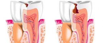
It is worth noting that in most cases, one of the most important stages in the treatment of pulpitis is the removal of the pulp or neurovascular bundle of the tooth. Many patients wonder: can a tooth hurt after treating pulpitis with this method? Despite the absence of a “nerve”, pain may be observed, this is a natural reaction to the intervention - removal of the pulp, treatment and filling of the root canals. It is important to know when it is normal and when pain is caused by a complication.
Natural pain after pulpitis treatment
Why does a tooth hurt after pulpitis treatment if there are no complications?
After the anesthesia wears off, pain may appear in the area of the causative tooth, and normally this condition is characterized by the following symptoms:
- the pain gradually subsides - an unpleasant sensation of a certain degree of severity may persist for some time, but it does not intensify;
- already 1–3 days after the procedure, the pain noticeably subsides;
- there are no other symptoms - bleeding, severe swelling and redness of the gums, increased body temperature, general weakness.
It is difficult to predict how much a tooth hurts after treatment for pulpitis; it depends on the individual characteristics, the specific tooth and the complexity of the root system, the presence or absence of concomitant diseases. The norm is that moderate pain persists for up to 7 days. Important: the intensity of pain becomes less over time.
It is difficult to talk about the norm if there are such “indicators” of complications as fever, swelling, too much pain, including increasing pain. There are several possible complications of endodontic treatment of pulpitis.
Cost of treatment
Dentistry Dentpremium is a center of expert medicine that provides professional services at affordable prices. For dental canal treatment, the cost is calculated taking into account several factors: the number of root canals, the method and materials used, the total volume and complexity of the specialist’s work.
On the clinic’s website you can see the price list, which indicates the average cost of root canal treatment. After an examination in the dentist’s office and receipt of diagnostic results, the attending physician will be able to announce what the final cost for root canal treatment will be.
On the website of the Dentpremium clinic you can find all the information you are interested in, make an appointment or a free consultation, and also read reviews about root canal treatment in our center. If you have any questions, you can contact us by phone.
Removing the filling material beyond the root apex
If, after treatment of pulpitis, the tooth hurts when pressed, perhaps we are talking about the removal of the filling material beyond the root of the tooth, into the adjacent tissues. The intensity of the sensation depends on how much material has gone beyond the apex of the root. Despite the fact that today a doctor has ample opportunities to control the accuracy of canal filling, as well as the use of high-quality materials, this can happen. It is worth noting that X-ray monitoring of treatment is intended to prevent the development of such a consequence - after the filling is completed, an image should be taken in which, in the event of a re-filling, the doctor will be able to notice excess material and choose the tactics for further action. Re-filling or removal of filling material beyond the root apex in itself does not pose a particular threat.
The exit of the filling material beyond the root apex can cause long-term pain immediately after the anesthetic wears off and persists for a long time - up to several months. This is a natural reaction of tissues to foreign material. Removal of material may be due to the following reasons:
- difficulties in determining the length of channels;
- incorrect selection of a pin from materials for the canal;
- lack of apical stop, etc.
However, this is not the only possible treatment difficulty that provokes pain.
Insufficient canal filling
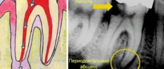
The opposite situation, in which a tooth may hurt after treatment of pulpitis, is insufficient filling of the root canals, the formation of voids at the apex of the root. In this case, often unpleasant sensations do not occur immediately, but as the inflammatory process develops - this can happen either after 1-2 weeks or after a longer period of time.
Here, painful sensations are associated with the proliferation of pathogenic microorganisms in the voids. This process leads to one of the diseases:
- periodontitis - inflammation of the peri-root tissues;
- tooth root apex cyst;
- granuloma.
Depending on the individual characteristics of the body, pain may appear immediately or make itself felt when the cyst or granuloma has reached an impressive size. Its intensity is often low, minor pain is more often observed when biting after treatment of pulpitis, and in the absence of mechanical impact on the tooth there is no discomfort at all.
Why does this complication occur?
Some patients find it difficult to imagine why dental material gets into the maxillary sinuses, and they often blame the doctor for this. However, the reasons for the appearance of this complication during treatment are usually completely different. Foreign material entering the maxillary sinuses occurs during the following dental procedures:
- Canal filling;
- Some methods of dental prosthetics.
To better understand the essence of the treatment process and see possible ways of penetration of dental material into the maxillary cavities, it is necessary to study the tooth filling scheme. Such a clear example will help you understand that if the layer of bone tissue between the gum and sinus is too thin, damage to the wall of the jawbone can occur. Dental material passes through this small hole. A similar complication usually occurs during dental treatment by a dentist. Another way for filling material to penetrate into the sinuses can be a hole formed when drilling the root canal of a tooth and jaw bone. A similar complication during root canal treatment occurs if the roots and paranasal sinuses are very close to each other. The third option for dental materials to enter the maxillary sinuses is bone tissue augmentation, which is carried out during dental prosthetics. Such additional intervention is necessary for high-quality tooth restoration, and when introducing materials for bone building, damage to the bone tissue may occur, through which pieces of dental preparations will fall.
How does damage to bone tissue in the maxillary sinuses occur?
Looking at the diagram of the anatomical location of the maxillary sinuses and upper teeth, you will notice that there is only a small area of tissue between them. The bones of the upper jaw are especially easily injured during the treatment of second premolars and first and second molars. The likelihood that such damage will occur increases in various pathological conditions and diseases:
- formation and suppuration of cysts in the upper jaw;
- osteomyelitis;
- dental injuries;
- periodontal acute inflammation of the maxillary sinus;
- process of installing prostheses;
- removal of a tooth in the upper jaw, complicated by a suppurative process of the tissues of the upper jaw;
- augmentation of bone tissue of the upper jaw;
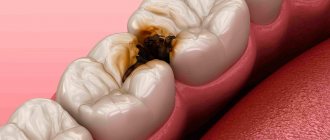
- caries complicated by pulpitis.
The severity of the complication associated with the entry of dental material into the maxillary sinuses is increased by the fact that bacteria from the oral cavity can also penetrate through the damaged opening. As a result, some microorganisms can provoke the development of severe purulent inflammation, which will lead to an even greater deterioration in the patient’s well-being and an increased risk of developing even more dangerous complications.
One of the root canals was not processed
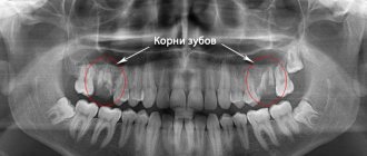
This is one of the rarest complications, since before starting endodontic treatment the doctor will definitely prescribe an x-ray diagnosis. It allows you to reliably determine the number of channels and evaluate their structure. But there are cases of abnormal location of root canals or their extremely small sizes, which makes them unnoticed in the image.
This leads to the following situation: the doctor removes the pulp in each canal, but one remains unattended - the inflamed pulp continues to hurt, and pathogenic bacteria continue to multiply. Such pain is difficult to confuse with other complications - the patient simply does not receive relief after treatment, and the pain characteristic of pulpitis itself remains. The sensations are pulsating in nature, the pain intensifies when eating, exposure to temperature on the tooth, and becomes unbearable at night.
The same symptoms can be observed with incomplete removal of the pulp in the diagnosed canal. In both cases, other symptoms may be observed:
- headache, sensations “radiate” to the ear, temple - depending on the specific tooth;
- increased body temperature;
- symptoms of general malaise.
Most complications are related to the complexity of the canal structure, and the following is no exception.
Instrument breakage in the root canal
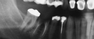
After treatment of pulpitis, the tooth may also hurt due to a broken instrument. Part of it remains in the root canal, and unpleasant sensations occur immediately or a few days after the visit to the dental office.
The nature of the pain may vary depending on the size of the fragment and other conditions.
It is worth noting that this complication is quite rare. This is explained by the fact that a good dental clinic uses modern, high-quality instruments from reliable manufacturers that are sufficiently durable. In addition, the rules for their use must be strictly observed, including multiplicity, force during exposure, etc. And finally, the doctor will notice the presence of a foreign body in the tooth on a control image, which he will prescribe after treatment.
Why is this complication dangerous?
The maxillary sinuses located above our upper jaw (their area extends from the inner corner of the eye to the outer) perform important functions. They can warm the air and thereby protect the respiratory tract from hypothermia and foreign particles. The sinuses are located under the bone tissue and if its thickness is too small, when cleaning the dental canals, filling masses can fall into these cavities. Once the filling is completed, it is no longer possible to remove the foreign material on your own, since this procedure can only be carried out by a specialized specialist who has the necessary tools. The doctor can perform this procedure without difficulty and it will not take much time. It is numbed with local anesthesia and after removing the foreign body from the maxillary sinus, the patient will need to stay in the clinic for several hours. In some cases, discharge occurs the day after this minimally invasive procedure.
Root perforation
Perforation is the creation of an artificial root hole. As a rule, in this case, the pain is sharp, unbearable and makes itself felt immediately after the anesthetic stops working. This can happen during instrumental processing of canals - preparing them for filling.
As in most other cases, the structure of the root system plays an important role here. Narrow, curved canals are a common cause of perforation. In this case, the tool does not move along the canal, but into the root wall.
If a tooth hurts for this reason after treatment of pulpitis, the symptom may be accompanied by bleeding. In addition, some patients report pain even with current anesthesia - subjectively it is perceived as the feeling of an injection into the gums. If the tooth was filled after perforation, the filling material may leak beyond the root. Severe pain persists for up to 3 weeks, and the main complication of this phenomenon is the inflammatory process.
Allergy
This complication of pulpitis treatment is easier to differentiate - an allergic reaction is often accompanied by tissue swelling. If it is a consequence of material moving outside the root canal, there may be swelling of the gums around the treated tooth. In some cases, it spreads to other areas - cheek, lip, depending on the specific tooth.
When pressure is applied, the pain becomes stronger, it is difficult to relieve with painkillers, and over time it only intensifies.
What to do?
What to do if your tooth hurts after pulpitis treatment? The most important recommendation if you suspect a complication after treatment of pulpitis is a return visit to the doctor. If this is not possible, you should contact another specialist and explain the situation.
If it is not possible to visit the clinic immediately, you can use a painkiller - give preference to the one that you have already taken, to which there have been no negative reactions. However, it is better to try to consult with a specialist, if not in person, then by phone.
It is difficult to say exactly how quickly the pain will subside in each case. However, on average, after visiting the dentist, relief comes quite quickly:
- When removing the filling material beyond the root apex: a visit to the doctor will allow the specialist to remove excess material, and after 2-3 days the discomfort will subside.
- If the filling is insufficient: the doctor will choose a tactic for further treatment; it is quite possible that several visits will be needed to eliminate the inflammatory process - for larger cysts or granulomas, surgical treatment is also required. This complication requires refilling to prevent recurrent complications.
- If the pulp or part of it is intact: the doctor will remove the remaining pulp and repeat the manipulations that were required to treat pulpitis. The pain in this case goes away within a few days after all measures are completed.
- If an instrument breaks off: the doctor will perform an X-ray diagnostic and, if the presence of a broken instrument is confirmed, will remove it. In rare cases, resection of the root apex is performed.
- In case of perforation: the dentist will treat the canals without affecting the hole. Using modern osteoplastic materials, he will close it, which will save the tooth. If a purulent process occurs, several visits will be required to eliminate the inflammation.
- In case of gum injury: contacting a doctor will allow you to receive recommendations regarding antiseptic and healing agents for treatment at home. Relief occurs on average within 1–3 days.
- In case of an allergic reaction: it will be necessary to refill the root canals using other materials. The doctor will perform the necessary manipulations and also prescribe decongestants that will quickly eliminate unpleasant symptoms.
Modern methods of treatment
Highly qualified and experienced specialists at the Dentpremium Dentistry Center use exclusively modern methods of treating inflamed tooth canals.
- Therapeutic or biological. This method allows you to save part of the fabric or the entire remote control. The biological method is effective only at the initial stage of the disease. Treatment consists of placing an anti-inflammatory drug into the pulp chamber with a temporary filling, or applying antibiotic dressings to allow the drug to flow through the dentin.
- Pulp extirpation. Treatment involves removing absolutely all neurovascular tissue, cleaning the cavity, treating with antiseptics and sealing. For treatment, filling, injection of gutta-percha (thermoplasticized) or filling with thermophiles are used.
In the most severe cases, when there is a cyst, granuloma or other formation, resection of the root apex is performed.
What not to do?
If, after treatment, a tooth hurts when pressed or without mechanical action, you should under no circumstances resort to folk remedies such as heating, hot compresses, heating pads - if there is an inflammatory process, this can greatly worsen the condition. In case of allergic reactions, heat will also increase swelling.
It is also not recommended to use folk remedies for rinsing, which can cause a burn to the mucous membrane - iodine, tinctures of alcohol or vodka, liquids with the juice of “scorching” plants, etc. Even if there is no gum damage, such measures can worsen the situation.
It is also not worth taking various painkillers uncontrollably - firstly, you need to see a doctor in any case, and the effect of analgesics will not allow you to fully evaluate the clinical picture. Secondly, it can be dangerous to health.
It is important to visit a doctor at the first opportunity and perform all the necessary procedures to eliminate unpleasant consequences and improve the condition.
Classical treatment methods
Dentistry has long been dealing with the problem of root canal inflammation. Treatment was carried out using two methods:
- opening the tooth chamber, placing arsenic paste, removing it after the tissue has died, and filling;
- opening the dental chamber, placing a resorcinol-formalin mixture, which blocked the inflammatory process, filling.
There are classical and modern treatment methods. When comparing, it can be clearly noted that classical therapy has a number of disadvantages:
- both compositions have a toxic effect on the body;
- risk of incomplete pulp death;
- risk of residual infection and relapse;
- painful procedure.
These methods are now practically not used, since modern means and drugs make it possible to completely clean the inflamed cavity and destroy the infection painlessly, with minimal trauma and without the risk of relapse.