Indications for installation of a cystostomy
A drainage tube is inserted in the following cases:
- Diagnosis of prostate adenoma, which, having increased in volume, begins to put pressure on the urethra, blocking the path for the free movement of urine. There are cases when the urethra narrows to such a size that it is impossible to insert a classic catheter into it. In this case, a cystostomy is performed as an emergency.
- If there is a need for long-term catheterization in people with an active intimate life.
- If the urethra is damaged, such injuries are often characteristic of difficult childbirth.
- During rehabilitation after certain gynecological and surgical procedures.
- When the patient is prescribed regular catheterization, which can cause various damage and contribute to infection. To eliminate such risks, the patient has a cystostomy installed.
- During the treatment of complex diseases of the genitourinary system that are bacteriological in nature, including scrotal gangrene, prostatitis caused by bacteria.
- Surgeries on the urethra or penis.
- When diagnosing benign prostatic hyperplasia.
- For pathologies of the patency of the bladder neck.
Cystostomy is often required in case of disturbances in the innervation of the bladder after a stroke, neurogenic dysfunction of the organ, or damage to the spinal cord or brain. The tube is also installed in people with psychiatric diagnoses who are not able to independently control the emptying process.
The procedure is also carried out when diagnosing neoplasms in the bladder area, which, due to compression of the organ elements, cause a narrowing of the lumen, which causes problems with urination. A cystostomy is installed in case of purulent processes in the ureter and bladder in order to carry out lavage. Drainage is used when the urethra is blocked by stones or other foreign bodies.
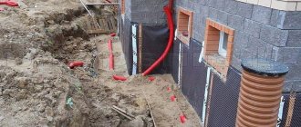
Suction drainage from the pleural cavity
Suction tube
For suction drainage of the pleural cavity, various rubber and synthetic tubes are used.
For the most commonly used drainage, a rubber tube about 40 cm long with several side holes at the end is used. This tube is placed along the lung (from the base to the apex) and passed over the diaphragm from the pleural cavity to the outside. The drainage is attached to the skin with a knotted U-shaped suture. When the suction drain is removed, the threads are tied again, thereby sealing the hole in the chest. A three-barrel suction catheter (Viereck) is advantageous, allowing free passage of the tube inserted inside.
Insertion of suction drainage
In the chest between the two pleural layers, the intrapleural pressure is lower than atmospheric pressure. If air or liquid gets between the pleural layers, then the normal physiological state can only be restored by long-term suction drainage. A closed drainage system is used to suction pleural fluid for recurrent pneumothorax and to treat empyema. This drainage is now usually inserted into the intercostal space through a trocar. The thickness of the drainage tube is determined in accordance with the consistency of the substance being sucked out (air, as well as watery fluid or serous, fibrinous, bloody, purulent fluid).
On the drainage, mark with paint or thread the place to which it will be inserted. The size of the trocar must correspond to the size of the drainage. It is advisable to have at least three trocars of different sizes with suitable tubes of 5, 8 and 12 mm in diameter. Before inserting the trocar, you must make sure that the selected drainage tube passes through it easily.
The site of the skin incision is filtered with novocaine to the pleura. A test puncture in the designated area makes sure that the desired air or liquid is really there. The assistant gives the patient the necessary position: the patient must sit and lean on the highly raised operating table so that the puncture area protrudes as much as possible, and the selected intercostal space is, if possible, expanded. A scalpel is used to cut the skin over an area slightly larger than the size of the trocar. Then the trocar is inserted with a strong movement along the upper edge of the rib into the pleural cavity. After removal of the trocar, unimpeded release of fluid or free entry and exit of air indicates its correct insertion. Drainage is performed and the trocar tube is removed. If you are not convinced that the drainage is in the right place, you should, in order to prevent the trocar from puncturing the lung, heart or large vessel, perform the puncture again, taking all measures to localize it under X-ray control.
Before closing each thoracotomy hole, a drainage is introduced into the pleural cavity, which is brought out above the diaphragm through a separate hole in the intercostal space. Through a hole measuring about 1-2 cm, a forceps is inserted into the pleural cavity under the control of the eyes and under the protection of the left hand to ensure the correct position of the drainage from the inside. The drainage is pulled through the chest wall with a forceps from the inside to the outside. Pay attention to the fact that the drainage section, free from holes, is at least 5 cm in the chest cavity. If the fixation of the drainage to the skin is broken, then it slips out, and the first side hole appears outside the pleural cavity above the skin. In this case, the closed system turns into an open one, suction becomes ineffective, and pneumothorax often occurs.
Suction systems
There are so-called individual (“bed side”) and centralized suction systems. The suction action due to the hydrostatic effect can be obtained by a tube lowered under water, a water or gas pumping device (in this case the action is based on the valve effect) or an electric pump. Both individual and central systems must ensure individual regulation. If the release of air from the lung is insignificant, then due to its simplicity, the Biilau drainage system is still successfully used today, which can be sufficient to straighten the lung. A glass tube immersed under water (disinfectant solution) is equipped with a valve made from a finger cut off from a rubber glove, which protects against reverse suction. The Biilau system uses the physical law of communicating vessels to move bottles under the bed to create a suction effect.
The Fricar air pump best meets modern requirements. This device can work for many days without interruption and without heating up. The strength of the suction effect can be precisely adjusted.
Central suction devices are triggered by an oxygen canister system or a powerful suction pump. The system of outgoing tubes, if necessary, supplies hospital departments located on different floors. Depending on the need, the required number of hospital beds can be connected. An oxygen-powered system has the advantage that the suction and supply of oxygen to individual hospital beds is provided by the same tubing system. The suction effect is provided by a valve tube mounted along the oxygen flow. In this case, however, the effect produced by the central suction pump is not achieved.
Individual adjustment can be carried out using a dosimeter tap connected to a well-functioning pressure gauge, or through the so-called. three bottle system. The latter can be easily prepared by yourself. This system also has the advantage that it can easily and reliably create a very low suction effect (from 10 to 20 cm of water column). It is rarely possible to achieve such low pressure values using factory pressure gauges.
Indications for suction drainage
Spontaneous and traumatic pneumothorax, hemothorax
Spontaneous pneumothorax occurs at a young age, more often as a result of rupture of single pulmonary alveoli in the apex of the lung, in older people - as a consequence of rupture of alveolar vesicles with diffuse emphysema. Due to the fact that the number of patients with emphysema is constantly increasing, the number of cases of spontaneous pneumothorax is becoming more frequent. The same applies to traffic accidents that result in closed injuries in the chest cavity, which often occur with pneumothorax or hemothorax.
Correctly performed pleural puncture for spontaneous pneumothorax is practically safe, and its benefits can hardly be disputed. If the flow of air from the damaged lung is completely stopped and the perforation site is closed, then it may be possible to completely remove the air that created the pneumothorax with a simple closed puncture. If pneumothorax recurs after puncture (even repeated), then drainage with long-term suction should be used. Recurrence of pneumothorax, even after prolonged drainage with suction, can only be reliably eliminated by surgery.
Traumatic pneumothorax most often results from rib fractures. When a rib fragment injures the lung, most often a significant amount of air comes out of it, and a tension pneumothorax occurs. At the same time, subcutaneous or even mediastinal emphysema may occur. Spontaneous pneumothorax can also occur when the pulmonary alveoli rupture or due to blunt force on an emphysematous lung. Therefore, in patients with pulmonary emphysema, chest injuries are often associated with the occurrence of pneumothorax, often severe tension pneumothorax. The principles of treatment for spontaneous and traumatic pneumothorax are the same.
If clinical symptoms indicate tension pneumothorax (severe respiratory failure, subcutaneous emphysema, mediastinal shift), then drainage of the pleural cavity should be performed immediately. If these symptoms are not present, then a closed puncture is performed and the air is sucked out. After this, the needle is left inserted into the pleural cavity, and its nozzle is connected to a pressure gauge and the pressure in the pleural cavity is determined (whether it is higher or lower than atmospheric). If the pressure in the pleural cavity is indicated by the pressure gauge needle in the positive direction, it means that air continues to be released into the pleural cavity, and, therefore, drainage is necessary. This issue can, of course, be resolved by X-ray examination. If there is a total pneumothorax, then drains are inserted in two different places. One of them runs along the posterior axillary line above the diaphragm in the VII-VIII intercostal space, the other is inserted along the midclavicular line between the 1st and 2nd ribs. In our experience, drainage inserted under the collarbone performs the task of straightening the apex of the lung better.
In case of encapsulated limited pneumothorax, drainage should be inserted locally, under X-ray control after a test puncture.
Empyema of the pleura
Pleural empyema is a disease for which treatment with suction from the pleural cavity is absolutely indicated.
The principle of treatment of empyema does not depend on the causative agent of the disease. It consists of gluing the pleural layers and eliminating the empyema cavity through early drainage and suction of fluid. Treatment with suction from the pleural cavity is combined with targeted local chemotherapy, based on the identification of the pathogen and its resistance to the drugs used. Most empyemas result from infection of the exudate. In this case, incorrect and insufficient suction from the pleural cavity plays a certain role. In cases where pockets with delimited fluid form in the pleural cavity, their complete emptying becomes increasingly difficult, more difficult, and infection is more likely. In such cases, complete recovery can only be ensured by surgery.
Suction treatment may fail for two reasons: one is the presence of pleural cords, the other is a bronchopleural fistula.
Pleural moorings are often the result of insufficient emptying of the pleural cavity. When moorings have already formed in the pleural cavity and the walls of the empyema cavity are thickened, there is little chance of eliminating the empyema by suctioning out the fluid. The ability to expand the lung in this case is also very controversial. In this case, drainage with suction is a preparatory measure before the inevitable operation. Radical surgery (decortication) is performed only after the patient’s general condition has improved by washing the pleural cavity and targeted antibiotic therapy.
Bronchopleural fistula reduces the effectiveness of suction and thereby the prospect of lung expansion. In cases where there is a large bronchial fistula and its closure is contraindicated (for example, a rupture of the cavity, tumor disintegration, rupture of a cystic, emphysematous lung that has lost its elasticity), success cannot be expected from the use of suction. On the other hand, suction can also be used in cases where surgery is indicated. In elderly patients, with low general resistance and the possibility of severe complications, surgery becomes impossible. Then all that remains is to leave the patient with permanent drainage.
In case of chronic pleural empyema, drainage should be inserted into the pleural cavity at its lowest point. Large-diameter drains are used so that the thick liquid does not close the lumen and it is easy to wash the pleural cavity. Often, in the area where the drainage will be introduced, a rib resection (2-3 cm) is performed.
Postoperative suction from the pleural cavity
In order to remove fluid that accumulates after thoracotomy from the pleural cavity and maintain normal intrapleural pressure, a suction drain should be available.
If during pleural operations and mediastinal, transthoracic interventions on the esophagus, stomach, heart and large vessels there was no damage to the lung, then the chest can be closed with the introduction of one perforated drainage into the pleural cavity. Drainage is carried out above the diaphragm along the midaxillary line with its pleural end installed at the level of the apex of the lung.
Two drains are inserted into the pleural cavity if the lung was damaged during separation of adhesions, as well as after resection or excision of lung tissue. In such cases, one of the drains is inserted along the anterior and the second along the posterior axillary line. The use of a third drainage may be considered relatively appropriate when it is brought to the site of anastomosis of the esophagus or bronchus or when thoracoplasty is performed in combination with lung resection (for suction from the subscapular space).
After removing the lung, one drain with a diameter of 12-15 mm is inserted into the pleural cavity and placed in the lower part of the cavity so that a piece of drainage 10-12 cm long is equipped with 2-3 side holes. Active suction through this drain is prohibited.
After median sternotomy, a drainage is inserted retrosternally and its second end is brought out in the epigastrium.
Intensity and duration of suction
The intensity of suction through drainage from the pleural cavity depends on the cause of the disease, the condition of the lung and the nature of the operation. The flow of air from the lung into the pleural cavity is of decisive importance. If this occurs, then more air should be sucked out of the pleural cavity per unit time than is supplied there. Only in this way can gluing of the pleural layers be achieved. In practice, however, this is often not feasible. If the connection of the bronchus with the pleural cavity is significant (for example, in the case of a bronchial fistula), then intensive suction cannot achieve the goal. If you increase the suction force, then at the same time the patient will experience increased respiratory failure due to “air theft” from the tidal volume. Despite this, the lung will not be able to expand. In such cases, surgery is inevitable.
When the lung is damaged or after lung surgery, air most often escapes from a hole the size of a pinprick. In this case, specialized suction is indicated. In children and adolescents, due to the fact that their lung parenchyma is healthy and not affected by fibrosis and emphysema, it does not matter with what force the suction is performed. It doesn’t matter whether they suction with an intensity of 25 cm of water. Art. or simple underwater drainage, the lung will expand in 24-48 hours. The drainage can be removed after 48-72 hours. This is the advantage of elastic tissue capable of lung retraction in young patients. With emphysematous lung in an elderly person, the situation is different. The pinprick holes become gaping holes in the lung because the surrounding tissue is unable to contract. If you try to reduce the flow of air coming from the damaged lung by increasing the intensity of suction, you can easily get a paradoxical effect. The flow of air from the lung will increase. Small holes, due to prolonged suction, stabilize and turn into fistulas.
What to do in such cases? Begin gentle suction from the pleural cavity (5-6 cm of water column) and pay attention to ensure that tension pneumothorax does not occur. Thanks to this, the resulting fibrin seals small holes in the lung. Within 24 hours, a decrease in the release of air from the damaged lung begins to be detected. The intensity of suction can be increased slightly. On the fourth day you can already suction with an intensity of 10 cm of water. Art., if no unforeseen complications arise, then the drainage can be removed on day 4-5.
The same principles are followed when treating spontaneous and traumatic pneumothorax with suction.
If there is a significant intake of air from the emphysematous lung, they begin to carefully perform suction with a gradual increase in its intensity. If, after many days of treatment with suction, the release of air from the lung does not stop, then it is recommended to immediately undertake surgery, without waiting for the development of infection in the pleural cavity. If suction from the pleural cavity continues for more than a week, the development of infection becomes real.
In cases where the patient does not undergo surgery due to low general resistance, suction from the pleural cavity remains to be continued. Prolonged and specialized suction under the guise of drug treatment may be more or less effective. The pleural layers are glued together completely or partially. Only small limited cavities remain that do not lead to complications. The drain can be removed.
In the treatment of pleural empyema, long-term use of suction drainage is a common method. The empyema cavity gradually becomes smaller and smaller, the amount of fluid decreases, and in the end it can become bacteriologically sterile. If the daily amount of fluid extracted from the pleural cavity does not exceed 10-15 ml, then the suction is stopped, the drainage is shortened, but left until the residual cavity is completely closed.
Contraindications for cystostomy placement
There are two types of contraindications to the installation of a urinary drainage tube: absolute, relative. It is strictly forbidden to perform cystostomy if:
- displacement of the bladder, which does not correspond to the standard physiological localization and is manifested by the inability to carry out full palpation even with maximum filling of the organ;
- with unclear contours of the urinary tract, which are determined by ultrasound;
- diagnosing MP oncology.
Relative contraindications include the following factors:
- surgery performed in the lower abdominal area or pelvic area;
- blood clotting disorder;
- the presence of various implants and orthopedic structures in the pelvic area.
The attending physician can independently determine the reasons that can worsen the patient’s condition if a drainage tube is installed.
Drainage for acute appendicitis
In 1979, O'Connor and Hugh, in an excellent review, concluded: “Intraperitoneal drainage is of little value in phlegmonous, gangrenous, or perforated appendicitis. However, drainage is indicated if there is a limited purulent cavity or a gangrenous stump that is imperfectly closed” (1).
I will not overwhelm you with details of all the available literature, since Petrowsky et al. recently released an excellent analysis of these studies (3). After presenting individual studies, including their own meta-analysis, the authors concluded that “drainage does not reduce the incidence of postoperative complications, and even appears to be harmful in terms of the formation of intestinal fistulas (the latter was observed only in drained patients). Drainage should be avoided in any form of appendicitis” (4).
Drainage after appendectomy for phlegmonous and gangrenous appendicitis is not needed. Most surgeons who took part in the survey understand this. What is said about perforated appendicitis with local formation of a purulent focus? Of our respondents, 22% will install drainage. As will be shown below, a “formed” or “unopened” abscess, according to most surgeons, is a good indication for installing drainage. But an abscess against the background of perforated appendicitis is not “unopened”: after the surgeon destroys its wall and evacuates the pus, the potential abscess space is filled with nearby loops of intestine, mesentery and omentum. Thus, the source of infection is removed, the abdominal cavity is cleaned by toileting. The peritoneal defense mechanism is then activated, supported by a short course of antibiotics, with complete eradication of bacteria without the presence of an irritating foreign body (4).
Uncertain closure of the appendiceal stump as a justification for drainage seems anachronistic. Safe closure is possible even in rare cases where perforation occurs at the base of the appendix, by placing a suture or stapler on the dome of the cecum.
Of our respondents, 23% use drainage for appendicitis complicated by diffuse peritonitis. However, as will become clear later, these are the same surgeons who advocate drainage for generalized intra-abdominal infection. And drainage in this situation - after controlling the source of infection - seems useless.
Advantages and disadvantages of cystostomy
The advantages of a cystoma include the following indicators:
- installation of a tube through the abdominal cavity does not cause injury to the bladder neck or urethra, unlike a classic catheter;
- after removal of the cystostomy, the incision heals quite quickly, the main thing during this period is to follow the doctor’s recommendations and carry out proper treatment;
- if the tube is clogged, urine can always be removed through the urethra;
- You can replace a cystostomy yourself, at home, although this is not recommended; it is better to seek help from the specialists of the “Your Health” clinic, who will carry out the procedure as safely as possible, without developing negative consequences;
- the catheter has a short duration of action, and the cystostomy can be installed for a long period, without the need for regular removal and reinsertion.
A cystostomy allows you to have a normal sex life and is quite easy to care for. The design itself is characterized by high reliability indicators.
Despite the large number of advantages, cystostomy has a number of disadvantages:
- the procedure is not recommended for obese patients, since care of the device in this case can be seriously complicated;
- the tube may cause bladder spasms;
- urine may leak in the area where the urinary element is installed, causing irritation and increasing the risk of inflammation;
- After a certain time, the tube must be replaced with a clean one.
In some cases, after removal of the cystostomy, it is necessary to treat the fistula. When installing a tube, the body may reject a foreign body. There is also a risk of spontaneous release of the device from the hole.
Moshe Shine
The history of abdominal drainage is as old as surgery itself (1). However, abdominal drainage is still a subject of debate and constant discussion. Even 100 years ago there were passionate proponents of drainage, like Robert Lawson Tait (1845-1899), who said: “When in doubt, drain!” There were also skeptics, like JL Yates (1905), who said: “Drainage in general peritonitis is physically and physiologically impossible”! There were also those like Joseph Price (1853-1911): “There are people who ardently defend drainage, and there are those who categorically deny it. Both are right in their own way.”
100 years have passed, during which operative surgery and supportive care have progressed continuously. But what about drainage? Are there fewer discussions and contradictions today? What awaits drainage tomorrow?
In this short post I try to answer these questions from the perspective of drainage for abdominal and abdominal infections. Elective surgeries will be mentioned only as part of the discussion. Percutaneous drainage, both primary and postoperative, is outside of our discussion.
How to prepare for surgery
Cystotomy does not require complex preparation; sometimes the operation is performed as an emergency, which generally excludes any preliminary measures. In most cases, surgical intervention is limited to the minimum collection of necessary data. Recommendations for preparation are most often based on general indicators of the patient’s condition, individual characteristics of the body, the nature and course of the disease.
Before the operation, the patient is prescribed laboratory and instrumental tests:
- general, biochemical blood test;
- X-rays of light;
- determination of the quality of blood clotting;
- ultrasonography;
- blood group and Rh factor analysis;
- analysis for HIV, hepatitis, syphilis.
14 days before surgery, you must stop taking blood thinning medications that can cause increased bleeding. Patients with diabetes must definitely visit an endocrinologist, who will adjust the current therapy to suit the current situation.
In some cases, antibiotics may be prescribed to prevent infection.
Before cystostomy, the patient needs to fill the bladder as much as possible by drinking large amounts of clean, still water. Such manipulations will help increase the efficiency of palpation and improve the image during ultrasound. If a cystostomy is installed due to urinary retention, there is no need to fill the bladder.
The operation can be performed either on an outpatient basis or in a hospital. The “Your Health” clinic offers to install a urological device in the comfort of your home, without the need to visit a medical center.
Answers to frequently asked questions about cystostomy:
- Calling a urologist to your home to replace a cystostomy
- The urological catheter is clogged
- Replacing a cystostomy at home
- The cystostomy is clogged
- How to properly rinse a cystostomy?
- How to remove a cystostomy?
- Which doctor will help with a cystostomy?
- Where to go for a cystostomy?
- Washing a cystostomy at home
- How to prepare for an appointment with a urologist?
- How to get tested for urological diseases?
- What tests can be done by a urologist?
- What diseases does a urologist treat?
- What symptoms should you consult a urologist for?
- What diagnostics can a urologist perform in your clinic?
- How to call a urologist at home?
Drainage classification
Drains are installed for therapeutic or preventive reasons.
Medicinal:
- To ensure the outflow of intra-abdominal fluid or pus (periapendicular abscess, diffuse peritonitis).
- To control the source of infection if it is impossible to remove it by other, radical methods; for example, with an external intestinal fistula (duodenal stump).
- Preventive:
- To prevent recurrence of infection - to evacuate residual serous fluid or blood, prevent the formation of an abscess.
- To monitor expected or probable leakage from the suture line (colic anastomosis, duodenal stump, cystic duct).
- To report complications (in the hope that drainage will work in case of bleeding or leakage of chyme from the anastomosis).
It is better, rather than discussing a rigid classification, to look at the problem of drainage through the eyes of a general surgeon. What are the common tactics? What is the practice for general surgery?
The literature is a poor source of information about how common abdominal drainage is in emergency surgery. Analyzing several publications from individual clinics or collective reviews on drainage for specific conditions, we cannot draw a conclusion about the dominant trends. Therefore, we present the opinions of general surgeons - members of SURGINET - and the results of an international Internet discussion (2) regarding their views on abdominal drainage. Of the 700 members, only 70 responded. Although this is quite small, it correlates with the frequency of Internet responses received for any online survey.
The survey involved 71 respondents, all general surgeons, many of them not academic specialists, but earning their bread and butter through practical work, from a total of 23 countries. Most of all (14) are from the USA, and in total from North America - 18, Western Europe - 10, Eastern Europe - 7, Asia - 15, including Israel and Turkey; Latin America – 15, Australia and South Africa – 3 each.
Surgeons who are active in the Internet survey are, as a rule, more interested, active, and familiar with the literature and modern practice in other activities. The survey results reflect inconsistencies and geographic differences in their surgical practices.
Common situations where drainage may be used
How is the operation performed?
Cystostomy is performed using two methods:
- in an open way;
- by trocar method.
The latter technology is more preferable, as it is minimally invasive and has minimal risks of complications. The second technique requires special stationary conditions.
Trocar cystostomy is a temporary measure that is often used in emergency cases when the bladder needs to be drained urgently. The trocar is made in the form of a hollow tube, which is complemented by an internal rod designed to pierce the skin. Using this design, a cystostomy is delivered to the MP area. Very often, this method is used for patients with chronic urine retention caused by prostate hyperplasia.
Trocar transportation of the drainage element cannot be carried out if there are adhesions in the area of the prevesical space, if stones, mechanical damage, or inguinal-scrotal hernia are detected. The procedure is also contraindicated in case of sclerosis of the bladder and protrusion of the organ.
The operation consists of the following steps:
- treating the skin in the area of cystostomy with an antiseptic;
- infiltration of soft tissues with painkillers;
- making an incision up to 1 centimeter in size through which the device will be inserted;
- piercing the wall of the bladder and inserting the end of the tube into the bladder;
- removal of the trocar;
- fixation of the cystostomy and treatment of the incision area with an antiseptic drug.
A bag is attached to the structure to collect the discharged liquid. To collect urine for laboratory tests, a specialist uses a cannula with a stopper located on the side. Using special divisions marked on the container, the doctor can determine diuresis for a certain period of time.
Cystostomy is often used for mechanical removal of dead tissue, small stones, and pus. The procedure is prescribed for cystitis of various natures.
Open cystostomy is used quite rarely. During the operation, general or spinal anesthesia is used.
Complication during the procedure
Cystostomy may be accompanied by the following complications:
- increased sensitivity and pain in the area where the tube is installed;
- suppuration and infection in the area where the device is inserted;
- inflammatory processes in the bladder;
- allergic reactions;
- injury to blood vessels;
- development of cystitis;
- bleeding;
- damage to some intestinal segments;
- development of acute pyelonephritis, prostatitis.
To avoid negative consequences, it is important to seek help from qualified, experienced specialists and carefully monitor your health, following all doctor’s instructions.
Prevention of postoperative complications
Prevention is primarily aimed at eliminating the development of complications, as well as restoring the storage function of the bladder. To do this, the patient must regularly carry out special training of the organ, which helps not to lose the contractility of the organ walls. The continuous outflow of urine disrupts the normal functioning of the bladder, so it is important to periodically fill the bladder.
The classes consist of the following simple exercises:
- the device is pinched until the urge appears;
- after the desire to urinate arises, the tube is released.
It is recommended to carry out training under the strict supervision of a doctor, after preliminary consultation. It is also worth knowing that such exercises are not recommended for all patients. Patients with vesico-paromintestinal fistulas, urethral injuries, inflammation, and bleeding are therefore strictly prohibited from exercising.
How to care for a cystostomy
Caring for the drainage device is based on general rules:
- it is important to constantly ensure that the urinal tube is not bent or twisted, and that the structure is free of cracks, ruptures and other defects;
- it is necessary to take seriously the condition of the skin around the cystostomy, the surfaces must be clean, without signs of inflammation, in order to exclude any negative manifestations, the skin must be treated with antiseptics;
- the urine bag should be below the bladder;
- To maintain personal hygiene, a shower is recommended; taking a bath is strictly contraindicated; you should also not visit the pool, sauna, or bathhouse;
- the patient is obliged to maintain a drinking regime and drink at least 1.5 - 2 liters of clean, still water per day;
- It is forbidden to independently introduce any solutions into the cavity of the MP; this should only be done by a doctor with the appropriate qualifications;
- if the catheter is clogged or obstructed, it is important to urgently contact a specialist; only a person who has undergone special training can carry out independent replacement;
- it is important to constantly monitor the filling of the urine collection container, avoiding overflow;
- The cystostomy catheter must be changed once a week, the urinal container must be emptied every 3-8 hours, depending on the volume of the container.
All manipulations associated with the drainage device must be performed with thoroughly washed hands wearing sterile gloves. The skin located in the area of the tube must be treated daily.
For home care of a cystostomy, it is recommended to keep in the first aid kit:
- several urinal containers;
- antiseptic solution: miramistin, iodine, alcohol, hydrogen peroxide;
- sterile gloves;
- new drainage.
Acute appendicitis
In table Figures 1 and 2 summarize the surgeons' responses. “Simple” or “phlegmonous” appendicitis was excluded. Only complicated forms are discussed: when the appendix is black, there is usually some fluid in the pelvis, but it is not obvious pus. As follows from the table. 1, only one respondent drains the cavity in this situation. Next, when the appendix is perforated, a laparotomic or laparoscopic surgeon removes the appendix and sucks out the pus present around the appendix. In these cases, the surgeon can destroy the barrier of the omentum or loops of the small intestine, exposing a small abscess of a few cubic millimeters, and the pus is also aspirated. Table 2 shows that 80% of respondents do not install drainage in this situation. However, there is no geographical pattern. Table Figure 3 illustrates advanced cases where perforated appendicitis is combined with a “pus everywhere” situation - in the pelvis, along the right lateral canal, even in the upper floor. And, although 80% of surgeons do not install drainage in this situation, there is a geographic dependence: they never or almost never drain the abdominal cavity in North and Latin America, and quite often in the Asian world. This depends on how the surgeon views the role of drainage in the treatment of diffuse peritonitis; complications will be discussed later.
Table 1. Do you place drainage after appendectomy for gangrenous appendicitis?
| Quantity | No | Yes | |
| North America | 18 | 18 | |
| Western Europe | 10 | 10 | |
| Eastern Europe | 7 | 7 | |
| Latin America | 15 | 15 | |
| Asia | 15 | 14 | 1 |
| Australia | 3 | 3 | |
| South Africa | 3 | 3 | |
| Total | 71 | 70 (98%) | 1 |
Table 2. Do you place drainage for perforated appendicitis, when there is little pus and it is present locally?
| Quantity | No | Yes | |
| North America | 18 | 14 | 4 |
| Western Europe | 10 | 8 | 2 |
| Eastern Europe | 7 | 6 | 1 |
| Latin America | 15 | 11 | 4 |
| Asia | 15 | 12 | 3 |
| Australia | 3 | 3 | 0 |
| South Africa | 3 | 2 | 1 |
| Total | 71 | 56 (78%) | 15 |
Table 3. Do you place drainage for perforated appendicitis with diffuse spread of pus?
| Quantity | No | Yes | |
| North America | 18 | 18 | — |
| Western Europe | 10 | 8 | 2 |
| Eastern Europe | 7 | 5 | 2 |
| Latin America | 15 | 14 | 1 |
| Asia | 15 | 7 | 8 |
| Australia | 3 | 2 | 1 |
| South Africa | 3 | 1 | 2 |
| Total | 71 | 55 (77%) | 16 |
Cleaning the cystostomy at home
Urological manipulation provides care for the cystostomy, which after some time of operation forms mucus, tar deposits and other contaminants that increase the risk of inflammation, infections and narrow the lumen, which negatively affects the quality of urine output.
To eliminate all unpleasant moments, you need to periodically flush the catheter. The procedure is recommended approximately twice a week and it is advisable that the service is provided by a qualified specialist.
Washing is carried out according to the following algorithm:
- the lower part of the patient is placed on a waterproof surface;
- the doctor performs hygienic measures in the patient’s genital area;
- a large-volume syringe is filled with furatsilin, boric acid or other disinfectant solution;
- the tube is disconnected from the urinal and connected to the outlet of the syringe;
- no more than 50 cm3 of rinsing agent can be administered at a time;
- When you disconnect the syringe, the tube tilts towards the drain tank.
The procedure is carried out until the liquid becomes clean and transparent. After completion of the event, the area where the drainage device is installed is treated with an antiseptic and covered with sterile material. A urologist will help you perform the rinsing; you can call him by dialing the phone number of the “Your Health” clinic.
Removing a cystostomy at home
The drainage tube is removed after complete completion of treatment, after the doctor gives official permission to do so. To remove the device, you need to call a specialist to your home.
The cystostomy is removed as follows:
- the doctor first assesses the condition of the fistula; if the situation is positive, any signs of an inflammatory process should be excluded in this area;
- the area around the incision is treated with an antiseptic solution;
- the urine container is disconnected;
- the tube is compressed with a special clamp, which prevents the outflow of urine;
- The cystostomy is carefully removed.
After all the main actions have been carried out, the skin is re-processed and a sterile bandage or other similar material is applied to the resulting hole. The incision heals spontaneously; in some cases, medical suturing is required.
Replacing a cystostomy at home
For the procedure, it is necessary to prepare in advance the following new, sterile materials:
- saline;
- a new catheter and urine collection container;
- antiseptic, it is recommended to use chlorhexidine;
- bandage;
- medical gloves;
- gauze, alcohol wipes;
- syringe.
The replacement of the drainage device should be carried out by a doctor and only in extreme cases can the action be entrusted to the patient. The tube is changed once a month, in some cases such manipulations are necessary a week after installation of the catheter. Replacing a cystostomy is similar to the previous procedure, only after the tube is removed, a new structure is installed in its place.
Replacing a urine bag at home
The algorithm for replacing a urinal is as follows:
- the patient is placed on an oilcloth covered with a diaper;
- wearing sterile gloves, disconnect the urine receiver tube from the drainage device;
- urine is poured into a pre-prepared container;
- a sterile urinal is connected to the cystostomy;
- the junction is wrapped in a prepared napkin and fixed with an adhesive plaster;
- the skin in the drainage area is treated.
The oilcloth and diaper are removed, the used material is sent to a special disinfectant solution. The urine is disposed of and the container is thoroughly washed. Replacement must be performed by a qualified medical professional. Any initiative can lead to serious consequences.
Types
Drains can be represented by strips of latex, made of rubber, silicone, vinyl chloride, fluoroplastic and Teflon. Sometimes drainages made of gauze folded in several layers are also used, but since they have a short operating time, they are used infrequently. Rubber drainage also has shortfalls. It is quickly delimited by fibrin formed in the wound, adhesions of the epidermis and deep tissues in which it is installed.
At the moment, surgeons prefer complex drainage systems, represented by multi-lumen, cuff, rubber-gauze, T-shaped and fan types. The general requirements for drainage are softness, smoothness, strength and transparency of the material. Also, all drains must be radiopaque.
Calling a urologist to your home to replace a cystostomy at home
The “Your Health” clinic in Moscow offers to call a urologist to your home for a fee, this will allow you to carry out the procedure in a comfortable, inviting environment. You can submit a request for the service by calling +7.
Having a doctor visit your home has a number of advantages:
- quality professional medical care at an affordable price;
- qualified approach to the problem;
- fast reaction;
- the ability to choose a convenient time;
- absence of extraneous distractions.
By contacting our clinic with such a delicate problem as cystostomy replacement, the patient receives a range of services without leaving home. The patient does not have to waste time and energy on the way to the clinic, wait his turn and contact other strangers, or experience discomfort.
At home, nothing distracts the doctor from the procedure, which improves its quality and reduces the risk of complications.
To call a doctor at home, you must leave a request. The operator will clarify personal data, diagnosis and ask several leading questions in order to correctly understand the essence of the problem, this will allow sending a properly trained medical team.
The clinic employs highly qualified, experienced medical staff who will help carry out all the necessary procedures related to cystostomy.
Obligate drainage
In what situations is drainage necessary? Each respondent answered this question according to his own understanding (Table 9). From a purely practical point of view:
1. In the first place, and this is true, is the possibility of leakage of bile or pancreatic juice. These fluids are easily collected and evacuated by drainage, which, when installed in cases of biliary and pancreatic leakage, can be life-saving and have a therapeutic effect.
2. The second indication is an abscess containing pus. Some surgeons believe that a well-formed cavity containing pus requires drainage. Many respondents attach importance to terms such as “undrained abscess” or “thick-walled abscess” as an indication for drainage. I'm surprised that such abscesses are actually found in the abdominal cavity!
3. The third general point is insufficient control over the source of infection. A variety of indications appear here, such as bile leakage, urination, the impossibility of forming a full-fledged external fistula of the duodenum or jejunum without an intermediate cavity, which is quite reasonable.
4. Difficult duodenal stump – danger of incompetence after gastric resection according to B-2.
5. Other situations such as the danger of urination, the danger of leakage of the esophageal suture line, which is also reasonable. Regarding the danger of possible bleeding, which, as a rule, is unnecessary and is rarely performed. Even with significant bleeding, this is just the tip of the iceberg.
Table 9. In what situation will you install drainage?
| Situations | Number of respondents | Comments | |
| 1. | High probability of leakage of bile or pancreatic juice | 37 | |
| 2. | Proven abscess containing pus | 31 | Many people attach importance to “thick walls” or “an abscess that has not collapsed” |
| 3. | Lack of satisfaction from complete “source control” | 11 | Some say: "when I expect expiration" |
| 4. | Difficult duodenal suture line | 6 | |
| 5. | High chance of urine leakage | 4 | |
| 6. | Esophageal suture line | 2 | |
| 7. | Expected bleeding | 3 | Typically drain for 24-48 hours |
When to contact a urologist with a cystostomy
It is recommended to immediately consult a doctor in the following cases:
- falling out, accidental pulling out of the device;
- the appearance of a large amount of blood in the urine;
- there is a high body temperature;
- inflammation appeared around the catheter;
- severe pain in the side or lower back;
- decrease in volume or complete cessation of urine outflow;
- regular leakage of urine;
- dark urine.
In any unclear or critical situation, you need to call a doctor who will help professionally solve the problem.
Recommendations for patients with cystostomy
To eliminate any negative consequences, you must follow a number of important rules:
- change the urinal in a timely manner and drain the contents without waiting for it to be maximally filled;
- regularly rinse the MP through the drainage tube;
- follow a special diet; during treatment, it is important to remove all foods that provoke the formation of stones;
- exclude all moments that may have a mechanical effect on drainage;
- observe the rules of basic hygiene;
- regularly carry out antiseptic treatment;
- wash the device on time;
- Do not engage in amateur activities, but completely trust the specialists.
Following simple recommendations will eliminate the risk of complications and blockage of the drainage structure. Also, a correct attitude towards your health will help you avoid the development of peritonitis and cystitis.
You can call a urologist at home and get qualified help by phone. The specialist will arrive at the patient’s address at a strictly appointed time.
Methods for diagnosing urological diseases:
- Calling a urologist to your home
- Diagnosis of kidney diseases
- Diagnosis of bladder diseases
- Diagnosis of prostate diseases
- Diagnosis of scrotal diseases
- Diagnosis of sexually transmitted infections
- Urine tests
- Tests for STIs
- PSA tests
- Ultrasound at home
- Kidney ultrasound
- Ultrasound of the prostate
- Ultrasound of the bladder
- Urological ultrasound at home
- Urethral swab
- Ultrasound of the penis
- Transrectal ultrasound
- Prostate juice tests
- Ultrasound of the genitourinary system
Kinds
Drainage can be passive, flow-flushing or active.
Types of drainage (source https://studfiles.net/preview/4603856/page:16/)
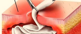
Passive
Passive drainage is currently carried out using perforated tubular systems made of polyvinyl chloride or thin tubes filled with gauze. Drains are positioned in such a way that they drain the liquid component from top to bottom. This mechanism of action is ensured by the force of gravity pressing on them.
Active
When performing active drainage of sealed tissue and epidermal damage, aspiration is used based on vacuum, provided by a special suction. This technique allows you to remove dead flesh, minimize the joining of wound edges, and reduce the likelihood of pathogenic microflora entering the cavity from the outside.
The drainage is installed in such a way that it removes the separated liquid from the bottom up, against the influence of gravity on it. It is necessary to take into account that this drainage is not used to remove growing hematomas.
Flow-flushing
Flow-flush type drainage is carried out using aspiration flushing with the installation of counter-perforated drainage systems. Medicine is injected into one of them, and transudate is removed from the damaged tissue through the second.
The drug can be administered into the drainage in a stream, drip, fractionally or continuously. Outflow is carried out according to the active or passive method. With the help of such drainage, pathogenic microorganisms do not enter the wound, transudate is completely removed from it, thus creating favorable conditions for healing and unfavorable conditions for bacteria.
Postoperative wounds are drained due to the high risk of developing inflammatory processes of purulent etiology. This situation is due to the fact that contamination occurs in the wound during surgery, represented by subcutaneous tissue and the impossibility of completely removing dead tissue.
Drainage must be installed by introducing counter-perforated systems into the cavity through the holes left from dialysis after surgery. Drainage is often prescribed in the case of removal of cancerous formations in the mammary gland, local hernias of the ventral type, amputation of the lower and upper extremities and surgical cleansing of a purulent focus in soft tissues.
After the purulent focus is opened, the doctor installs passive drainage, which is always done in such cases. The technique of installing drains is studied by operating nurses in the dressing room or operating room when working with patients with various types of wounds.
Which clinic's services do you prefer?
The best way to choose the right medical institution is to visit an information site that provides information on location, qualifications of personnel, types of diagnostics performed and treatment methods performed.
Our Help Desk for private clinics in Moscow “Your Doctor” has such a database, which makes choosing a medical center quick and convenient; in addition, the site’s capabilities include making an appointment with a specialist or calling a urologist at home, online.
Publication date: 2019-05-29