Causes of eye asymmetry
Facial asymmetry can develop immediately after birth. This means that the condition began while still inside the womb. Another type of disease is pathological, and occurs several months or years after birth.
Anomalies of the skull structure
Incorrect skull structure is inherited. Defects may include various irregularities and depressions:
- the right side is larger than the left, or vice versa;
- the affected area is excessively large;
- sloping forehead or back of the head;
- one cheekbone is larger than the other.
All these factors change facial features, making them asymmetrical.
Developmental disorder of the lower jaw
The most harmless condition is malocclusion. A person develops a jaw that is pushed forward, a disruption in the normal development of wisdom teeth, and a receding or overly large chin. The lower jaw is one of the largest parts of the face, so its violations affect a person’s appearance.
Muscle and connective tissue problems
If a person has weakness in the facial muscles, drooping of the eyelids, cheeks, and eyebrows occurs. Reduced quality of connective tissue contributes to sagging skin. If this condition is common on one side of the face, it will be asymmetrically lower than the other.
A typical picture of muscle and connective tissue disorders is one eye wide open, the other narrow.
If the muscle tissue disorder can be not only on the face, but also on the neck. If it is very tense, the person's head will lean in this direction.
Violation of the structure of the temporomandibular region
If a person has a disorder in the joint between the lower jaw and the temporal bone, movement is blocked when opening the mouth. It is formed unevenly; a person can open first to the right side, then to the left, and vice versa. If the condition is severe enough, the person must first move it, then open it.
Strabismus
Strabismus develops in one or both eyes. Defects form immediately after birth or after some time. The causes are various neurological and ophthalmological primary diseases. The coordinated work of the eyes is disrupted. If one organ of vision decreases in acuity, it gradually turns off its function.
Vision defects
There are various diseases that reduce vision function. Many of them form external changes. A person develops a cataract, a cataract, or a scar on one eye. At the same time, the second eye remains healthy, this forms asymmetrical facial features.
Mechanical damage and inflammatory conditions of nervous tissue
Such diseases lead to swelling. If the nerve is inflamed on only one side, it increases sharply in size. Additionally, a pain syndrome is formed. This is observed with mechanical damage, the motor activity of one side is impaired. A person retains facial expressions only on the healthy side.
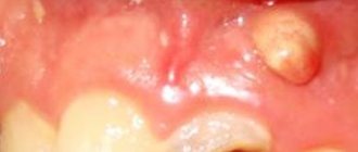
Absence of a large number of teeth
If the changes are only on one side, the cheek is pressed inward. The healthy part of the face remains normal. A person's face changes when talking. A lisp and a lack of pronunciation of certain sounds appear.
Bad habits
These include:
- squinting of one eye;
- Constantly chewing gum or foreign objects;
- sleep on one side.
Defects develop gradually. A bad habit affects muscle, connective and bone tissue. The location of muscles and bones relative to each other changes. If a large area is affected, the defect can only be corrected through surgery.
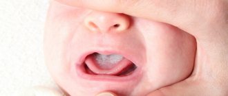
Reasons for the formation of anomalies in children
Often, an orthopedic doctor immediately after birth notes asymmetry of the skull in newborns. It is determined by the factors of the child’s development in the womb and the conditions of birth (the position of the fetus in the uterus, its passage through the birth canal).
Usually after 2-3 days the newborn’s skull takes the correct shape. If this does not happen, a thorough examination of the baby is necessary to identify the causes of the deformities.
In addition to the birth anomaly, most children under one year of age have facial disproportion due to infants being in one position for a long time.
Birth and infant asymmetry are natural types, and in most cases do not require medical intervention, except perhaps massage.
The pathological anomaly is more often observed in preschool children. It can be congenital or acquired, right-sided or left-sided.
Congenital asymmetry is a consequence of genetic factors and abnormalities in fetal development caused by the condition of the mother and embryo during pregnancy (intrauterine infections, abnormal position of the fetus, asphyxia, lack of nutrients, etc.).
These factors lead to disruption of the formation of soft tissues, cartilage and bones of the skull, uneven healing of sutures, etc.
The specific, most common congenital causes include the following:
- Hypertonicity of the masticatory muscles.
- Muscular dystonia (involuntary muscle contractions leading to impaired development of the TMJ and, often, crossbite).
- Crossbite (due to muscular dystonia or accelerated/slow development of one of the jaws).
- Cleft lip. An anomaly manifested by the presence of 1 or 2 clefts in the upper lip, leading to disruption of the shape of the nose and a depression in the middle area of the face.
- Stigmas of embryogenesis. Various etiological malformations of the fetus. They disrupt the symmetry of the dentofacial apparatus, nose, and skull.
- In addition to pathological factors associated with pathologies of the dentofacial apparatus, there are many other congenital diseases that lead to disruption of facial symmetry.
- These are torticollis, Sturge-Weber syndrome, craniofacial microsomia, etc. The dentist’s task is to differentiate them from pathologies of the dentofacial apparatus, and refer the patient to a doctor of appropriate specialization.
Acquired jaw asymmetry develops in the postnatal period under the influence of various external and internal factors:
- Malocclusion. The most common cause is crossbite, which develops due to bad habits and incorrect positioning of the child in the crib, stroller and at the table. A crossbite can result from slower or faster development of the upper or lower jaw.
- TMJ pathologies.
- Odontogenic and non-odontogenic tumors in the jaws (hard and soft odontoma, osteoma, salivary stones, etc.)
- Impacted teeth.
- Injuries to bone (fracture of jaw bones, skull and face) or muscle tissue.
- Birth injury.
- Periostitis.
- Periodontal inflammation.
Among the non-dental acquired disorders that can lead to an anomaly, the following should be mentioned: diseases of the ENT organs, eye pathologies, curvature of the spine, paresis of the facial nerve, stones in the salivary glands, tumors in the nasal cavity and paranasal sinuses.
How to recognize pathology?
The main goal of diagnosing ptosis of the upper eyelid is to establish the cause that led to the development of this disease.
- Assess the position and mobility of the eyelid.
- Assess the symmetry of eye movements.
- Assess eyebrow mobility.
- Determine the size of the eyelid fold.
- Determine the strength of the muscle that lifts the upper eyelid.
- Determine the presence of strabismus, amblyopia.
- Check your vision.
- Measure intraocular pressure.
If ptosis is caused by mechanical damage, the doctor should check the bone structures for damage. To do this, you need to conduct a survey x-ray. If there is a suspicion that ptosis has appeared due to problems with the nervous system, then a computer or magnetic resonance imaging of the brain is performed, and a referral to a neurologist and neurosurgeon is given.
- General clinical blood test. It will show leukocytosis and accelerated erythrocyte sedimentation characteristic of inflammation.
- Biochemistry of blood. This laboratory test will help identify cytolytic enzymes. Their level increases if there is destruction of cells and tissues in the body.
- General ophthalmological examination. As part of it, visual acuity is measured using Sivtsev tables.
- Perimetry. This is a method for determining peripheral vision and the degree of its impairment.
- Ophthalmoscopy. Using a slit lamp, the technique examines the condition of the fundus of the eye, the exit point of the optic nerve, the vascular network, and the anterior and posterior chambers of the eye.
- Magnetic resonance and computed tomography of the head. These instrumental methods help to identify hemorrhages, foci of destruction or neoplasms in the cranial cavity.
- Measurement of cerebrospinal fluid pressure. At the same time, cerebrospinal fluid should be collected for microbiological examination.
We invite you to familiarize yourself with the instructions for using Diklo-F eye drops
start of treatment
Treatment of facial nerve neuritis in our center begins with an appointment with a vertebroneurologist. The initial appointment is conducted by the candidate of medical sciences, chief medical doctor Lyudmila Ivanovna Mazheiko.
At the appointment, the vertebroneurologist will conduct a special diagnosis to establish the level of damage to the facial nerve, determine the type of disorder in the surrounding tissues, prescribe additional examinations if necessary, and develop an individual treatment plan.
Appointments with a vertebroneurologist are carried out by appointment. To begin treatment, make an appointment.
Symptoms
With asymmetry, the right and left sides are different. If they are not natural, the change is observed by 1-2 mm, no more. If the condition is caused by a pathological factor, the asymmetry is visible to the naked eye. Many patients are not satisfied with this, so they turn to surgeons.
The following asymmetrical features are distinguished:
- violation of the location of folds on the face (the area between the lips and nose, folds on the eyelids);
- when smiling, lips curl downwards rather than upwards;
- one-sided smile;
- greater width of one eye compared to the other;
- impaired muscle mobility on one side of the face;
- the formation of discomfort and pain in the damaged area;
- speech defects, lack of pronunciation of certain sounds;
- if a prolapse of connective muscle tissue occurs, the person looks exhausted and tired;
- abnormal location of the palpebral fissure.
Facial asymmetry can not only appear externally, but also lead to pain. In this case, urgent treatment is required. Timely consultation with a doctor reduces the risk of complications.
One side of the face is larger than the other: normal or pathological
If you notice that the sides of your face are different, first of all, you need to determine whether it is pathological or normal. To do this, you need to know the symptoms of the pathology.
If there is facial asymmetry, parchment skin may be present. This symptom is associated with a genetic disease.
Asymmetry, which is considered normal, should not be too obvious. If this is a normal facial structure, then the difference is noticeable only upon careful examination.
Symptoms of abnormal facial asymmetry:
- The affected side is swollen and the tissue may be hard;
- The corners of the mouth and cheek on the affected side are drooping and drooping;
- The affected half of the face loses mobility;
- The folds on the forehead and nose suddenly smooth out sharply;
- The eye on the affected side becomes narrower, or, conversely, larger;
- The eyes, nose and lips on the affected side lose mobility;
- There are speech disorders;
- The affected part is painful.
Even one of these signs suggests that asymmetry is the result of a serious problem. In this case, it needs to be treated.
How to recognize pathology?
It is not difficult to notice the asymmetry of the folds of the face. But recognizing the reason why one eye is smaller or larger than the other is not so easy. In newborns, asymmetrical palpebral fissures are congenital anomalies of the development of facial muscles and skin. But the cause can also be intrauterine and birth injuries to the head, in which hemorrhage occurs in the intracerebral cavities.
Therefore, to diagnose pathology, it is important to know the general history and protocol for pregnancy management. As for adults, the difference in eye size is visible to the naked eye. It is accompanied by concomitant clinical symptoms. It includes headaches, loss of consciousness, neurological disorders, unsteadiness when walking, and the inability to read text or look at pictures.
Preventive measures
Prevention of any dental diseases consists of following a number of rules that are relevant for almost every person. And although, unfortunately, they do not provide a complete guarantee of preventing dental diseases, they reduce the risk of their development. And if they have already appeared, they speed up and facilitate treatment.
This is the fight against bad habits of children, teaching them proper oral care. Quitting smoking and foods that are harmful to teeth. Using mouthguards when playing sports. And, perhaps most importantly, regular visits to the dentist for preventive purposes.
In the video, experts talk about the causes of asymmetry of the face and jaws.
Diagnostics
Diagnosis of the patient’s condition consists of several stages:
- Anamnesis collection. This is data obtained from the patient or his close relatives. Based on them, the doctor suggests a diagnosis.
- General inspection. Assessment of the condition of skin surfaces and muscle tissue. Doctors identify neurological symptoms, the degree of mobility of facial tissue, the presence of injuries and diseases.
- Lab tests. With the help of testing, an infectious focus and inflammation are identified. A general blood and urine test and blood biochemistry are prescribed.
- Assessing facial symmetry using measuring instruments. The doctor reveals the exact proportions, damage and deformations are detected. If a person has pathological asymmetry, it is formed when the location of parts of the face is disturbed by 3 mm or more.
Based on diagnostic data, the doctor makes a reliable diagnosis. Next, treatment is carried out.
our treatment
Our center provides treatment for peripheral neuritis, which is caused by pinching of the facial nerve in the area where it exits the cranial cavity.
We resolve dense scars, eliminate tissue degeneration and relieve muscle spasm around the pinched facial nerve. This leads to its release and restoration of normal conduction of nerve impulses.
To treat neuritis of the facial nerve, we use a special technology using the method of muscle mesotherapy.
If necessary, a course of nutraceuticals and osteopathy sessions are prescribed.
The main condition for successful treatment is early seeking help.
Why does facial asymmetry occur?
There are two main reasons for the development of such a visual deviation: congenital or acquired. These include: • defect in the formation of the temporomandibular joint; • abnormal structure of the skull; • pathology of the formation of the neck muscles (on one side); • general underdevelopment of the lower jaw; • various defects in connective tissue or muscles.
Acquired asymmetry can occur as a result of: • inflammation, injury or pinching of the endings in the facial nerve; • visual defects due to strabismus; • bite pathologies, problems with teeth or jaw; • in the absence of teeth on one half of the jaw; • various facial and jaw injuries, facial bone fractures;
In children, this defect develops with muscular or neurogenic torticollis, congenital shortening on one side of the sternocleidomastoid muscle.
Facial asymmetry also occurs with aging of the entire human body or with age-related diseases.
Symptoms
In the presence of natural asymmetry, the difference in size between the right and left sides of the face is not pronounced (about 2-3 mm), only the overall proportionality is slightly disturbed.
Most often, it is the right half that is larger and wider, and the left half is delicate and has a smoother appearance, but these deviations are not pronounced, so there is no reason to worry.
If there is neuropathy of the facial nerve, then the indicated differences in the symmetry of the face are more visible, and their clinical picture is more pronounced: • in the affected half of the face there is weakness in the facial muscles and it becomes like a mask; • nasolabial and frontal folds are less noticeable; • the palpebral fissure increases;
• the corners of the mouth fall down; • the affected part acquires a pained or crying expression; • it is difficult to move the facial muscles, close your eyes, wrinkle your forehead; • speech and articulation are impaired, it is difficult to eat, because it just falls out of your mouth; • painful sensations are noted in the affected nerve.
Facial asymmetry in children occurs due to muscular torticollis or when lying in a crib on one side for a long time. Most often, part of the face is smoothed, the jaw angle is smaller, the head is tilted towards the affected side, and parts of the face (in the area of the cheek and head) are flatter.
The diagnosis is based on identifying facial asymmetry using a visual examination (presence of pathology of muscles, teeth, nerves), asking the patient about heredity and possible injuries.
Additionally, facial proportions are measured with special instruments, which are expressed in millimeters and degrees. A deviation of more than 3 mm or 5 degrees is considered pathology.
In order to prove the presence of asymmetry in the face, an image of a person’s face is taken, composed of its two right or left halves. The result is two absolutely symmetrical portraits, which often differ from the person’s real face.
We will describe the methods that are used to correct the main parts of the face.
1. Eyebrow asymmetry. The defect develops when the facial nerve, its frontal branch, is damaged. Also, its manifestation is facilitated by hyperfunction of the levator brow muscle. In order for symmetry to be restored, drugs are injected into the muscles (frontal, corrugator brow): Lantox, Botox, Dysport.2. There are numerous muscles around the mouth that work in different directions. Asymmetry around the mouth is corrected by introducing Botox, Lantox into the area of the muscle that depresses the lower lip and the area of the mouth
3. For asymmetry of the eye slits in the presence of the “big eye versus small eye” symptom, medications Lantox, Botox, and Dysport are used. A small dose of botulinum toxin can also be used; the drug is injected into the lower part of the orbicularis oculi muscle, 1 mm away from the edge of the eyelashes.
We all understand that the body of any person cannot be absolutely symmetrical, so there is no need to worry about this. But when eye asymmetry occurs, this already indicates the presence of pathology and you should definitely consult a specialist for advice.
When eye asymmetry occurs, the reasons may be the following: 1. The presence of infectious eye diseases, in this case with slight swelling of the eyes becoming larger (for example, barley or adenoviral conjunctivitis). The tumor occurs due to an attack by pathogenic bacteria, which leads to inflammation of the mucous membrane of the eye.
We invite you to familiarize yourself with Prenacid - instructions for use, description, reviews, analogues
Therapy and diagnosis are carried out only by an ophthalmologist, because self-medication can lead to worsening of the condition. 2. Injuries. It should be borne in mind that even a small injury or bruise in the eye area can cause swelling. Treatment of injuries is carried out depending on their formation, but only by a specialist. 3.
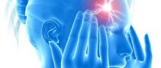
If swelling of the eye and a change in its size occurs without a reason, this situation is considered the most dangerous. This defect can lead to neurological disease or even more serious illness. 4. Bulbar syndrome is a dangerous pathology that is associated with the condition of the brain.
At the initial stage of development of the disease, asymmetry of the eyes is noted. It is at this moment that you should consult a doctor to prevent the development of a disease when one of the eyes does not function properly (for example, paralysis). Most often, in parallel with a change in the size of the eye, deformation of the eyelid, incomplete closure of the eyes, and a change in the shape of the eye occur.
Let's draw conclusions: if you have a visually visible difference in the size of your eyes, you should pay attention to the accompanying symptoms (swelling of the eyelid, discharge with purulent contents, redness of the eye mucosa). If the deformity is accompanied by attacks of pain, most often a diagnosis of “neuralgia” can be made.
All pathologies must be diagnosed by a specialist, so do not delay a visit to an ophthalmologist.
Diseases
The cause of acquired asymmetry can be various diseases. “First of all, dysfunction of the facial nerve,” says Irina Ivanova. — With this disease, weakness of the facial muscles develops, the corner of the mouth droops, the upper eyelid droops, the palpebral fissure becomes wider, the nasolabial folds smooth out, and the face on the affected side acquires a pained expression. Injuries, fractures, especially displaced ones, and the consequences of unsuccessful plastic surgeries are also often the reason why the two sides of the face look different.”
According to experts, you can encounter the problem of facial asymmetry at any age. But over time, unfortunately, it becomes more and more noticeable. Simply due to the fact that bad habits live with us longer. “However, do not despair,” says Irina Ivanova. — Asymmetry, as a rule, can be corrected. First of all, it is necessary to eliminate the causes: contact a neurologist, dentist, or orthodontist. There is physiotherapy, gymnastics for facial muscles, massage. To correct violations of proportions and volumes, cosmetologists use contour plastic surgery. Botulinum therapy is also used successfully. In the most severe cases, surgical correction is possible.”
Treatment
There are various treatment methods. They are selected by the doctor individually for each patient. He, in turn, chooses the most appropriate method:
- Facial correction using cosmetics. If it is necessary to narrow the area, it is darkened. If you expand it, it brightens it up. Corrective foundation creams and pencils are used for this.
- Maxillofacial or plastic surgery. They contact a plastic surgeon, who individually selects the type of operation for the patient.
- Visiting the dentist. He can install crowns or new teeth.
- The use of gymnastics and massage complexes for reduced muscle tone.
- Treatment by a neurologist to eliminate the inflammatory condition of the nervous tissue.
Facial asymmetry can be minor or severe. In the latter case, patients want to change external parameters. To do this, they turn to doctors of various specialties: dentist, neurologist, ophthalmologist, plastic or maxillofacial surgeon. After proper treatment, facial features become symmetrical.
Treatment methods
The variety of causes and nature of the disease determines a wide range of possible treatment methods - from cosmetic to surgical.
Depending on the root cause, cosmetic, conservative, orthodontic and surgical treatment can be used. As well as massage and therapeutic exercises.
Massage
Massage has multiple therapeutic effects:
- Relaxes and tones muscles, relieves hypertension.
- Activates blood and lymph circulation in the affected area, relieves swelling and inflammation.
- Stimulates nerve fibers.
- Reduces pain.
Manual therapy in children is more effective than in adults due to the plasticity of children's muscles. The earlier the anomaly is diagnosed, the more effective manual therapy is.
Cosmetology procedures
Cosmetic treatment consists of getting rid of hypertonicity of the masticatory muscles, which causes facial asymmetry. Technologically, the procedure involves injections of Botox into the masticatory muscles.
The drug relaxes overstrained muscle fibers, straightens the face, returns it to harmonious proportions, and gives a calm and confident expression.
Conservative therapy
Conservative therapy is determined by the nature of the pathology and provides for the treatment of pulpitis, caries, periodontitis, periodontitis and other diseases of the teeth and periodontium, which make it possible to do without surgical intervention.
Analgesics (for pain), antibiotics (for infections), antiseptics (for local inflammation and wounds on the mucous membranes), NSAIDs (for severe inflammation) may be prescribed.
Orthodontic correction
Orthodontic treatment is used for crossbites, narrowing of the jaws, and other dental anomalies. Treatment is carried out with the help of splints, mouthguards, extra- and intraoral devices, braces and other fixed and removable orthodontic appliances.
Surgical intervention
The type of surgical intervention is determined by the nature of the disease and is divided into:
- for dental (removal of tumors and cysts, removal of teeth, opening of abscesses);
- maxillofacial (lavage, arthroscopy, phlegmonectomy, TMJ endoprosthetics, rhinocheilognatoplasty);
- eliminating the consequences of injuries (splinting and regeneration of jaw bones, tying with a ligature);
- otolaryngological (ectomy of paranasal sinus cysts, etc.).
If asymmetry is caused by damage to the brain and meninges, according to indications, ectomy of tumors and abscesses, transcranial or endoscopic removal of hematomas, areas of the brain crushed by TBI, and other surgical protocols are performed.
Stroke: treatment
Therapy for circulatory disorders should begin as early as possible, since early initiation increases the likelihood of successful restoration of muscle function, including facial muscles. Therapy directly depends on the type and cause of the stroke.
For cerebral ischemia, drugs are prescribed that resolve the blood clot and restore normal blood circulation (acetylsalicylic acid, thrombolytic therapy).
In case of hemorrhagic stroke, it is necessary to stop bleeding in the brain tissue, which is achieved with the help of antifibrinolytic therapy (alpha-aminocaproic acid).
However, it is not only drug therapy that plays a role. If the patient’s condition is satisfactory, physical therapy and massage should be started as early as possible to restore muscle function.
Stroke: symptoms
If your face becomes distorted during a stroke, you should immediately consult a doctor, since time plays a very important role here. Therefore, you need to know the following signs that will help you suspect the development of a stroke:
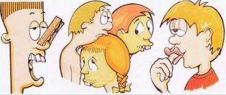
- In the vast majority of cases, only the lower half of the face is characterized by asymmetry, which is manifested by a drooping corner of the mouth, smoothness of the nasolabial fold, deviation of the tongue to one side, and the inability to show teeth or stick out the tongue completely.
- Paralysis (complete immobilization) or paresis (weakness) of a limb, typically involving unilateral damage to the arm and/or leg. It is difficult for the patient to raise his arm or he cannot stand up; with leg paresis, walking is possible, but difficult.
- Speech impairment, and the patient may have damage to both the speech understanding center, located in the frontal lobe of the brain, and the pronunciation center, located in the temporal lobe. In the first case, the patient pronounces words and sentences absolutely normally, but does not completely understand the meaning of what is being said to him. In the second case, he understands everything, but is either completely unable to say anything, or speaks separate incoherent words.
If you notice any of the listed symptoms in yourself or those around you, do not hesitate, call an ambulance immediately!
Facial nerve paralysis: treatment
What to do if your face is distorted due to facial paralysis? There are a number of drug and non-drug treatment methods that will help restore the previous appearance and function of the facial muscles:
- corticosteroids - relieve inflammation of the facial nerve;
- antiviral or antibacterial agents, if the presence of an infectious process is confirmed;
- surgical intervention for a diagnosed brain tumor;
- massage;
- a set of exercises for the facial muscles;
- physiotherapy;
- moisturizing eye drops on the affected side, antibiotic ointments to prevent infection.
Blepharospasm: causes and symptoms
Another condition that can cause the face to become distorted is blepharospasm, an involuntary contraction of the orbicularis oculi muscle surrounding it.
Main reasons:
- Facial paraspasm is a disease characteristic mainly of older people. Its exact cause is unclear, but it is believed that paraspasm occurs due to an imbalance of parts of the nervous system.
- Parkinson's disease.
- Meningitis.
- Multiple sclerosis.
- Inflammation of the eyes (keratitis, conjunctivitis).
- Sinusitis is inflammation of the paranasal sinuses.
With blepharospasm, only the upper part of the face is characterized by asymmetry: the palpebral fissure gradually narrows, and sometimes the eye may suddenly close its eyes. This brings a lot of inconvenience to the patient.