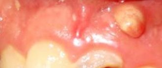
Causes
The main cause of the disease is infection with the Candida fungus, an opportunistic pathogen that is widespread among humans and animals. Candida is a yeast-like unicellular fungus that is part of the normal microflora in a significant proportion of healthy people. Most often it can be found on the surface of various mucous membranes of the body, for example, in the intestines, mouth, pharynx and tonsils. Today, about 150 varieties of this microorganism are known, 20 of which are capable of causing diseases in humans. The destructive effect of Candida on the mucous membranes and other tissues of the human body is due to the release of a large number of enzymes that break down proteins, fats and other cell components. As a result of this, symptoms characteristic of candidiasis such as burning, dryness, and soreness occur.
When unfavorable conditions occur, Candida becomes covered with a special protective shell, which helps the fungus survive in the external environment and travel from host to host. Infection can occur in various ways - airborne, household contact, intrauterine. Newborn children are infected, as a rule, from medical personnel, or during childbirth, when passing through an infected birth canal.
At an early age, the child’s body does not have a developed immune system, if only for the simple reason that he has had virtually no contact with infectious agents and has not developed the appropriate antibodies. This is why children under one year of age are especially prone to developing candidiasis. However, this disease also occurs in older children. In this case, its appearance and development is often preceded by the formation of multiple carious lesions of the teeth. Caries, which is a constant source of infection, contributes to the weakening of local and general immunity, resulting in the rapid and active proliferation of opportunistic and pathogenic microflora. Various fungi of the genus Candida also fall into this category.
Factors that can provoke the development of candidiasis in a child’s mouth are also various concomitant diseases, such as diabetes mellitus, diseases of the gastrointestinal tract, acute or chronic infections, and other somatic ailments. Poor oral hygiene, taking antibiotics, hormones and immunosuppressants, and insufficient or unbalanced nutrition also increase the risk of developing candidiasis.
For active reproduction of the fungus, certain conditions are necessary - in particular, this microorganism is most active at temperatures from 30 to 37 degrees Celsius. The level of acidity that is optimal for the growth of Candida occurs in a person’s mouth when consuming large amounts of sweets, flour and other foods containing many simple carbohydrates.
Recurrent urogenital candidiasis: features of diagnosis and treatment
A.M. SAVICHEV
, Doctor of Medical Sciences, Professor,
E.V.
SHIPITSYNA , Doctor of Biological Sciences,
Research Institute of Obstetrics, Gynecology and Reproductology named after.
BEFORE. Otta, St. Petersburg Vulvovaginal candidiasis is the most common manifestation of urogenital candidiasis. The disease is widespread among women of reproductive age. In 5-10% of cases the disease has a recurrent form. The article discusses the issues of diagnosis and treatment of urogenital candidiasis, with special attention paid to the problems of recurrent vulvovaginal candidiasis.
Urogenital candidiasis is a widespread disease that can occur in both sexes, but most often affects women of reproductive age. According to the international classification of the X revision (2007), there are:
• B37.3 Candidiasis of the vulva and vagina. • B37.4 Candidiasis of other urogenital locations.
Experts suggest the following clinical classification:
• sporadic urogenital candidiasis (observed in patients with normal immunity and characterized by a clinical course of moderate severity), • recurrent urogenital candidiasis (characterized by the presence of 4 or more episodes of urogenital candidiasis within 1 year).
Candidiasis of the mucous membrane of the vulva and vagina - vulvovaginal candidiasis (VVC) is the most common form of urogenital candidiasis. The frequency of registration of VVV has almost doubled over the past 10 years and currently accounts for 30-45% of the structure of infectious lesions of the vulva and vagina. According to researchers, 70-75% of women have at least one episode of VVC during their lives, while in 5-10% of cases the disease becomes recurrent. CVC is rare in girls before menarche, but by the age of 25, about 50% of women, and by the beginning of menopause, about 75% of women have at least one episode of the disease diagnosed by a doctor. It is also known that VLE almost never occurs in postmenopausal women, with the exception of women receiving hormone replacement therapy [1–3].
Along with the clinically pronounced disease, there is asymptomatic colonization of the vagina with yeast-like fungi. It should be emphasized that about 20% of healthy women are carriers of yeast-like fungi in the vagina, which does not require treatment. Candidiasis of the urinary system (urethritis, cystitis, pyelonephritis) is also possible. According to modern classifications, urogenital candidiasis does not apply to sexually transmitted infections, which, however, does not exclude the possibility of candidal balanoposthitis in men who are sexual partners of women with CVV [1–3].
Etiology
Yeast-like fungi of the genus Candida are widespread in nature. In humans, they often contaminate the skin and mucous membranes. Risk factors contributing to the development of candida inflammatory process are hormonal imbalance, disorders of systemic and local immunity, changes in the normal microbiocenosis of cavities due to irrational use of antibacterial drugs, etc. Fungi of the genus Candida
They predominantly colonize the organs of the gastrointestinal tract, in different parts of which several species of yeast-like fungi are found in 50-60% of observations.
Candida is found in the oral cavity in 30% of adult women. It is believed that orogenital sexual intercourse contributes to the colonization of the male genital organs by fungi of the genus Candida
[2, 3].
When examining the vaginal discharge of patients with vulvovaginal candidiasis, the species most often found is Candida albicans
(about 70-90%).
Other species of the genus Candida - C. tropicalis, C. parapsilosis, C. kefir, C. krusei, C. lusitaniae, C. guilliermondii, C. glabrata, C. lambica
are isolated in 10-30% of observations.
In recent years, C. glabrata has been described as a causative agent of nosocomial infections. The fungi C. glabrata
, characterized by the absence of the ability to form pseudomycelium, can also be commensals of human mucous membranes.
Proportion of isolation of C. glabrata
among other
Candida spp
.
from the oral cavity, vagina, intestines and feces reaches 9% of observations. Candidiasis infection associated with C. glabrata
is predominantly endogenous, but exogenous infection from the external environment is also possible: soil, water, excrement, some food products, etc. [2, 3].
Clinical picture
VVV is characterized by the formation of a whitish coating on the hyperemic mucous membrane of the vulva and vagina. A characteristic crumbly “curdled” white discharge appears. Patients are bothered by painful itching and burning. There may be a burning sensation in the vulva when urinating and pain during sexual intercourse. Infection of the vulva and vagina by yeast-like fungi is characterized by great persistence and a tendency to relapse. With recurrent VVV, dryness, atrophy, lichenification in the affected area, and scanty whitish vaginal discharge may be observed.
Complications of urogenital candidiasis in women include the development of PID, and possible involvement of the urinary system in the pathological process (urethrocystitis). Against the background of urogenital candidiasis, the incidence of complications during pregnancy increases, and the risk of ante- or intrapartum infection of the fetus increases. Fetal candidiasis can lead to intrauterine death and premature birth.
In newborns, candidiasis can occur in the form of a localized infection (conjunctivitis, omphalitis, lesions of the oral cavity, larynx, lungs, skin) and disseminated lesions that develop as a result of candidemia. In the postpartum period, women may develop candidal endometritis [1–3].
Diagnostics
To diagnose urogenital candidiasis infection, microscopic and cultural methods are used with the isolation of yeast-like fungi, their species identification, determination of the sensitivity of fungi to antimycotic drugs, and molecular biological methods (PCR) [3]. It is necessary to remember the possibility of rapid proliferation of the fungus and begin the study as soon as possible after aseptic collection of the material. A vaginal swab or 10 µl inoculation loop is used to collect material. The material is collected from the vaginal vault and the lateral walls of the vagina. For microscopic examination, the material is placed on two glass slides; for cultural and molecular biological diagnostics, it is placed in a special transport medium. Microscopic method
is preferable for diagnosing urogenital candidiasis, since 20% of healthy women have candida in the vagina, which will grow during culture, which can lead to an unfounded diagnosis. For microscopy, unstained preparations are used, as well as preparations stained with Gram, Romanovsky-Giemsa, and methylene blue. The diagnosis is based on the detection of fungal elements: single budding cells, pseudomycelium, and other morphological structures (blastoconidia, pseudohyphae).
Culture method
necessary for chronic relapsing course of the disease, for identifying yeast-like fungi (especially for identifying species not related to C. albicans), when studying the effect of medicinal antifungal drugs, in the atypical course of the disease, when the etiological role of other possible pathogens is excluded. The need for species identification of the pathogen in practical terms is due to the resistance of some Candida species to antimycotic drugs. To confirm the diagnosis of recurrent CPV, it is necessary to isolate the microorganism in culture with identification of rare species, especially C. glabrata. C. glabrata and other non-C. albicans Candidia species are isolated from 10% to 20% of patients with recurrent VVC, but C. glabrata does not form pseudohyphae or hyphae and is therefore difficult to visualize microscopically. Traditional treatment regimens are not as effective against these species compared to C. albicans [3–4].
Molecular biological methods
(PCR) are aimed at detecting specific fragments of DNA and/or RNA and can be used for species identification of fungi of the genus Candida. The need for species identification of the pathogen in practical terms is due to the resistance of some Candida species to antimycotic drugs. Highly sensitive and specific. They have limitations due to the possible presence of yeast-like fungi in normal conditions.
Treatment
To prescribe rational treatment, it is necessary to take into account the clinical form of candidiasis, its prevalence and identified predisposing factors (general and local). If there are obvious clinical manifestations of this disease, treatment can be prescribed without additional laboratory testing. In case of recurrence of the process, it is necessary to conduct a laboratory test to determine the sensitivity of candida to antifungal drugs.
The principles of treatment of urogenital candidiasis should be the following: eradication of the pathogen, elimination of risk factors, elimination of allergenic factors, strengthening of the body's nonspecific immunological reactivity. Treatment regimens for urogenital candidiasis are presented in the table. In sporadic VVV, antifungal azole derivatives (fluconazole, clotrimazole, etc.) and polyene antibiotics (natamycin) are often used externally in appropriate forms: suppositories, vaginal balls, vaginal tablets and cream with a special applicator [1, 2, 4].
| Table. Treatment regimens for urogenital candidiasis | |
| Treatment of candidiasis of the vulva and vagina | — Natamycin vaginal suppositories 100 mg once a day for 6 days or — clotrimazole vaginal tablet 200 mg once a day before bedtime for 3 days or 100 mg once a day before bedtime for 7 days or — clotrimazole 1% cream 5 g once a day intravaginally before bedtime for 7-14 days or - itraconazole vaginal tablet 200 mg once a day before bedtime for 10 days or - miconazole vaginal suppositories 100 mg once a day before bedtime for 7 days or — butoconazole 2% cream 5 g once a day intravaginally before bedtime for 3 days or — itraconazole 200 mg orally once a day for 3 days or — fluconazole 150 mg orally once |
| Treatment of candidal balanoposthitis | — Natamycin 2% cream 1-2 times a day for 7 days or — clotrimazole 1% cream 2 times a day for 7 days or — miconazole 2% cream 2 times a day for 7 days or — itraconazole 200 mg orally 1 once a day for 3 days or - fluconazole 150 mg orally once |
| Treatment of recurrent urogenital candidiasis | After the main course of therapy, including systemic and local antimycotics, maintenance therapy is recommended for 6 months. one of the drugs: - fluconazole 150 mg orally once a week or - clotrimazole 500 mg vaginal tablet once a week - |
| Treatment of candidiasis not caused by Candida albicans | — Local therapy with azoles for 7–14 days — boric acid 600 mg in gelatin capsules vaginally once a day for 2 weeks — if necessary — maintenance therapy with nystatin in vaginal suppositories 100,000 units once a day |
| Treatment of pregnant women | Locally acting antimycotic agents are used: - natamycin vaginal suppositories 100 mg once a day for 3-6 days (approved for use from the 1st trimester of pregnancy); or - clotrimazole vaginal tablet 100 mg once a day before bedtime for 7 days or 1% cream 5 g once a day intravaginally before bedtime for 7 days (approved for use in pregnant women from the 2nd trimester) |
For recurrent VVC, a longer course of intensive therapy with fluconazole is used - local therapy for 7-14 days or 3 doses of 150 mg orally with an interval of 3 days [1, 2, 4]. Also, for recurrent vulvovaginal candidiasis, maintenance therapy is recommended for 6 months: 150 mg of fluconazole orally every week. The effectiveness of this approach for the prevention of recurrent VVC was confirmed in a recent meta-analysis [5]. If oral administration is not possible, local therapy is prescribed in intermittent courses. It is believed that 30–50% of women experience relapses when maintenance therapy is stopped [4].
For the treatment of recurrent VVV associated with yeast-like fungi other than C. albicans
, therapy using boric acid is proposed [4, 6].
Literature data confirm that boric acid is a safe, inexpensive alternative drug for the treatment of recurrent VVC caused by non- C. albicans candida
[6].
The effectiveness of routine treatment of sexual partners is controversial. Scientific studies conducted on the basis of the principles of evidence-based medicine have established that the frequency of relapses of urogenital candidiasis in women does not depend on the preventive treatment of sexual partners. If a woman’s sexual partner develops symptoms of candidal balanoposthitis and urethritis, it is advisable to conduct an examination and, if necessary, treatment [1, 4].
Conclusion
The recurrent form of VVV occurs in 5-10% of cases. To confirm the diagnosis of recurrent VVV, it is necessary to isolate the microorganism in culture with identification of rare species, especially C. glabrata, since traditional treatment regimens are not so effective against these species. When treating recurrent VVC, a longer course of intensive therapy and maintenance therapy are required.
Literature
1. Russian Society of Dermatovenereologists and Cosmetologists. Clinical recommendations for the management of patients with sexually transmitted infections and urogenital infections. Moscow: Business Express, 2012, 112 p. 2. Savicheva A.M. Diagnosis and treatment of urogenital candidiasis. Difficult Patient, 2006, 4(9): 28-32. 3. Savicheva A.M., Kisina V.I., Sokolovsky E.V. and others. Vulvovaginal candidiasis. Methodological recommendations for doctors. St. Petersburg: Publishing House N-L, 2009, 88 p. 4. Centers for Disease Control and Prevention. Sexually Transmitted Diseases Treatment Guidelines, 2010. MMWR, 2010, 59 (RR-12). 5. Rosa MI, Silva BR, Pires PS, et al. Weekly fluconazole therapy for recurrent vulvovaginal candidiasis: a systematic review and meta-analysis. Eur J Obstet Gynecol Reprod Biol., 2013, 167(2): 132-136. 6. Iavazzo C, Gkegkes ID, Zarkada IM, Falagas ME. Boric acid for recurrent vulvovaginal candidiasis: the clinical evidence. J Womens Health (Larchmt), 2011, 20(8): 1245-1255.
Source:
Medical Council, No. 9, 2015
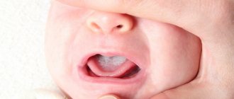
Symptoms of the disease
As a rule, the highest concentration of fungus is found on the surface of the inside of the cheeks, tongue, pharynx and tonsils, and palate. At an early stage of the development of the disease in children, redness of the mucous membranes is observed; they become swollen, the sensitivity of the mucous membrane increases, which is why children are often irritable, capricious, and their appetite and sleep are disturbed. The primary episode of the disease usually occurs in a more acute form; Patients may experience general symptoms: fever, headaches and dizziness, nausea or vomiting, deterioration in general health.
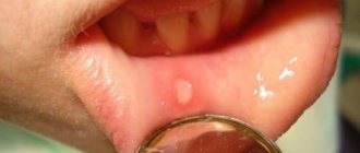
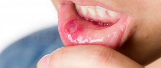
Some time after the onset of the disease, white grains begin to appear on the surface of the mucous membranes of the oral cavity, which gradually increase in size and merge together, forming plaques, and then plaque covering large areas of the mucosa. During this stage, patients suffer from severe dryness and swelling in the mouth. The mucous membranes become painful. Burning, itching and other unpleasant sensations are most often associated with the development of an allergic reaction caused by the proliferation of pathogenic microorganisms and the release of toxins during their life processes. The pain intensifies when eating and swallowing food, when eating hot, sour, spicy foods and drinks. Infants often refuse to eat.
The plaque formed during candidiasis has a very characteristic appearance and resembles milk films or remains of cottage cheese. It consists of destroyed cells of the mucous membranes, food debris, bacterial mass, fibrin and keratin. Flakes, scales and plaque can also appear outside the mouth - on the edges of the lips. The spread of infection to the lips is called candidiasis. Because of this disease, the skin in the corners of the lips dries out and cracks; In some cases, the infection affects the entire surface of the lips, resulting in cracking and peeling.
In mild and moderate forms of the disease, plaque is easily scraped off, and underneath it, areas of erosion (ulceration) or maceration (softening) of the mucous membrane are found. A longer course of the disease and the lack of adequate treatment leads to deep damage to the mucous membranes, as a result of which blood appears on their surface, turning the plaque brown or brownish.
How to avoid thrush in a child:
- the most important thing is strict adherence to child and personal hygiene;
- you also need to make sure that you yourself do not suffer from thrush;
- timely hygiene of the child after defecation;
- do not lick baby pacifiers (as the risk of fungal infection increases), you should always have several sterile pacifiers in stock;
- do not bathe your baby in a shared bathroom (this can even lead to the development of other diseases);
- You should avoid keeping your child in a diaper or nappy for a long time (this will prevent the growth of fungi);
- regularly check the condition of the child’s genitals: there is no redness or discharge;
- For preventative purposes, it is necessary to regularly sterilize baby bottles and nipples.
If a pathology caused by fungi is suspected, in particular if there is damage to the skin, hair, or nail plates, skin and nail scales are taken for examination in our medical center (the procedure is painless and safe).
To clarify the diagnosis, other types of studies (blood, feces, urine) may be required. Treatment of any disease should be carried out strictly under the supervision of a doctor. It is very important to control the treatment process, since fungi grow slowly in the human body and therefore the positive effect is not immediately visible. And the disappearance of the manifestations of the disease does not mean complete recovery, because the remaining elements of the fungus can give rise to a new growth of the disease.
Treatment of candidiasis
The main method of treating the disease is the use of local antifungal and antibacterial agents, as well as antibiotics. Antimycotic (antifungal) drugs can not only eliminate the symptoms of the disease, but also destroy the fungus in the oral cavity, as well as in other organs and tissues. For this purpose, antibiotics of the polyene series (levorin, nystatin, etc.), as well as clotrimazole, econazole and other imidazoles, are used. Fluconazole, Diflucan, and Nizoral also have a pronounced antifungal effect.
A significant part of antibiotics has a number of side effects and has a negative impact on the immune system. Therefore, when treating children, antibiotics are used only in the most severe cases, as well as in cases where there is a risk of complications and generalization of the process. At the initial stages of the disease and with a mild course of the disease, preference is given to local remedies. We recommend using ASEPTA series rinses, which contain chlorhexidine and other active ingredients that have antimicrobial and anti-inflammatory effects. To remove plaque, you can use special ASEPTA Baby wipes. The individually packaged finger wipe is made of hypoallergenic materials and allows you to carefully clean the mucous membranes without the risk of damaging them or causing additional infection. It is also recommended to regularly treat the oral cavity with Lugol or silver solution. This procedure must be repeated every three hours.
For the prevention and treatment of candidiasis in children, diet and oral hygiene are also of great importance. It is recommended to exclude or strictly limit the amount of food containing simple carbohydrates - these are, first of all, confectionery and flour products. Make sure your child brushes his teeth regularly - it is best to use a special toothpaste for this, for example - ASEPTA Baby, Kids, Teens. The components contained in these pastes help prevent caries and, as a result, reduce the infectious load on the child’s immunity.